Suppurative osteomyelitis in pig carcasses
During post-mortem inspection of pig carcasses suppurative lesions in the bones of the spine are relatively frequently observed:
This lesion is known as suppurative osteomyelitis which means purulent inflammation of the bone and vertebral bone marrow (both structures are always involved).
Other nomenclatures associated with this type of lesion are:
- Osteitis: generic term referring only to inflammation of the bone.
- Spondylitis: Inflammation affecting only the vertebral body.
- Diskospondylitis: inflammation of the intervertebral disc and the vertebra.
- Periostitis: inflammation that affects only the periosteum.
This type of lesion, of suppurative character, is associated with a bacterial etiology. In pigs the most frequently isolated agent is Trueperella pyogenes (formerly known as Arcanobacterium pyogenes, before that as Actinomyces pyogenes and even before as Corynebacterium pyogenes) but other bacteria have also been described as causative agents such as Erysipelothrix rhusiopathiae or different species of staphylococci and streptococci. Brucella suis can also cause diskospondylitis.

Suppurative lesion in the vertebral body. Microbiological culture was performed from 5 samples submitted to SESC, and in all cases Trueperella pyogenes was isolated. Samples were taken with a cotton swab directly from the lesion.
The pathogenesis of these lesions consists of an hematogenous entry of the bacteria which settle somewhere in the bone generating a suppurative inflammatory reaction. The unique anatomy of blood vessels in the bones facilitates this phenomenon. This inflammation can destroy and weaken the bone to the point that the affected vertebra can end up releasing purulent material and fractured bone fragments in the spinal canal affecting spinal cord. If there is no fracture, inflammation can spread to adjacent tissues: spinal musculature, pleura or peritoneum or protrude into the spinal canal. On the other hand, inflammation can become chronic and be contained, with no clinical signs and be detected only as an inceidental finding at slaughter. Another consequence of osteomyelitis can be the occurence of secuestra: necrotic bone fragments surrounded by inflammatory tissue or, in more chronic cases, fibrous tissue.
Most cases appear to be a result of a hematogenous spread of the infection to the bone during the neonatal period in which animals have inadequate passive immunity . Bacteria can enter through the umbilical cord, through the respiratory tract, digestive tract or through the placenta moments immediately before delivery. In the case of pigs it is believed that the tail cutting and the presence of tail biting problems can be predisposing factors facilitate the entry of bacterial agents and generate localized lesions in the caudal part of the column. Other pathways can be wounds, fractures, traumatisms or even punctures which directly inoculate bacteria into the bone or periosteum.
Within the differential diagnosis of bacterial osteomyelitis one can include: bone fractures, neoplasms, ischemic bone necrosis and other less common causes of osteomielitis such as tuberculosis, fungal or viral infections.
Regardless of the route of entry of bacteria, during meat inspection, it is important to determine whether it is a focalised process or, on the contrary, it is a more extended process which has to be considered as a generalized disease.
In the following table, a set of criteria are defined to help determine which of the two situations are we dealing with in a carcass:
|
Focalised lesion |
Widespread disease |
||
| Ante-mortem inspection | - Absence of antemortem changes.
- Presence of skin lesions near the site of osteomyelitis may explain a local entry of bacteria (eg. tail biting lesions). |
- The presence of fever (rectal temperature above 40°C) apathy, prostrated animals... may indicate that the animal has septicaemia. | |
| Post-mortem Inspection | Lymphadenopathy | - Absence of lymph node lesions.
- Lesions restricted only to the lymph nodes closest to the bone injury indicate that it is a focalised process. |
- A general increase in size, redness or purulent lesions in lymph nodes are indicators of septicaemia. |
| Other lesions | - Presence of skin lesions near the site of osteomyelitis may explain a local entry of bacteria (eg. tail biting lesions).
- Abscence of other lesions in the carcass.. |
- Presence of other lesions such as petechial hemorrhages in different serous and mucous membranes, skin or muscle tissue are indicative of septicaemia.
- Presence of abscesses in other parts of the carcass (muscles, serous membranes) or viscera that indicate the presence of bacterial emboli (pyaemia). |
|
| Number and location of osteomyelitis lesions. | - Single or multiple lesion but in close proximity (in the same bone or adjacent vertebra). | - Multiple lesions affecting bones separated from each other. | |
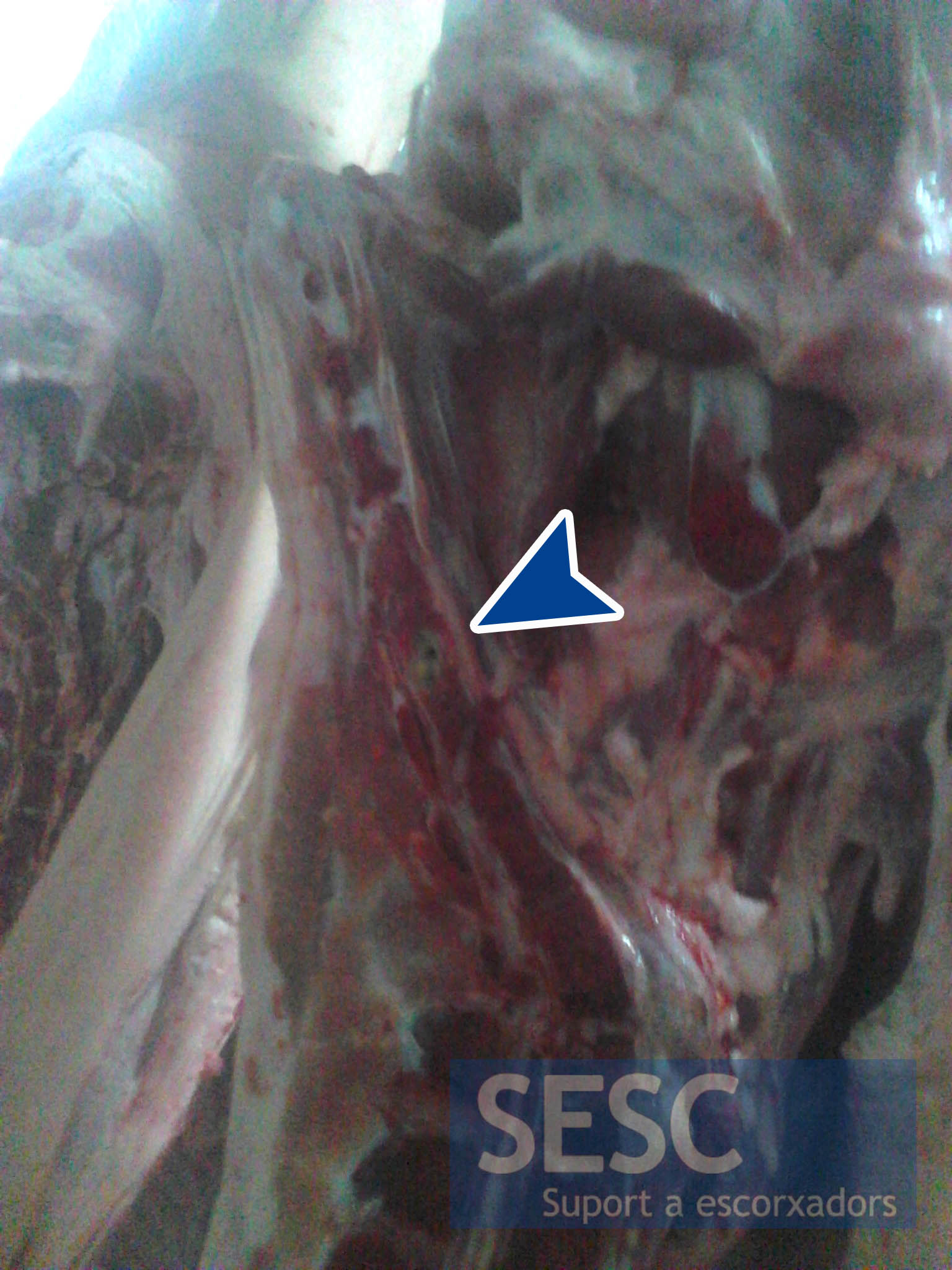
Example of vertebral osteomyelitis in the caudal region.

Case of vertebral osteomyelitis in the cervico-thoracic region.
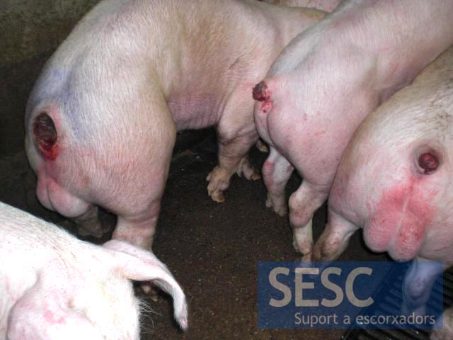
Examples of animals in antemortem inspection with lesions associated with tail biting.

8 comment(s)
Hello. How can i refer this article on a work for school?
Hello Daniel,
Thanks for reading us, you just need to mention the website “SESC Case Archive” and the url to the particular entry you are referring to.
FYI SESC is an acronym for Slaughterhouse support network in our languge (Catalan). You can also use the images provided they are not modified and the source (URL) is mentioned.
Good luck with your work!
Osteomyelitis, staph bone infection, may be treated with a new model discovered in previous research. Previous research found a new form of Treatment For Osteomyelitis that involves testing with a new mouse model.
Ficha de la FAWEC sobre mordeduras de colas en cerdos:
http://www.fawec.org/es/ficha/mordedura-cola-cerdos.htm
More comments from Animal Health forum:
W.G.: The occurrence at post mortem inspection level might not be as high in abattoirs as abattoirs mostly slaughter commercial pigs that are produced under hygienic and controlled environments. However in South-Africa there is a lot of “backyard” pigs in rural areas and informal settlements that are slaughtered at home for own consumption, I surely think the incidence of this type of pathology would be much higher under these conditions where the animals are more exposed to injury and are fed swill. This might be something to look at for all those aspiring researchers out there.
Comment form the Animal health forum in LinkedIn:
A.Z.: I believe I observed these lesions in 2002 – 2004 on a Louisiana, USA swine farm in which postmortem examination of sows euthanized or found dead often revealed vegetative endocarditis, pleural adhesions, and chronic peritonitis, all characterized by the sort of yellowish color seen in the photos included with the present report. Occasional sows displayed posterior paresis or paralysis, and necropsy revealed suppurative spinal lesions similar to those shown. Vaccination for H. parainfluneza was instituted, resulting in disappearance from the herd of all of the above lesions, along with marked improvement in animal health, productivity and welfare.
More commetns:
S.L: Very well discussed. I think the common route of entry is via infected wound that progressed to septicemia.
R.S.P.: No doubt sepsis, when not in back part where lymfvessels may lead the bacteria to the boon. But the crusial discussion is: do you condem the carcas on basis of number of processes or on basis of evaluating if the proces is still acute (no connective tissue bordering the bacteria). Risk accesment basis? In Denmark more processes is OK, if bordered and no other pyaemic alterations. But cutting up must then be under supervision.
Comment form Veterinary Pathology group in LinkedIn: Are the carcasses condemned? In Denmark they will be condemned, if there are no fibroses around the purulent processes. If judged cronic, the carcas will be cut under supevision by an instructed worker to secure, there are no more abscesses from the former pyæmia.