Download presentation
Presentation is loading. Please wait.

1
phylum:Nemathelminthes class:Nematoda order:Ascaridoidea
Ascaris lumbricoides

2
Ascaris lumbricoides Disease : Ascariasis (round worm infection)
Reproduction: dieceious sexual.(sexes are separated) Diagnostic stages: egg (fertilized, unfertilized), adult worm. Usual habitat: small intestine. Life cycle: simple (occures in single host). Definitive host: Man Infective stage: embryonated egg containing the larva Route of infection: ingestion of food or water contaminated with the embryonated eggs (that contain laryae) Diagnostic test : detection off eggs by microscopic stool examination.
 Diagnostic stages: egg (fertilized, unfertilized), adult worm. Usual habitat: small intestine. Life cycle: simple (occures in single host). Definitive host: Man . Infective stage: embryonated egg containing the larva Route of infection: ingestion of food or water contaminated with the embryonated eggs (that contain laryae) . Diagnostic test : detection off eggs by microscopic stool examination.")
4
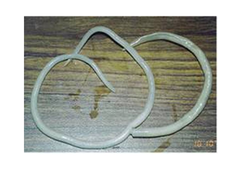
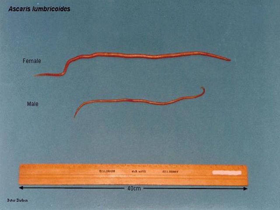
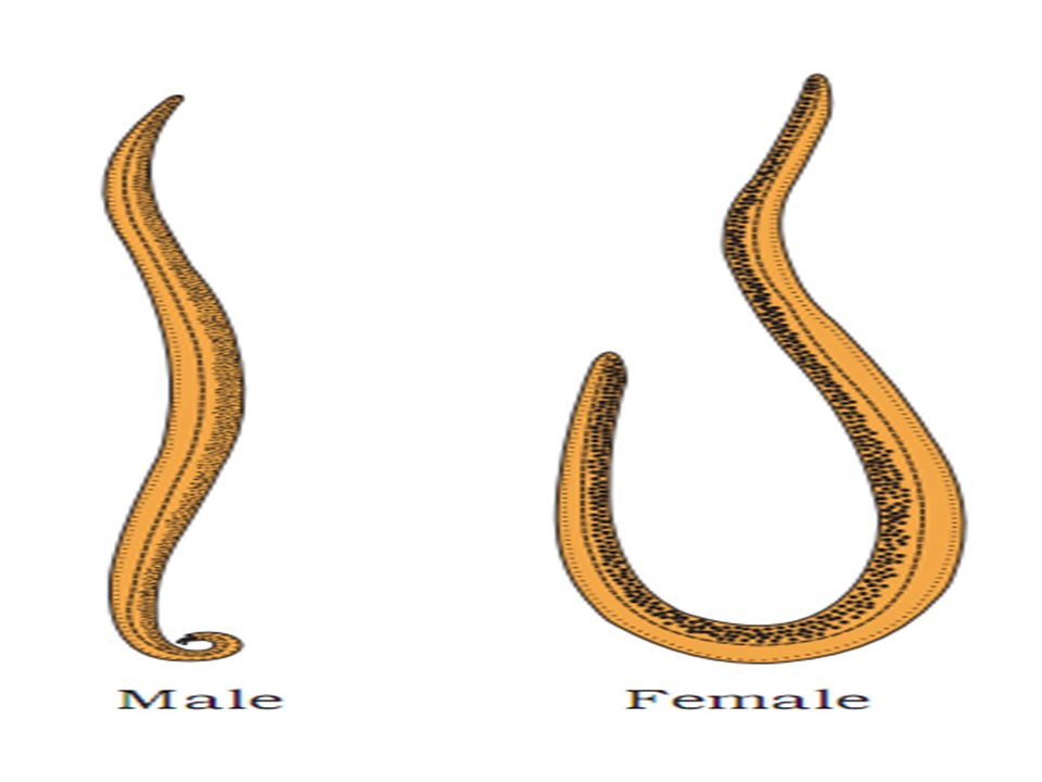
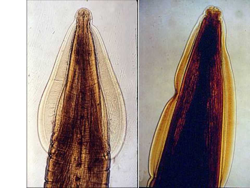
Ascaris lumbricoides adult worm
The female 22 – 35 cm, the male 10 – 31 cm this worms like earth worm Male and female anterior end, with three lips Ascaris lumbricoides adult worm Note: white, brown redish or light brown or pink . The posterior end of the male, curved with 2 spicules, the female with straight end

10
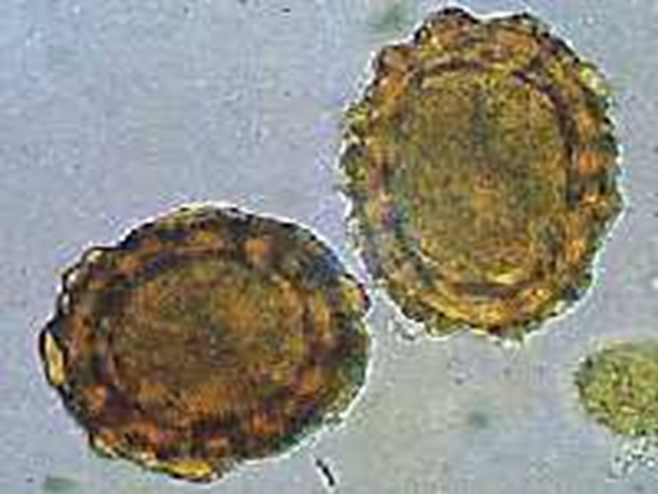
Ascaris lumbricoides: -Unfertilized Egg stool smear
In Iodine s. In Saline s. Elongated oval, no semilunar space with irrregular albuminous layer 88 – 94 X 40 – 50 µm Ascaris lumbricoides: -Unfertilized Egg stool smear

11
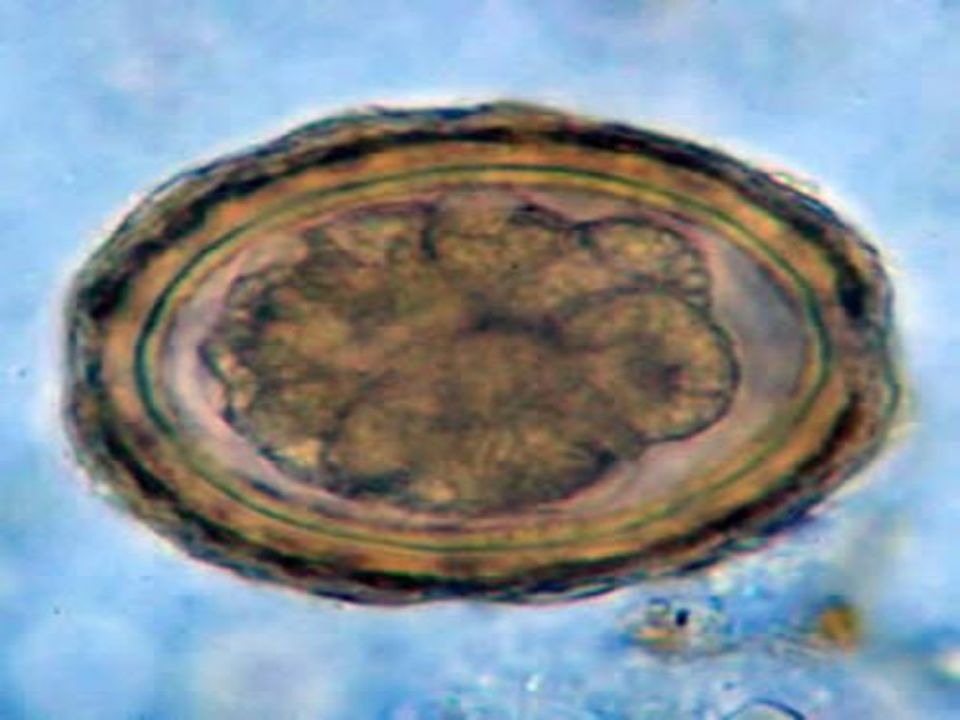
Ascaris lumbricoides: -Fertilized Egg In Saline s.

12
Its fertile ovum, but the outer coat is sometime lost
Fertile, with Fertile, with larva one-cell stage Iodine Stain. Semilunar space Vitelline layer (1st) One cell stage Decorticated egg Its fertile ovum, but the outer coat is sometime lost 2nd layer (thick) 3rd layer outer coarse albuminous layer (regular) Ascaris lumbricoides: Fertilized Egg In Iodine s. stool smear ( golden brown in colour ) 60 – 75 X 40 – 50 µm, is spherical or oval with semilunar space and regular albuminous layer
 One cell stage. Decorticated egg. Its fertile ovum, but the outer coat is sometime lost. 2nd layer (thick) 3rd layer outer coarse albuminous layer (regular) Ascaris lumbricoides: Fertilized Egg In Iodine s. stool smear ( golden brown in colour ) 60 – 75 X 40 – 50 µm, is spherical or oval with semilunar space and regular albuminous layer.")
13
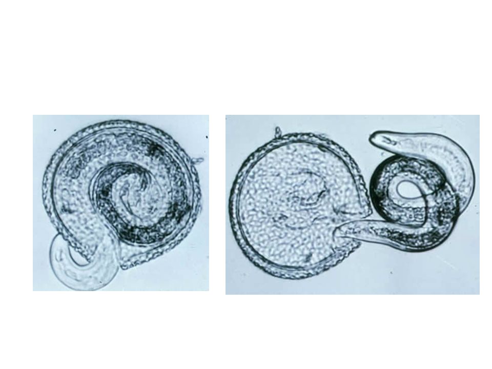
Ascaris lumbricoides egg hatching

14
Ascaris lumbricoides Larva in Lung Section
Note : the larva also detected in sputum Hematoxylim - eosin s.

15
life cycle of Ascaris lumbricoides
280 Ascaris lumbricoides life cycle of Ascaris lumbricoides

16
Ascaris lumbricoides in Intestine
Case of death because, of the No. of adult worms of Ascaris lumbricoides making blockage of lumen of intestine Ascaris lumbricoides in Intestine

17
phylum:Nemathelminthes class:Nematoda order:Ascaridoidea
parascaris equorum

18
Female 50 c m Male cm

19
Parascaris equorum

20
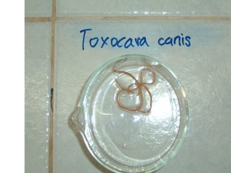
Toxocara Canis

21
Introduction Dog Round Worm - Phylum: Nematoda Zoonotic Disease
T. cati is the feline form

22
Infection Geographic Range: Worldwide Definitive Host: Dogs
Intermediate Host: None Accidental Host: Humans and other mammals Children more susceptible than adults

23
Infection Dogs Humans Found in Intestines Ingest Egg Transplacenta
Transmammary Puppies Born Infected with T. cannis Puppies less than 5 weeks Humans Can be found in liver, lung, brain, heart, muscle, or eye

24
Morphology Adult Female 5 to 18 cm long Adult Male 4 to 10 cm long

28
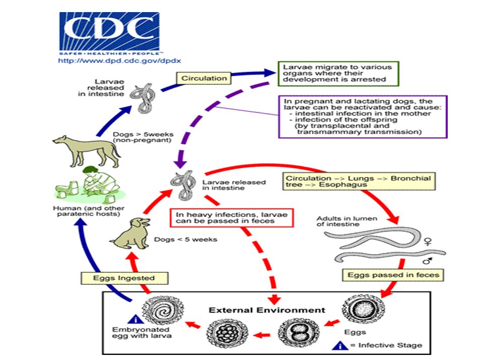
Life Cycle Eggs must be present in external environment for 2 weeks to bi infective Ingestion by dog Eggs hatch and larvae penetrate the gut wall Migrate into various tissues; encyst if dog older than 5 weeks Younger dogs larvae migrate through lungs, bronchial tree, esophagus, and move back into the small intestine

29
Life Cycle Older Dogs Encysted Stages reactivate during pregnancy
Infection spread by transplacental and transmammary routes Infective eggs spread through lactating bitches

30
Life Cycle Accidental Host Infected by ingestion of infective eggs
Eggs hatch and larvae penetrate the intestinal wall Carried by Circulatory System to various tissues Larvae don’t undergo further development but can cause reactions in tissue (toxocariasis)
")
32
Symptoms In dogs usually asymptomatic
Heavy infections can result in death In Humans Abdominal Pain Decreased Appetite Restlessness Fever Other symptoms vary with site larvae infection

33
Ocular Larvae Migrations (OLM)
Caused by larva migration to the retina Inflammation Scar formation Retinal Detachment Partial to Full Vision Loss 10,000 Infections per year 700 permanent vision loss

34
Visceral Larvae Migrations (VLM)
Caused by movement of worm larvae throughout various organs of the body Dependent on organ infected Fever Coughing Asthma Pneumonia Wheezing Hepatosplenmegaly

35
Treatment Use anti-parasitic drugs in combination with anti-inflammatory medications Albendazole Preferred Choice Mebendazole Thiabendazole Ocular Larvae Migrations Require Surgery

Similar presentations


>")


