Download presentation
Presentation is loading. Please wait.

1
CARDIAC CATHETERIZATION
AND INTERVENTIONAL TECHNIQUE

2
I- Cath. Room Equipments

3
Cardiac Cath. Machine

4
1- Catheter Table 2-C-Arm Shaped X-Ray Machine 3- Operative Dual Monitor 4- Injector 5- Control Room 6- Imaging Processing Camera & DVD production

5
1- Catheter Table -Similar to the operative table
- Table top X-Ray transparent - Supported only on one leg at the patient caudal side -Changeable level capability

6
Cath. Table & C-arm &Dual Monitors

7
2- C-arm Shaped X- Ray Machine
The X-Ray tube and the X-ray detector ( called the tower ) are supported at the ends of C-arm holder to allow easily movement around the catheter table The X-Ray tube is relatively small sized compared to the tower , so to facilitate different exposure positions , the tube mostly lie below the table in the narrow space and the tower ( Large size ) freely mobile above in relatively more wide space
 are supported at the ends of C-arm holder to allow easily movement around the catheter table. The X-Ray tube is relatively small sized compared to the tower , so to facilitate different exposure positions , the tube mostly lie below the table in the narrow space and the tower ( Large size ) freely mobile above in relatively more wide space.")
8
3- Operative Dual Monitors
The operative dual monitors essential for the Operating team to guide them during the technique Usually one monitor used for the fluoroscopic assessment during the act The other one used for illustration of Rout Mapping and to review the previous images

9
Operating Monitors

10
4- INJECTOR The injector is the tool responsible for contrast injection usually synchronized with the machine connect to it ( Ct & MR & Angiogaraphic or catheter machine )
")
11
Injector

12
ROLE OF INJECTOR IN ANGIOGRAPHIC IMAGING
It allow powerful – fast injection of large contrast bullous with a control rate usually ml/ sec producing a homogenous continuous intra vascular contrast column which can be followed to produce angiographic imaging - it is very difficult to produce the same result with manual injection specially in case of flash aortography which need very high pressure - In case of selected and super selected angiography manual injection is prefer because a small amount of contrast is needed & and The small selected vessel cannot tolerate the high injection pressure produced by injector )
")
13
ROLE OF INJECTOR IN MULTIPHASIC CONTRAST STUDY
The synchronization with the radiographic machine , allow injection of the contrast at the a defined timing to get contrast image of the target organ at a certain phase of circulation ( arterial phase & capillary phase & Venous phase & Portovenous Phase &Delayed phase ) - Timing Control : ( Its a capability of the imaging machine ) so the software used named according to the mother company of the machine eg , Smart prep. & Sure Start ….
 - Timing Control : ( Its a capability of the imaging machine ) so the software used named according to the mother company of the machine eg , Smart prep. & Sure Start ….")
14
INJECTORE HAZARS The injector is a tool allow very fast and powerful injection of the contrast bullous , so Precautions most be taken before use - The selected vessel for injection most be tested manually by rapid injection with saline to examined its pressure tolerance so , the injector is avoided during the selected and super selected vessel injection ( as in coronary arteries ) to avoid vessel rupture , in this condition manual contrast injection is preferred as the capacity of the vessel is limited and the pressure can be easy overcome
 to avoid vessel rupture , in this condition manual contrast injection is preferred as the capacity of the vessel is limited and the pressure can be easy overcome")
15
Dual Head injector …. For what ?

16
Injector syringe set

17
5- Control Room

18
II- Cath. Interventional Technique

19
Per cutaneous Arterial Catheterization Seldinger Technique

20
Seldinger Technique A- Needle ( Arterial puncture needle ) inserted into the artery ( At a defined site see diagram ) B- Suitable Guide wire passed through needle into the artery C- needle withdrawn leaving guide wire in the artery ( then the tract is dilated with facial dilator and dilated sheath is applied ) D- catheter passed over the guide wire into the artery E- Guide wire withdrawn leaving the catheter in the artery
 inserted into the artery ( At a defined site see diagram ) B- Suitable Guide wire passed through needle into the artery C- needle withdrawn leaving guide wire in the artery ( then the tract is dilated with facial dilator and dilated sheath is applied ) D- catheter passed over the guide wire into the artery E- Guide wire withdrawn leaving the catheter in the artery")
21
Seldinger Technique Instruments
Arterial puncture needle Guide Wire

22
Dilator with sheath Facial Dilater

23
Examples of Guide wire variety
Straight tip GW Curved tip

24
Examples of Catheter Variety
Flash aortic catheter Curved selected visceral catheter

25
SELECTED LT CORONARY CATH
( long wide curve ) SELECTED RT CORONARY CATH ( small curve )
 SELECTED RT CORONARY CATH. ( small curve )")
26
Diagram show suitable site of arterial cath.
Brachial puncture Femoral puncture

27
Selective Angiography
The aim is to select one branch of the aorta ( eg RT or LT coronary arteries & Renal artery & coeliac artery & carotid artery ) Curved end radiopaque catheter with a single tip hole is used like ; ( Visceral catheter or Preshaped cath. eg RT & LT coronary catheters or hook shaped cath. or head hunter cath. Or many other named catheters ) Technique : Under image with minimal contrast injection the site of the orifice of the target artery is identified with rotation of the outer end of the catheter its curved internal tip jump into the orifice of the target artery ( the catheter is a flexible structure difficult to push it ) so the guide wire repassed into the selected catheter to reach the target artery , then the catheter pushed over it easily )
 Curved end radiopaque catheter with a single tip hole is used like ; ( Visceral catheter or Preshaped cath. eg RT & LT coronary catheters or hook shaped cath. or head hunter cath. Or many other named catheters ) Technique : Under image with minimal contrast injection the site of the orifice of the target artery is identified with rotation of the outer end of the catheter its curved internal tip jump into the orifice of the target artery ( the catheter is a flexible structure difficult to push it ) so the guide wire repassed into the selected catheter to reach the target artery , then the catheter pushed over it easily )")
28
Super Selective angiography
The aim is ; More advanced angiography to select a branch of the selected artery eg ( LAD & LCX & PDA & OM & AM ) Technique ; A super selected catheter is used which has a narrowed diameter can passed within the selected catheter , rotate again to selected the target branch then pushed forward over the guide wire as the rule
 Technique ; A super selected catheter is used which has a narrowed diameter can passed within the selected catheter , rotate again to selected the target branch then pushed forward over the guide wire as the rule.")
29
Coronary Angioplastic Techniques
1- Balloon dilatation of the narrowed lesion ; Balloon catheter is used ( catheter with a balloon at its tip inflated from outer end of the catheter ) Technique : Guide wire with straight tip is pushed hopping to pass within the narrowed segment , if the trail success the balloon catheter is push over the guide wire until the balloon tip fit at the narrowed segment then the balloon inflated from out side to dilated the segment
 Technique : Guide wire with straight tip is pushed hopping to pass within the narrowed segment , if the trail success the balloon catheter is push over the guide wire until the balloon tip fit at the narrowed segment then the balloon inflated from out side to dilated the segment.")
30
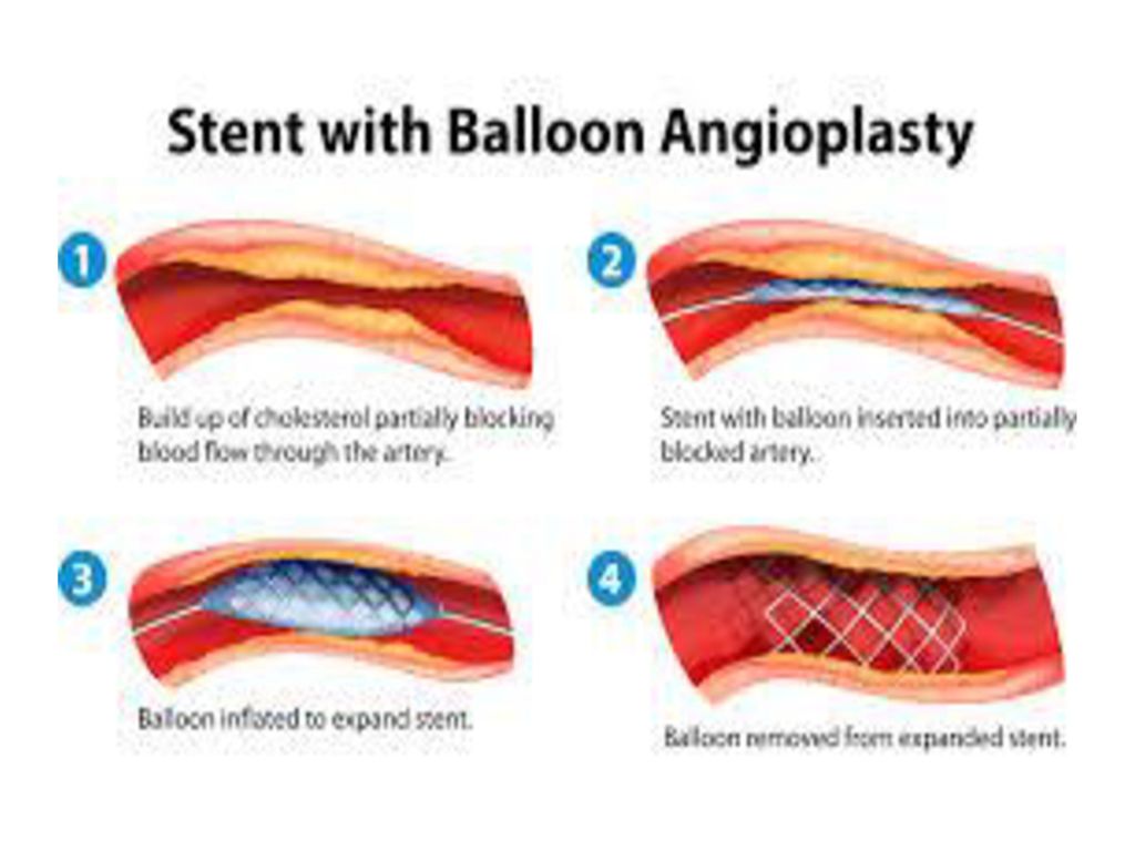
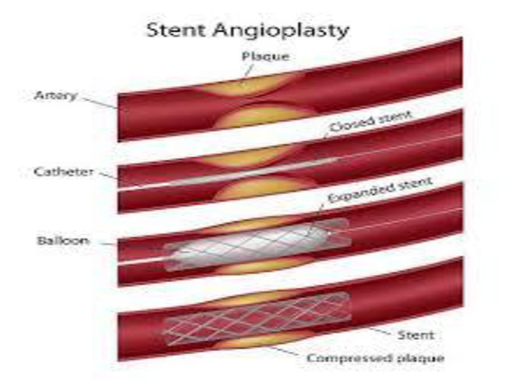
2- Stent with Balloon angioplasty just dilatation of the narrowed segment with balloon has a high rapid recurrence , so the technique is advanced with application of a stent balloon when the balloon inflated the outer stent is expanded at the narrowed segment to produced more stable dilatation

33
III- Imaging Positions
The patient lie flat in supine position while the C-arm were moved in certain directions to obtain ideal views for different segments of the coronary arteries

34
RT Coronary system Projections

35
1- LAO ( Straight ) Dead Lateral view of the heart The Target : The mid RCA
 Dead Lateral view of the heart The Target : The mid RCA")
36
2- LAO ( Cranial ) The Target ; Proximal RCA & PDA
 The Target ; Proximal RCA & PDA")
37
LT Coronary system Projections

38
1- LAO ( Cranial ) Target : Prox. LAD and Prox. LCX.
 Target : Prox. LAD and Prox. LCX.")
39
2- LAO ( Caudal ) Target ; Distal LM & Prox LAD and LCX
 Target ; Distal LM & Prox LAD and LCX")
40
3- AP ( Cranial ) Target ; Distal LAD
 Target ; Distal LAD")
41
4- AP ( Caudal ) Target : LM
 Target : LM")
42
5- RAO ( Cranial ) Target ; LAD – Septal - Diagonal
 Target ; LAD – Septal - Diagonal")
43
6- RAO ( Caudal ) Target ; LCX
 Target ; LCX")
44
Digital Sub traction Angiography
The aim of technique is to produce angiographic image of the selected artery alone without the surrounding and overlapping structures ( Soft tissue , bones …….)
")
45
Digital Subtraction Image

46
Technique : Scout film : the imaging position is adjusted properly and the catheter is in suitable location for injection , one imaging shot is obtained ( Before contrast injection ) the base line image called scout film 2- Negative Film : Immediately the computer of the digital machine reverse the scout film to a negative configuration and stored it Contrast images : obtain during the contrast injection of the target artery 4- Angiographic sub traction images : The computer of the digital machine subtract the contrast images from the saved negative image of the scout film the result is isolated arterial images without the back ground structures
 the base line image called scout film 2- Negative Film : Immediately the computer of the digital machine reverse the scout film to a negative configuration and stored it 3- Contrast images : obtain during the contrast injection of the target artery 4- Angiographic sub traction images : The computer of the digital machine subtract the contrast images from the saved negative image of the scout film the result is isolated arterial images without the back ground structures")
Similar presentations


>")
>")


 examines the ECG potentials generated along the three-dimensional axes of the body; that is, the x, y, and z planes. The.>")


 Irfan-ullah(EE-01083-247)>")
