
Berghia marcusi, Domínguez, Marta, Troncoso, Jesús S. & García, Francisco J., 2008
|
publication ID |
https://doi.org/ 10.1111/j.1096-3642.2008.00390.x |
|
DOI |
https://doi.org/10.5281/zenodo.5747215 |
|
persistent identifier |
https://treatment.plazi.org/id/4615878D-443F-670C-4FD7-711CFC49FDEC |
|
treatment provided by |
Carolina |
|
scientific name |
Berghia marcusi |
| status |
sp. nov. |
BERGHIA MARCUSI View in CoL SP. NOV.
Berghia coerulescens Marcus, 1957: 477–481 View in CoL , figs 237, 240, 241, 245.
Type material: Holotype: one specimen, 12-mm long, 0-m depth at Praia de Armação (Buzios, Rio de Janeiro), 22 June 2000, deposited in the Museo de Zoologia de la Universidad de São Paulo, Brazil (catalogue number MZUSP 64150 View Materials ).
Paratype: one specimen, same data as for holotype, 11-mm long, deposited in the Museo de Zoologia de la Universidad de São Paulo, Brazil (catalogue number MZUSP 64151 View Materials ) .
Other material: Praia dos Ossos (Buzios, Rio de Janeiro), 21 March 1998, two specimens, 0-m depth, 5–7-mm long . Praia de Armação (Buzios, Rio de Janeiro), 22 March 1998, one specimen, 0-m depth, 16.6-mm long . Praia de Armação (Buzios, Rio de Janeiro), 23 March 1998, one specimen, 0-m depth, 7-mm long . Praia dos Ossos (Buzios, Rio de Janeiro), 26 March 1998, two specimens, 0-m depth, 6–7-mm long . Praia dos Ossos (Buzios, Rio de Janeiro), 27 June 1999, one specimen, 0-m depth, 11-mm long . Praia de Armação (Buzios, Rio de Janeiro), 25 June 2000, one specimen, 0-m depth, 13-mm long .
Etymology: This species is named after Ernst Marcus, who first described these specimens. Marcus contributed considerably to the knowledge of the Brazilian opisthobranchs.
External morphology: The general body colour is whitish. The oral tentacles are elongate, their bases are translucent with orange pigment, and the apical two-thirds of each is white ( Fig. 1F View Figure 1 ). The rhinophores bear papillae on their posterior faces. Some specimens have papillae arranged in oblique rows on the anterior and lateral faces. Approximately, the apical twothirds of the rhinophores are white–cream, and the bases are reddish. An orange band is on either side of the head, between the oral tentacles and rhinophores. The cerata are organized into between five and seven pairs of groups ( Fig. 6A View Figure 6 ). The cerata have translucent tips, below which there is an orange ring, and the digestive gland is brown. Below the orange ring there is a whitish area that is more visible in some specimens. The cerata are arranged in horseshoe-shaped arches ( Fig. 6B View Figure 6 ). The cerata of the middle part are larger than those at the ends. The first group contains ten or 11 cerata, and the number diminishes towards the tail. The posterior groups consist of two or three cerata, not in arches. There are oblique bright orange lines on the borders of the insertions of the cerata, especially on the first group. Near the cerata the dorsum is translucent white, and extending posteriorly from the base of the rhinophores there is an undulating opaque white band. This band may be broken, or can extend uninterrupted to the tip of the tail. On the largest specimens the dorsal band is broken on the pericardium, and there are some spots of the same colour on this area. The genital opening is among the cerata of the anteriormost group on the right. The anus is located below the second right arch. The foot is translucent, with elongated foot corners curved posteriorly ( Fig. 1G View Figure 1 ).
Internal morphology: The radula is uniseriate with between ten and 12 bi-arched teeth ( Fig. 7A View Figure 7 ). Each tooth has a triangular median cusp with a very fine denticle on each side (in some teeth it only appears on one of the sides). In a 5-mm-long specimen the median teeth have 17–21 denticles on each side ( Fig. 6C View Figure 6 ), and the smallest tooth has 12–13 denticles on each side ( Fig. 6D View Figure 6 ). In an 11-mm-long specimen the smaller teeth have 19–23 denticles on each side, and the larger teeth have 24–28 denticles.
Some specimens have radular teeth with denticles that are piled up ( Fig. 6E View Figure 6 ), or with denticles ending in two or more tips ( Fig. 6F View Figure 6 ).
The masticatory border of each jaw plate shows a long row of small rounded denticles. ( Fig. 7B View Figure 7 ).
Remarks: The presence of the European species B. verrucicornis and B. coerulescens in the Western Atlantic has been an object of discussion. The Western Atlantic specimens of Berghia were assigned to a very variable species, B. coerulescens ( Engel, 1925; Marcus, 1957; Edmunds, 1964, 1966; Marcus, 1976), although some of them were re-examined and subsequently assigned to B. verrucicornis by Tardy (1962). However, several authors have doubted the amphiatlantism of these Berghia species, and have recognized the need for future investigations to clarify this question ( Edmunds, 1968; García-Gómez & Thompson, 1990; Muniain & Ortea, 1999).
The study of the specimens from the Brazilian coast allows us to observe several characters, mainly related to the coloration, that differ from the specimens of the Mediterranean and Eastern Atlantic ( Table 1 View Table 1 ). The animals from Rio de Janeiro present orange markings at the bases of the ceratal clusters, which are especially visible in the anterior groups. This characteristic is present in all examined specimens, but is not present in the descriptions of B. verrucicornis from the Eastern Atlantic ( Tardy, 1962; Edmunds, 1968; Ballesteros, 1977; García-Gómez & Thompson, 1990; Garcia-Gomez, 2002). To corroborate this affirmation, we have examined specimens of B. verrucicornis from the Atlantic coast of Huelva (south-western Spain). Variations of colour are shown in Fig. 8A and B View Figure 8 . The back of one specimen is mostly orange ( Fig. 8A View Figure 8 ). The other specimen has a whitish back with a translucent orange mid-dorsal band, and the pericardium has further orange pigmentation ( Fig. 8B View Figure 8 ). These animals differ from the Brazilian Berghia specimens in the lack of an orange line on the ceratal insertion, and the opaque white band on the dorsum is also absent. Furthermore, the Western Atlantic specimens possess fewer pairs of ceratal groups than the Eastern specimens. The Brazilian animals are 5–16.5-mm long, and have between five and seven groups. Ballesteros (1977) described a 13-mm-long specimen from the Mediterranean Sea with ten ceratal clusters, and Edmunds (1968) studied a 15-mm-long specimen from Ghana with eight groups.
Internal characters of Brazilian material were also compared with those from Huelva. The radular teeth of B. verrucicornis ( Fig. 7C View Figure 7 ) have more elongated and numerous denticles than those of B. marcusi sp. nov. The teeth of B. verrucicornis have a vertical crack below the median cusp, and the lateral denticles become large gradually ( Fig. 7D View Figure 7 ). Berghia marcusi sp. nov. radulae have larger and stouter denticles than those of B. verrucicornis , but a very fine denticle on one or both sides of the median cusp may be present. Furthermore, B. marcusi sp. nov. has the masticatory border of the jaws with rounded denticles, whereas B. verrucicornis has either smooth jaws ( Thompson, 1980; Garcia-Gomez, 2002) or jaws with hair-shaped denticles ( Tardy, 1962; Marcus, 1972).
Our specimens from Rio de Janeiro show great similarity with those examined from Ubatuba by Marcus (1957). Other authors such as García-Gómez & Thompson (1990) reported special features for these animals, which differed from the other specimens from the Western Atlantic. They stated that the specimens from Brazil identified as Spurilla coerulescens by Marcus may belong to another (new) species. Small differences exist between our material and the Ubatuba specimens (orange pigment in our animals and red pigment in those of Marcus), but both animals have orange markings on the dorsum, have oral tentacles and rhinophores that are yellow-tipped and have red bases, have red marks on the head, and have cerata with red rings.
Edmunds (1968) described three specimens of Berghia from Jamaica, called A, B, and C, that showed orange markings at the ceratal insertions. He compared these animals with those from Ghana, and suggested two possibilities: first, a very variable species is present in the Western Atlantic; second, two or more new species of Western Atlantic Berghia exist. According to García-Gómez & Thompson (1990), specimen B is distinct from any described species, and it can be distinguished from B. verrucicornis by the orange marks at the base of the cerata, and the oral tentacles with orange bases. The description of specimen B from Jamaica agrees with the description of our animals, except for the pale yellow ring and the white ring below the cnidosacs. Our specimens possess cerata with orange rings.
Specimens A and C from Jamaica also possess orange markings at the base of the cerata, but differ from the Brazilian animals in that specimen A lacks the whitish dorsal band, and specimen C has the front half of the body orange, and the rear half grey.
Thompson (1980) assigned to B. verrucicornis two specimens from Jamaica with orange lines at the ceratal insertions, and a mid-dorsal white band. However, these specimens are different from our animals because the cerata have three bands (white, yellow, and white) inside the orange ring, and the masticatory border of the jaws is smooth (our specimens possess denticles). The animals assigned to B. verrucicornis from Florida by Marcus (1972) are very similar to the Brazilian ones. Both have orange markings at the ceratal insertions, and a mid-dorsal white band, but the specimens from Florida have greyish coloration, and they have more ceratal groups (nine groups in a 12-mm-long specimen). Furthermore, the cerata lack rings on cnidosacs. The radulae possess more teeth and more denticles per tooth (19 teeth and 30–38 denticles on each side).
The species Berghia columbina ( García-Gómez & Thompson, 1990) found in south-western Spain has a similar colour pattern to our specimens ( Fig. 8C View Figure 8 ). Berghia columbina has orange markings at the ceratal insertions and on the head, orange or reddish rhinophores with white tips, and translucent orange oral tentacles with white tips. However, B. columbina possesses more pairs of ceratal groups (between nine and 11 groups according to Garcia-Gomez, 2002), whereas B. marcusi sp. nov. presents between five and seven groups. Furthermore, a mid-dorsal orange line exists in B. columbina inside a white band, and the cerata do not have orange rings. The radula presents more teeth and more denticles per tooth ( Fig. 7E View Figure 7 ), and the jaw masticatory border is smooth ( Fig. 7F View Figure 7 ).
Muniain & Ortea (1999) described a new species from the Western Atlantic, Berghia rissodominguezi , which is similar to the Brazilian specimens, but there are also several differences. The specimens are larger than B. marcusi sp. nov., having an orange triangle on the head, and a mid-dorsal reddish-brown line from the third cerata group (our specimens have a cream or opaque white spot on the head and dorsal band). The rhinophores of B. rissodominguezi are cream–white in colour, with yellow on the apical portion inside a red base and yellow tip. The translucent oral tentacles have a yellow area covering two-thirds of them (the oral tentacles of B. marcusi sp. nov. have white tips). Finally, the masticatory border of the jaw in B. rissodominguezi is smooth ( Muniain & Ortea, 1999; MD, JT & FG, this paper, pers. observ.), differing from the denticulate border of B. marcusi sp. nov.
For all of these reasons, we consider that the Eastern Atlantic and Mediterranean specimens of B. verrucicornis are different from the Western Atlantic Berghia specimens, and possibly that there are two or more West Atlantic species assigned to B. verrucicornis . Our animals from Rio de Janeiro are very similar to the specimens cited as B. coerulescens from São Paulo ( Marcus, 1957), and Berghia specimen B from Jamaica ( Edmunds, 1968). Therefore, we suggest that our Brazilian specimens, Berghia specimen B by Edmunds (1968) and B. coerulescens by Marcus (1957), belong to the same new taxon, which is different from B. verrucicornis . The distinguishing characteristics of the species studied in this work are compiled in Table 2 View Table 2 .
Distribution: Jamaica ( Edmunds, 1964).
Brazil: Ubatuba, São Paulo ( Marcus, 1957), Praia de Armação, Praia dos Ossos (Buzios, Rio de Janeiro) (present paper) .
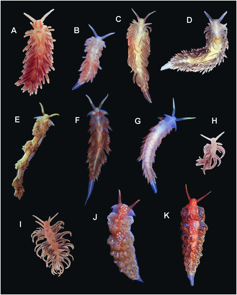
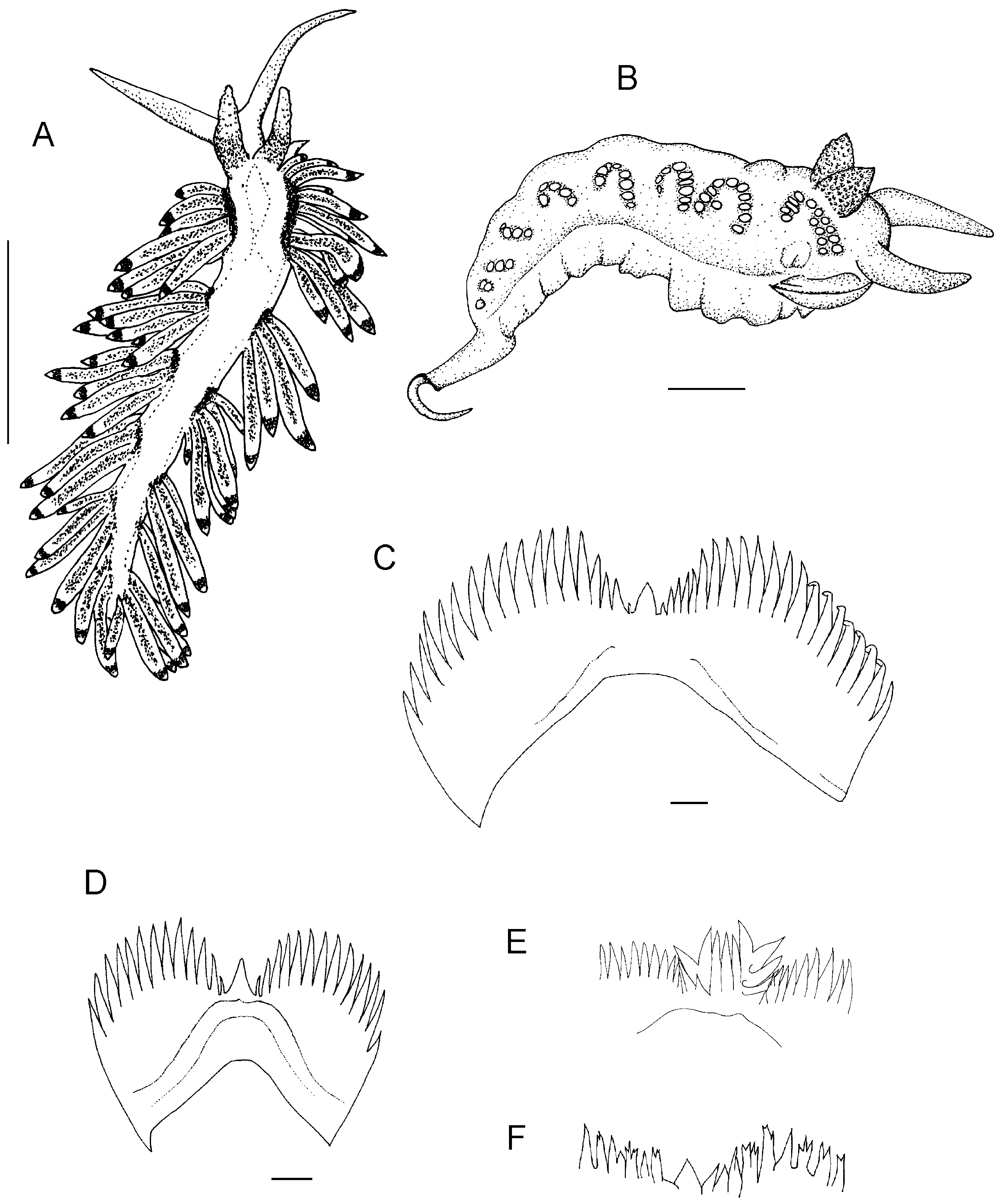
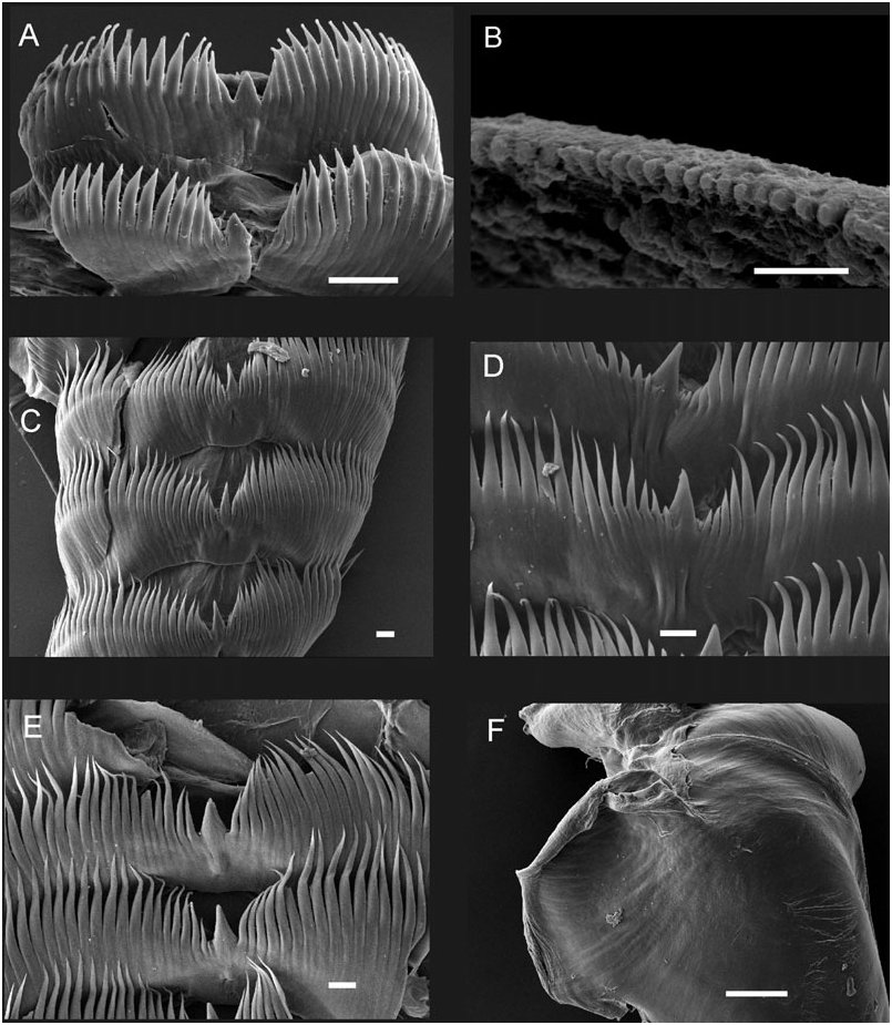
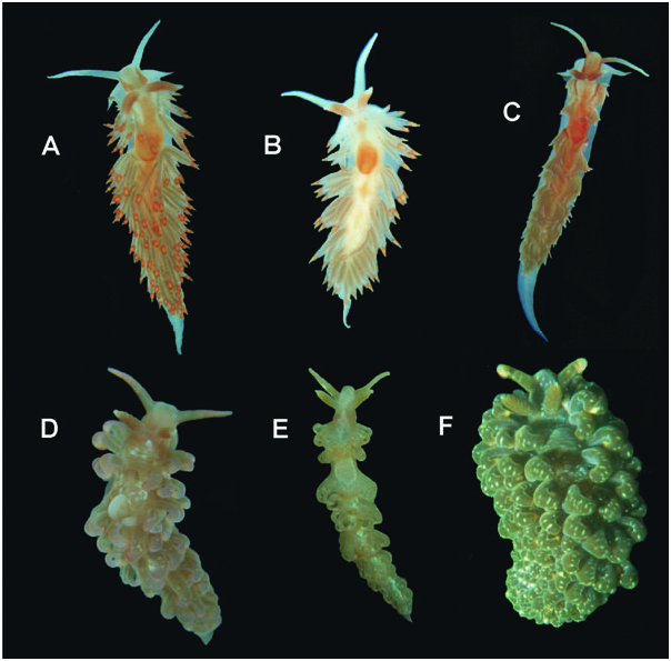
Table 1. Distinctive features of our Berghia marcusi sp. nov. specimens, of Berghia marcusi sp. nov. specimens described by other authors, and of Berghia verrucicornis and Berghia columbina specimens
| Length | N° of | Orange line | N° of | N° of | ||||||
|---|---|---|---|---|---|---|---|---|---|---|
| alive | ceratal | Marks on | on ceratal | Colour of | Colour of | radular | denticles on | Masticatory | ||
| Distribution | (mm) | groups | dorsum | insertion | the cerata | rhinophores | teeth | radular tooth | border | |
| Berghia marcusi | Buzios (Rio | 5–16 | 5–7 | Mid-dorsal | Present | Translucent | Distal two | 10–12 | 12–21 on | Small denticles |
| de Janeiro) | white band | tip, below | thirds white | (specimen | either side | |||||
| (present | orange ring | or cream; | 5 mm) | (specimen | ||||||
| paper) | and whitish | reddish base | 5 mm); 28 | |||||||
| zone | (specimen | |||||||||
| 16 mm) | ||||||||||
| Berghia marcusi | Ubatuba | 12–20 | 6–10 | Yellow | Present | Red ring | Pink base; | – | Oldest tooth | Minutes denticles |
| cited as | (Sao Paulo) | mid-line and | (red | around | yellow tip | 10–22 on | ||||
| B. coerulescens | (Marcus, | yellow triangle | coloured) | cnidosac | either side; | |||||
| 1957) | between eyes | a big tooth | ||||||||
| with 21–23 | ||||||||||
| Berghia marcusi | Jamaica | 12 | 8 | Mid-dorsal | Present | Pale buff or | Orange papillae | 16 | 40–64 | With denticles |
| cited as | (Edmunds, | white line | greyish yellow | in the basal | per tooth | |||||
| Berghia B | 1968) | digestive gland. | half; creamy | |||||||
| Pale yellow | white tip | |||||||||
| ring and | ||||||||||
| white band | ||||||||||
| ‘ Berghia | Jamaica | 18 | 10 | Mid-dorsal | Present | Brown digestive | Orange, with | 22 | About 30 on | Smooth |
| verrucicornis ’ | (Thompson, | white streak. | gland. White, | lemon-yellow | either side | |||||
| 1980) | Orange patch | lemon yellow | tips | |||||||
| over the | and white | |||||||||
| pericardium, | bands. | |||||||||
| and triangles | ||||||||||
| behind the | ||||||||||
| rhinophores | ||||||||||
| Berghia | île de Ré | 20–30 | 10 | Mid-dorsal | Absent | Yellowish brown | Red, with | – | 44–50 | Pectinate |
| verrucicornis | (French) | white band | digestive gland; | white tip | per tooth | |||||
| ( Tardy, 1962) | red-orange ring | |||||||||
| Berghia | Tema ( Ghana) | 15 | 8 | Mid-dorsal | Absent | Pale brown | Vermilion on | 13 | 44–48 | With denticles |
| verrucicornis | (Edmunds, | white line | digestive gland; | sides and | per tooth | |||||
| 1968) | vermilion ring | posterior | ||||||||
| below tip. | papillae; | |||||||||
| Distal third | cream tip and | |||||||||
| white | distal papillae | |||||||||
| Berghia | Florida | 12 | 9 | Opaque white | Present | There is no | Colorless | 19 | 30–38 on | About 350 |
| verrucicornis | (Marcus, | pattern | orange ring | rhinophore; | either side | hair-shaped | ||||
| 1972) | orange base | denticles |
No known copyright restrictions apply. See Agosti, D., Egloff, W., 2009. Taxonomic information exchange and copyright: the Plazi approach. BMC Research Notes 2009, 2:53 for further explanation.
|
Kingdom |
|
|
Phylum |
|
|
Class |
|
|
Order |
|
|
Family |
|
|
Genus |
Berghia marcusi
| Domínguez, Marta, Troncoso, Jesús S. & García, Francisco J. 2008 |
Berghia coerulescens
| Marcus Er 1957: 481 |