

Clavicle
What, to you, is the most distinguishable feature about the girl in the picture presented as the featured picture? Some of you may say her cheeky smile; some may say her gentle cheekbones; but of course, the ones with the medical and curious mind may look at this picture and think, “That is a really well-defined clavicle man! Whoo!”
Okay, maybe not (or maybe), but that is what we’ll talk about today, very briefly, – the clavicle.
Gross Anatomy of The Clavicle
Let us start off by picking out what we identify from a very superficial view of the clavicle.
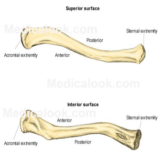
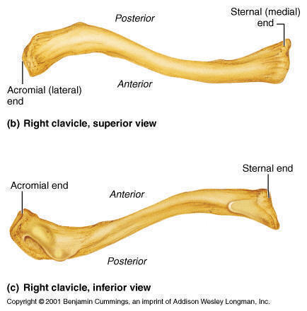
First of all, we can appreciate that the clavicle has the appearance of an elongated capital S.
Why is this?
The capital S shape is due to the anterior and posterior curvatures of the clavicle.
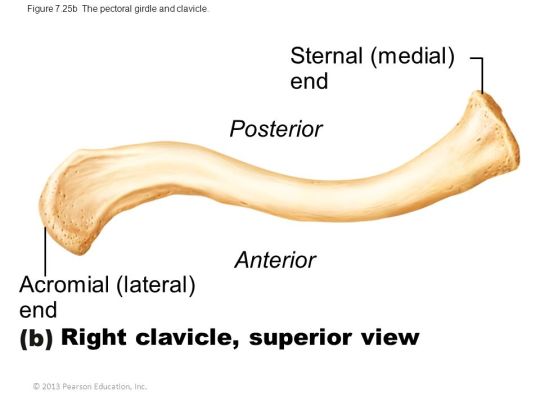
Just to refresh this point, remember that a convex surface is one that protrudes out, while a concave surface is one that “invaginates” in, like a cave. Looking at the diagram above then, we can appreciate a massive anteriorly directed convexity closer to the medial end, known as the anterior curvature, and a much smaller, posteriorly facing convexity, closer to the lateral end. Where the anterior and posterior curvatures meet is where the clavicle can be divided into a lateral and medial end.
Thus, the medial 2/3 of the clavicle is marked by the anterior curvature, and the lateral 1/3 of the clavicle is marked by the posterior curvature. The anterior curvature has an extremely important function of surrounding anteriorly and protecting the brachial plexus of nerves and the apex of the lungs.
The clavicle itself is considered a bone of the upper limb, due to one very important reason.
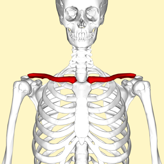
Look at where the clavicle is. The clavicle is considered a bone of the upper limb because it connects the axial skeleton at the sternum, to the appendicular skeleton, at the acromion of the scapula, part of the upper limb. This has several consequences which will be explained later.
For now, this means that the medial end of the clavicle attaches to the sternum, and is thus called the sternal end of the clavicle. The lateral end attaches to the acromion of the scapula and is thus called the acromial end of the clavicle.
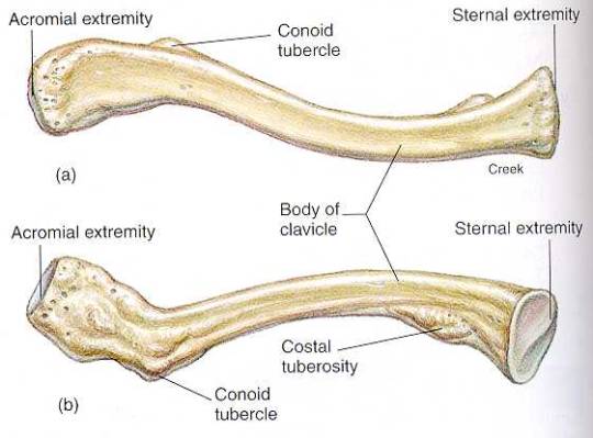
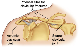
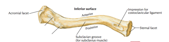
The sternal end of the clavicle is large and pyramidal in cross-section, as seen in the diagram above, and articulates with the clavicular notch of the manubrium sterni to form the sternoclavicular joint (SC joint). This SC joint is a saddle type synovial joint, that allows primarily elevation and depression of the shoulder joint and essentially the whole arm (try moving your SC joint!) Furthermore, the SC joint also allows minor anterior and posterior movements.
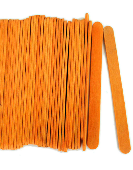
The acromial end, on the other hand, is much flatter, as seen in the cross-section of the same diagram above. The much flatter cross-section leads to the acromial end being shaped as a flat orange craft stick.

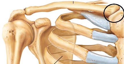
This forms an acromioclavicular joint (AC joint) with the acromion of the scapula. This AC joint is a plane synovial joint that serves to support the function of muscles that move the clavicle.
Now we can talk about the very specific features of the medial 2/3 and lateral 1/3 of the clavicle.
Medial 2/3 Of Clavicle:
Because the medial 2/3 of the clavicle is a quadrangular in cross section, it means that the medial 2/3 can be essentially expressed as a cuboid.

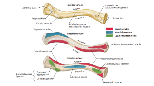
We can therefore see that the medial 2/3 has 4 surfaces, a superior, inferior, anterior and posterior, each with its own features.
Generally, the superior surface of the clavicle is much more smooth than the inferior surface, and the reason is due to the number of attachments:
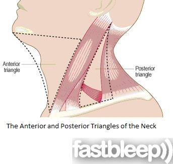
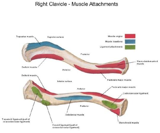
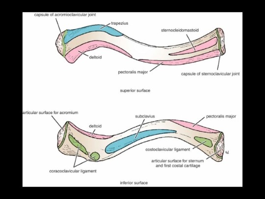
Superior Surface: The superior surface of the medial 2/3 of the clavicle simply gives origin to the clavicular head of the sternocleidomastoid. Notice in the diagram below where the sternocleidomastoid originates from.
Inferior Surface: The inferior surface of the medial 2/3 of the clavicle gives 2 features:
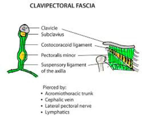
- A rough surface known as the subclavian groove, that attaches the subclavius muscle. The margins of this groove give attachment to the clavipectoral fascia, which covers the pectoralis minor.
- A rough impression for costoclavicular ligament, or the costal tuberosity attaches the costoclavicular ligament, which connects the clavicle to the first costal cartilage. This costoclavicular ligament prevents excessive elevation that may damage the sternoclavicular joint.
Anterior Surface: The anterior surface of the medial 2/3 of the clavicle gives origin to the pectoralis major muscle.
Posterior Surface: The posterior surface, in conjunction with the inferior surface of the medial 2/3 of the clavicle gives origin to the sternohyoid muscle.

Lateral 1/3 of Clavicle:
Looking back at the diagram of the orange stick to show how flat the acromial end of the clavicle is, we can easily say that the clavicle has 2 surfaces and 2 borders.
2 Borders: Anterior and Posterior Border
2 Surfaces: Superior and Inferior Surface
Superior Surface: The superior surface of the lateral 1/3 of the clavicle contains no well distinguishable features.
Inferior Surface: The inferior surface of the lateral 1/3 of the clavicle gives two important rough features.
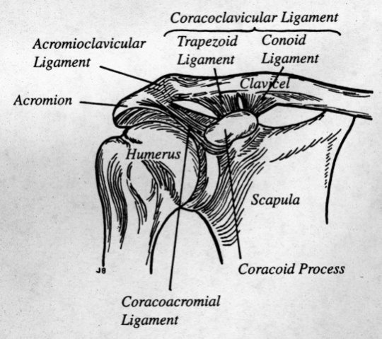
- Conoid Tubercle: The conoid tubercle is a small bony prominence on the inferior surface of the lateral 1/3 of the clavicle, closer to the midline of the clavicle. It gives attachment to the conoid ligament, which attaches the clavicle to the coracoid process of the scapula, nearly vertically. It is an upside down fan shaped structure with the wide base resting on the clavicle, and the sharp apex inferiorly projecting to the root of the coracoid process.
- Trapezoid Line: The trapezoid line is another prominence on the inferior aspect of the lateral 1/3 of the clavicle, further away from the midline of the clavicle. It gives attachment to the trapezoid ligament, a nearly horizontal ligament that connects the apex of the coracoid process to the clavicle.
Both the conoid ligament and the trapezoid line together are called the coracoclavicular ligament.
Anterior Border: Anterior border of the lateral 1/3 of the clavicle gives origin to the deltoid muscle.
Posterior Border: Posterior border of the lateral 1/3 of the clavicle gives insertion to the trapezius muscle.
Ossification of Clavicle:
The clavicle is an extremely cutaneous bone. It is the only long bone that is so superficial in the body and is very easily palpated.
It is the first bone to ossify in the fetus from membrane, and it does so from 2 centres, which ossify at the 5th week and rapidly fuse in the midline. Their site of fusion in the midline, is, as expected, the most easily fractured region of the clavicle.
The clavicle is sometimes pierced through and through by the supraclavicular nerve, and thus, the clavicle is referred to as a dermal bone, or a membranous bone, and is unique in being the only bone outside the skull to be a membranous bone.
That’s all guys! Hope this helped, as usual!
VIDEOS:

Awesome presentation I found it very helpful.
LikeLike
👏🏾👏🏾👏🏾👏🏾👏🏾
LikeLike