
Fortuynia smiti Ermilov, Tolstikov, Mary & Schatz, 2013
|
publication ID |
https://doi.org/ 10.11646/zootaxa.3957.4.1 |
|
publication LSID |
lsid:zoobank.org:pub:90175CBC-5C93-44CD-B13C-B17516643317 |
|
DOI |
https://doi.org/10.5281/zenodo.6113660 |
|
persistent identifier |
https://treatment.plazi.org/id/03B687D9-2826-F466-D3EE-FE1CD1BB9C34 |
|
treatment provided by |
Plazi |
|
scientific name |
Fortuynia smiti Ermilov, Tolstikov, Mary & Schatz, 2013 |
| status |
|
Fortuynia smiti Ermilov, Tolstikov, Mary & Schatz, 2013 View in CoL
Supplementary morphological information and first description of juvenile stages.
Although the original description of F. smiti ( Ermilov et al. 2013) provided excellent morphological information, details about the important van der Hammen’s organ and about some aspects of the legs are missing and were presented here.
Description of certain adult features. Adult ( Figs 14 View FIGURE 14 A–B). Females (N=1), length: 589 µm, width: 465 µm; males (N=4), length: 552–577 µm (mean 564 µm), width: 409–434 µm (mean 424 µm).
Lateral aspect ( Fig. 14 View FIGURE 14 C). The van der Hammen’s organ consists of sejugal channel branching off anterior dorsal circumgastric scissure, passing the bothridium posteriorly and running ventrad to the area between acetabulum II where it branches into a short anterior canal reaching acetabulum II and a short posterior canal running to acetabulum III; another canal connects the sejugal channel with acetabulum IV, it is larger and shows internal transversal cuticular ribs in transmitted light; a prodorsal canal branches of the sejugal channel at the posterior border of the bothridium, passes the bothridium and runs rostrad where it ends short before the insertion of the lamellar seta.
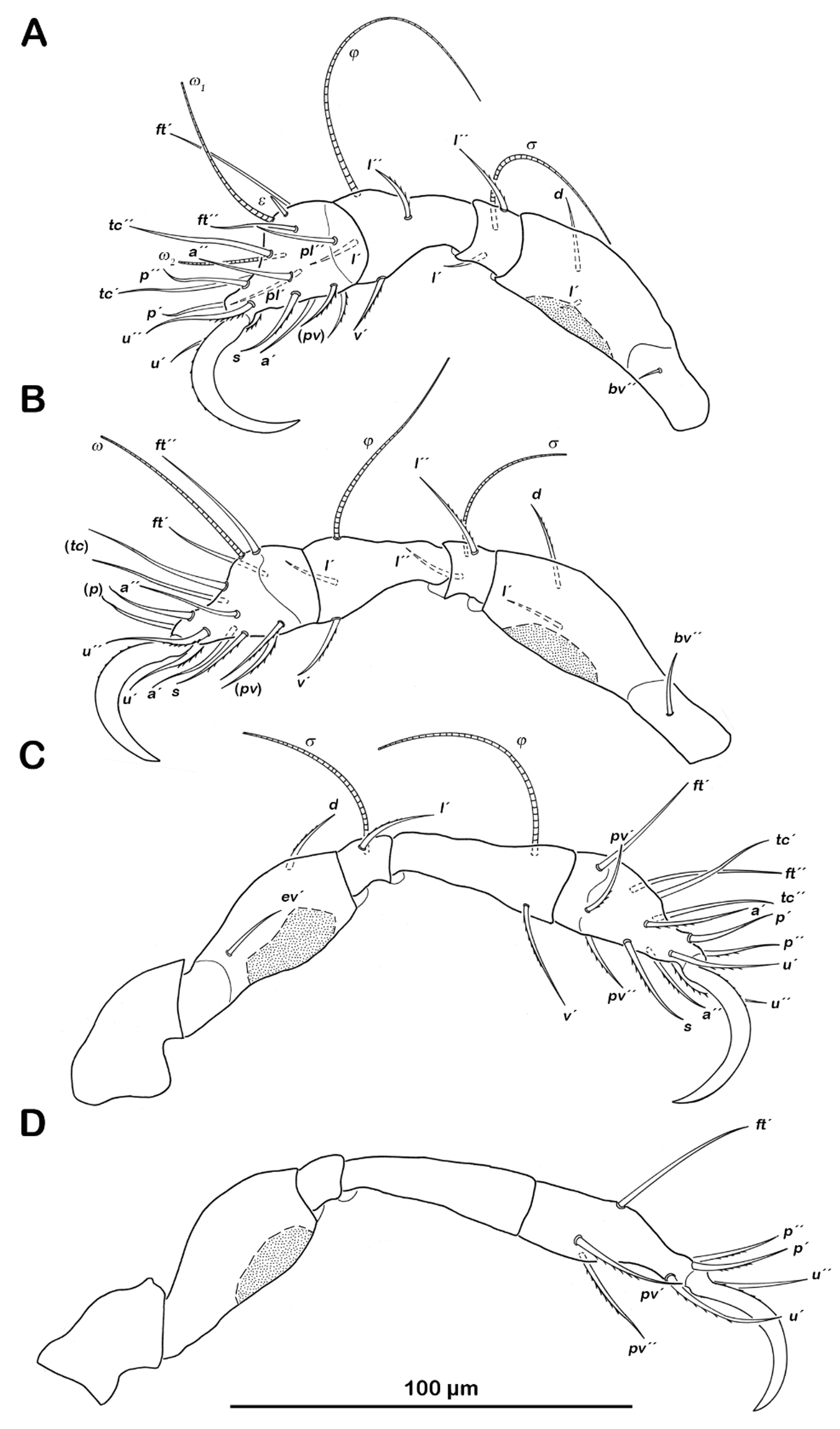
Legs ( Fig. 15 View FIGURE 15. F ). Tarsal lyrifissures present on all legs. Large porose areas with irregular borders on paraxial ventral side of all femora. Smaller circular porose areas on trochanter III and IV. Chaetome and solenidia see table 4.
Common features of juvenile stages. Apheredermous. Colour dark brown. Integument plicate and soft, except for centrodorsal plate. Prodorsum triangular, rostrum rounded, cerotegument overall finely granular, lighter longitudinal median area. Rostral setae (ro) robust and lamellar setae (le) also robust, slightly shorter. Interlamellar (in) and exobothridial setae (ex) minute. Bothridia small cups, laterally opened. Sensilla very short, smooth, clavate. On posterior border of prodorsum, adjacent to anterior border of hysterosoma, groups of small pores. Gnathosoma no differences from adult stage. Centrodorsal plate finely granular, except for lighter median area resembling an inverted Y. Large folds with granular surface, framing centrodorsal plate. Circular porose areas associated with bases of notogastral setae. Within certain lateral folds series of pores aligned longitudinally, leading into tracheal tubes. Orifice of opisthonotal gland gla situated in posterior third of lateral folds. Ventral aspect showing specific pattern of ventral folds, typical for juveniles of this genus. Cerotegument finely granular, slightly larger granules present in furrows and acetabular regions. Pores leading into tracheal tubes located along ventrosejugal furrow and furrows framing genital and anal orifice. Legs monodactylous with large hook-like claws. Large porose areas on the same leg segments and position as in adults, whereas porose areas on trochanter III and IV hardly discernible.
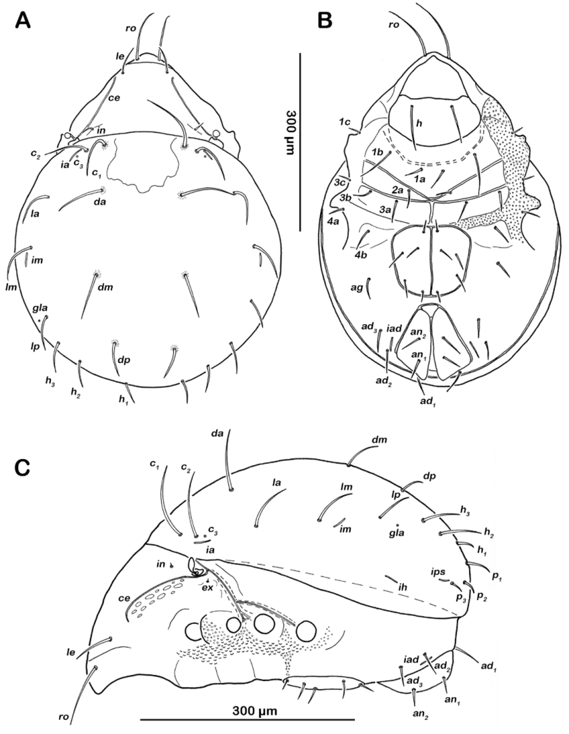
Larva ( Fig. 16 View FIGURE 16. F ). (N=3): length 280–299 µm (mean 288 µm).
Gastronotic region. Notogastral setae long, 11 pairs: c1-3, da, dm, dp, la, lm, lp, h1-2. Transversal ridge on centrodorsal plate passing posterior line of setae lm and dm.
Ventral region of idiosoma. Epimeral setation 2-1-2. Claparède’s organ globular medially covered by epimeral cuticle; no protective setae discernible. Aggenital, genital, adanal and anal setae not developed.
Legs. Chaetome and solenidia see table 4.
Protonymph. (N=2): length 353–366 µm (mean 360 µm).
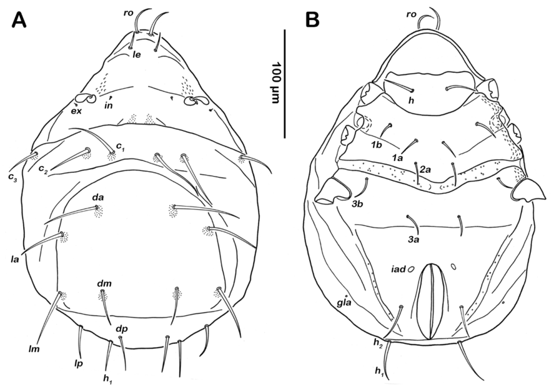
Gastronotic region. Notogastral setae long, 15 pairs: c1-3, da, dm, dp, la, lm, lp, h1-3, p1-3.
Ventral region of idiosoma. Epimeral setation 3-1-2-1. One pair of short genital setae. Adanal and anal setae not developed.
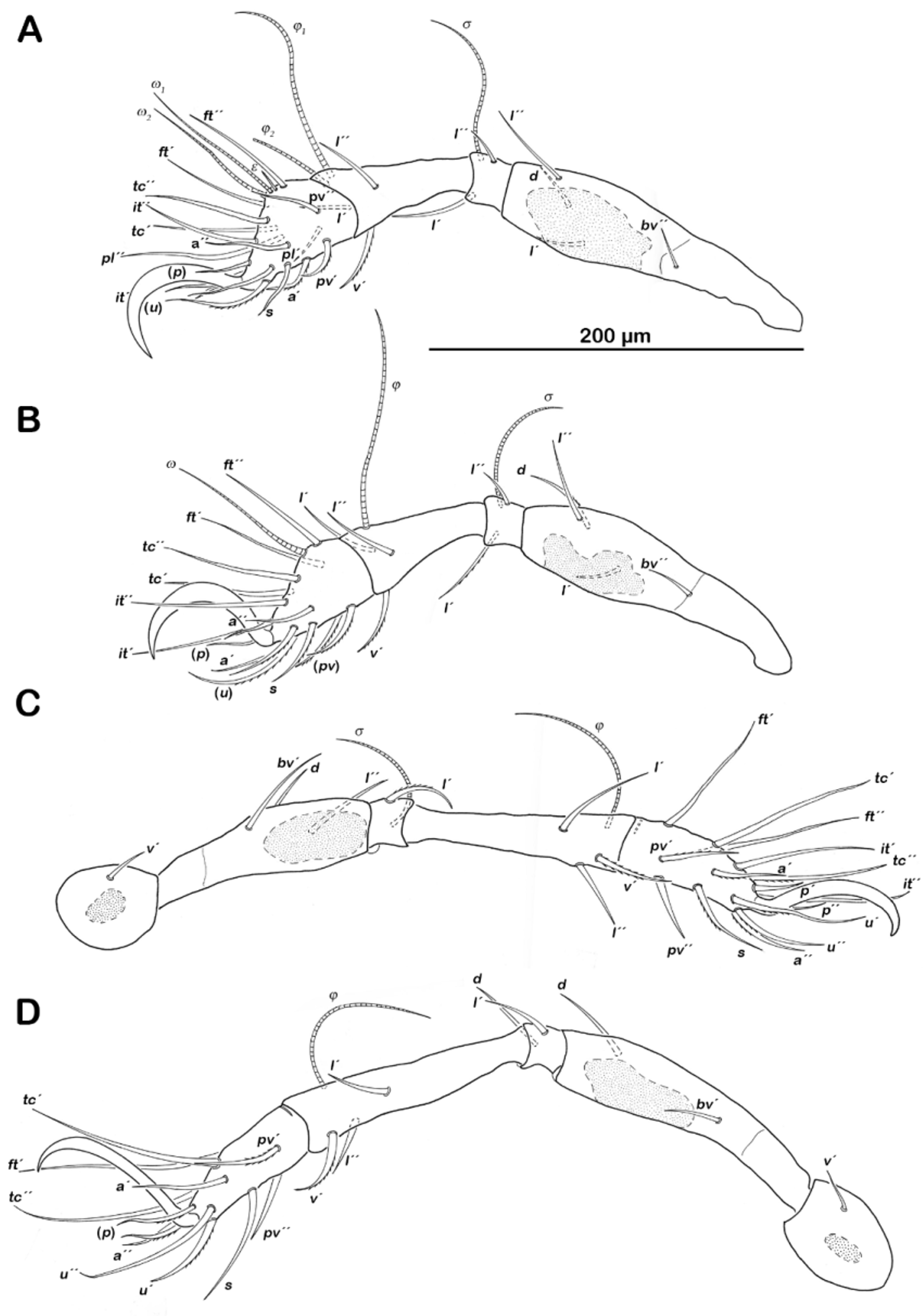
Legs ( Fig. 17 View FIGURE 17. F ). Chaetome and solenidia see table 4.
Deutonymph. (N=1): length 440 µm.
Gastronotic region. Notogastral setae 15 pairs, same positions and shapes as in protonymph.
Ventral region of idiosoma. Epimeral setation 3-1-2-2, seta 4b added in this stage. Genital setae short, 2 pairs; aggenital setae 1 pair, adanal setae 3 pairs, ad1-3, flanking anal orifice; anal setae vestigial, 2 pairs.
Legs. Chaetome and solenidia see table 4.
Tritonymph. (N=1): length 533 µm.
Gastronotic region. Fifteen pairs of notogastral setae, no difference to deutonymph. Ventral region of idiosoma. Epimeral setation 3-1-3-2, seta 3c close to trochanter III. Genital setae 4 pairs, aggenital setae 1 pair, adanal setae 3 pairs, anal setae 2 pairs.
Legs. Chaetome and solenidia see table 4.
No known copyright restrictions apply. See Agosti, D., Egloff, W., 2009. Taxonomic information exchange and copyright: the Plazi approach. BMC Research Notes 2009, 2:53 for further explanation.