
Phyllopodopsyllus pseudokunzi, Björnberg, Tagea & Kihara, Terue C., 2013
|
publication ID |
https://dx.doi.org/10.11646/zootaxa.3718.6.1 |
|
publication LSID |
lsid:zoobank.org:pub:4ED0B842-AFD9-486F-A01F-01B633972B4A |
|
persistent identifier |
https://treatment.plazi.org/id/03BA87BB-FFD8-FFA4-35C7-ADA2FBA8F984 |
|
treatment provided by |
ImsDioSync |
|
scientific name |
Phyllopodopsyllus pseudokunzi |
| status |
sp. nov. |
Phyllopodopsyllus pseudokunzi sp. nov.
( Figs. 11–16 View FIGURE 11 View FIGURE 12 View FIGURE 13 View FIGURE 14 View FIGURE 15 View FIGURE 16 )
Type material. Collected in Parcel da Praia Grande, São Sebastião Island (23 º 49 ’02”S 45 º 24 ’ 19 ”W). Four females (2 ovigerous), 14 Feb. 2007, Oliveira, J. M. coll. Holotype and paratypes deposited under same registration number - MZUSP 28029). The probable nauplius of the species collected in 23 º 50 ’00”S 45 º 26 ’ 82 ’W. The nauplii separated from the sample were very damaged and covered with debris, after a period of very rough sea.
Type locality. Parcel da Praia Grande, São Sebastião Is. (23 º 49 ’02”S 45 º 24 ’ 19 ”W).
Diagnosis. A Phyllopodopsyllus with foliaceous caudal rami inserted vertically (like the caudal fin of a fish) in the last urosomite, antennules with first segment very elongated; hook-like processes turned posteriorly on the second segment; leg 4 endopod 2 -segmented; female caudal rami 2.5 times longer than anal segment, almost oval in shape, dilated distally and straight at insertion point in anal segment.
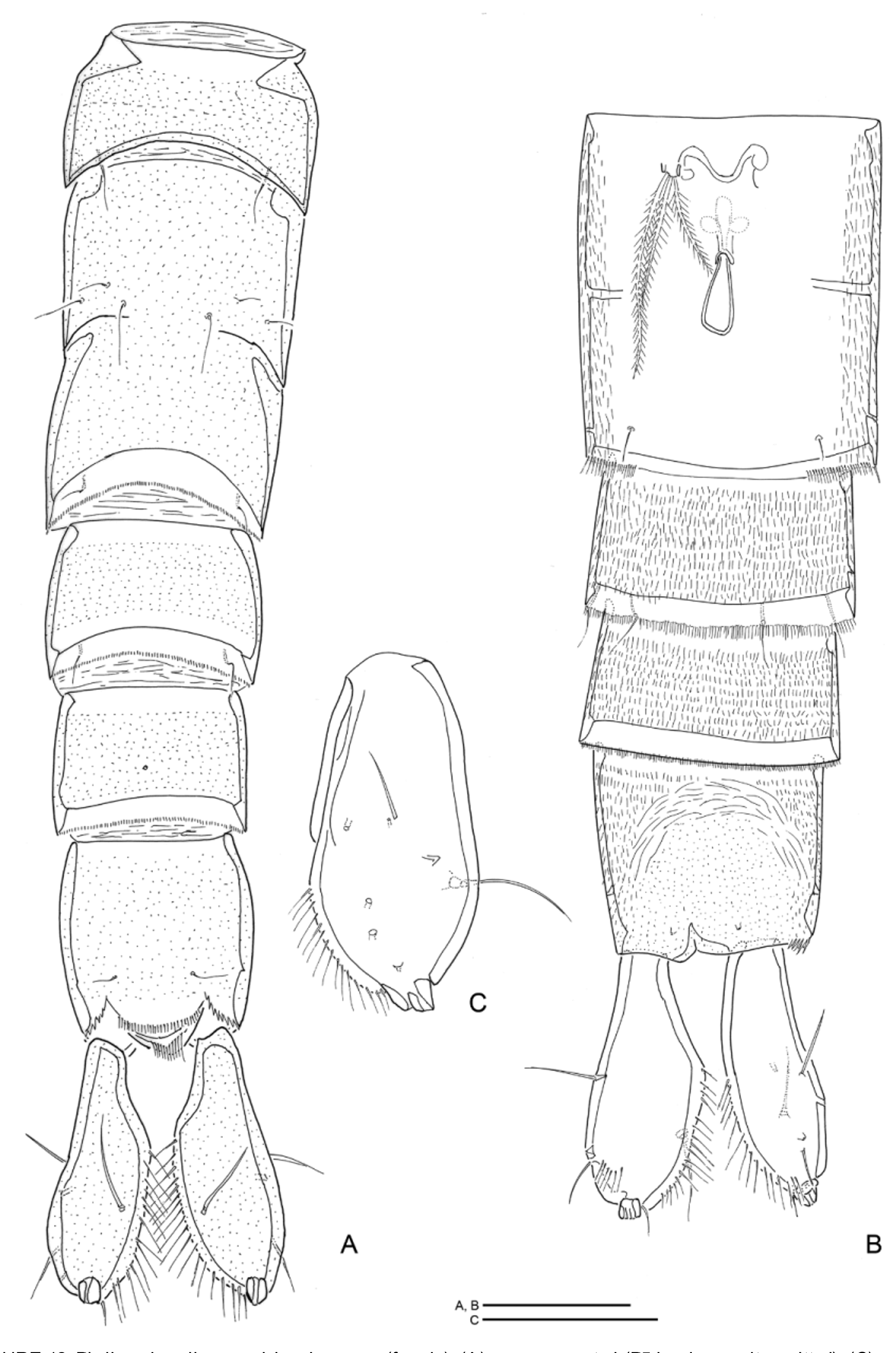
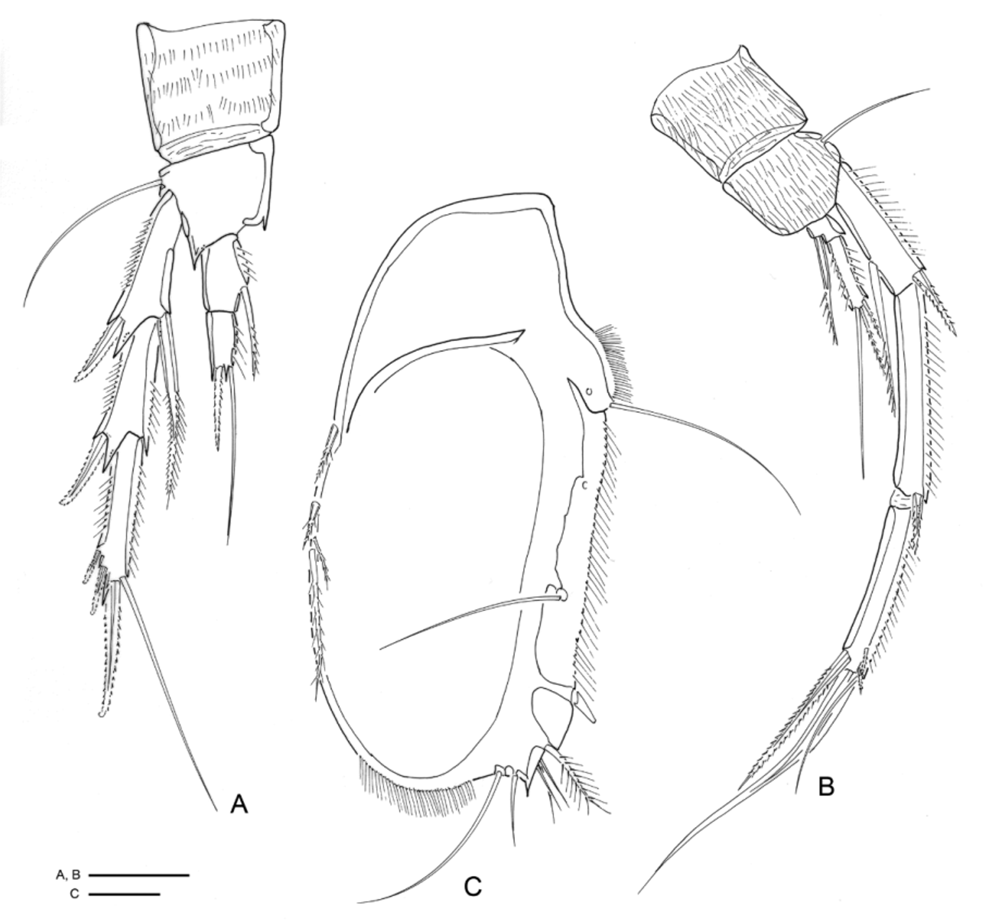
Description of adults. Female ( Figs 11 View FIGURE 11 A–C): Total body length from 680–830 µm long (n = 4, X = 751 µm). Color when alive light yellow-brown. Body with 10 somites, with hyaline frill on 2 nd to 4 th posterior urosomite margins. Very fine setules cover the whole body, rami included. Genital double somite with subdivision line visible dorsally and laterally. Genital area as in Fig. 12 View FIGURE 12 B. Anal operculum with 2 minute sensilla laterally, one on each side, but inserted at a distance anteriorly to the operculum which has a setulose margin ( Fig. 12 View FIGURE 12 A).
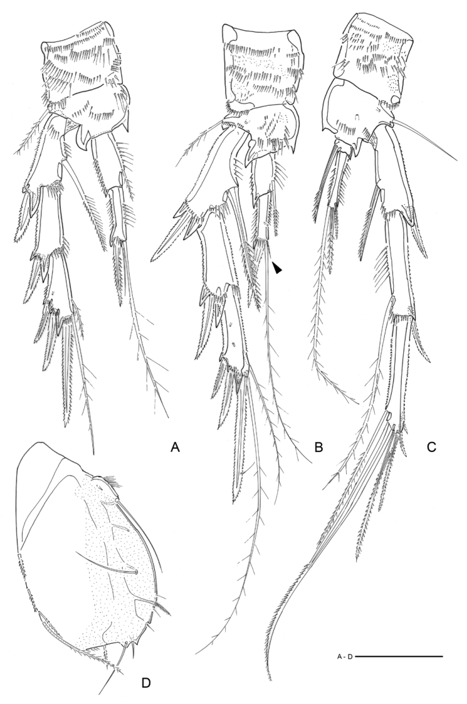
Caudal rami ( Figs 12 View FIGURE 12 A–C) 2.5 times longer than anal segment, almost oval in shape, dilated distally and straightened at insertion point in anal segment. Seta IV and V not observed in any of the 4 specimens examined, excepting for a small triangular process which seems to show 2 terminal insertion points located where the insertion points of the setae should be ( Fig. 12 View FIGURE 12 C). Two setae are present marginally (seta I or II and seta III); dorsal articulated seta present.
Rostrum flat. Defined at base, with two frontal sensilla.
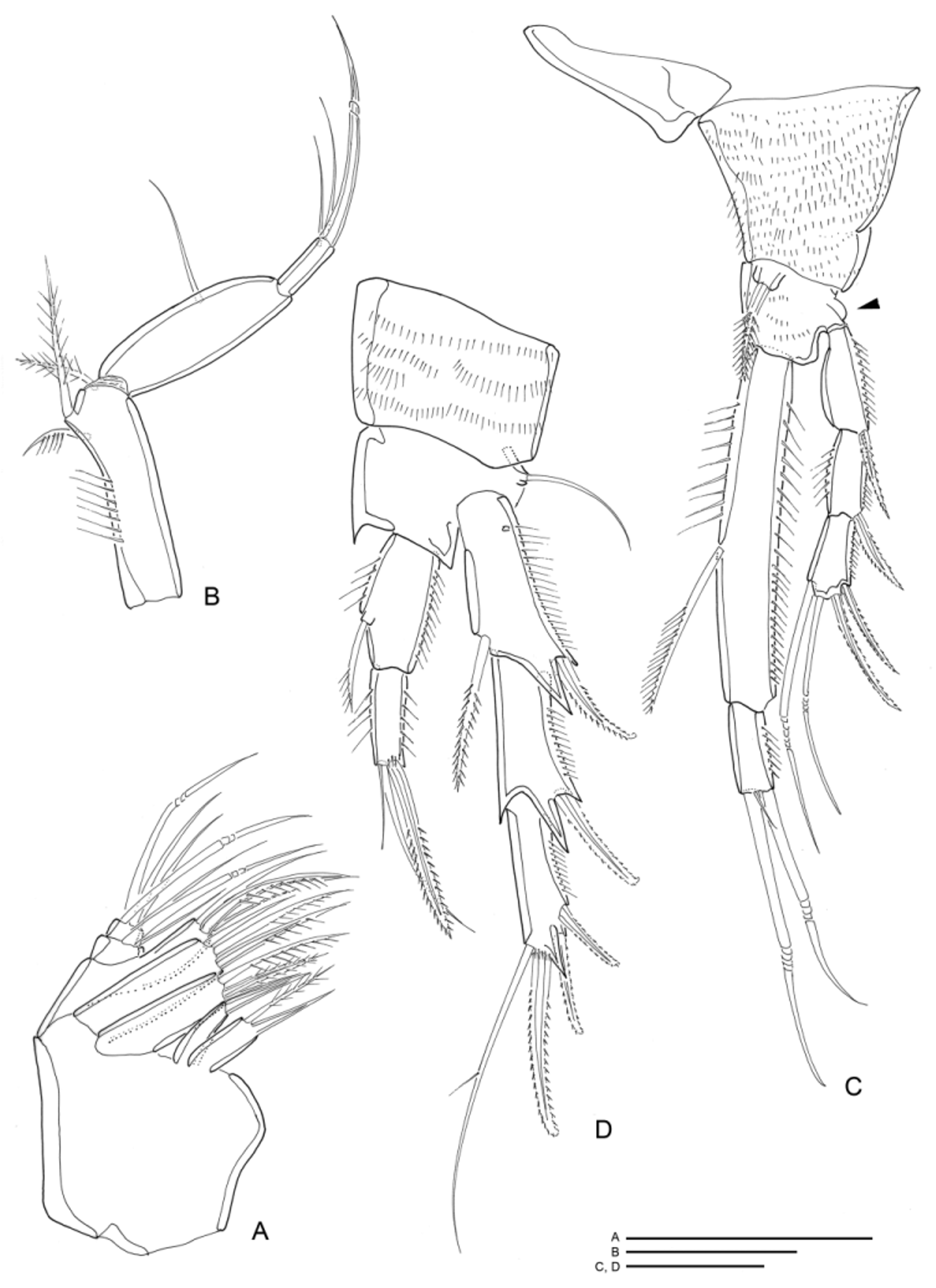
Antennule ( Fig. 13 View FIGURE 13 A) 8 -segmented. Segment- 1 very long, 4 times longer than wide, about as long as 5 following segments summed together. Segment- 2 with prominent pointed hook turned posteriorly. Setal formula: 1, 9, 7, 4 + 1 ae, 2, 3, 4, 7 + 1 ae.
Antenna ( Fig. 13 View FIGURE 13 B) biramous. Basis unarmed with row of short spinules along inner margin. Exopod 1 - segmented with 3 bipinnate setae (one of which confluent with exopod). Endopod 2 -segmented, segment- 1 unarmed with row of short spinules along inner margin, segment- 2 with 7 terminal setae (one unipinnate), 3 preterminal setae (two lateral) and rows of spinules (1 median and 2 marginal).
Mandible ( Fig. 13 View FIGURE 13 C) coxal gnathobase with 2 tooth-like processes, row of spines and 1 pinnate seta. Basis with tuft of spinules and two bipinnate setae. Exopod 1 -segmented with 3 setae (1 lateral and 2 apical). Endopod 1 - segmented with 2 medial and 6 terminal setae.
Maxillule ( Figs 13 View FIGURE 13 D–F) damaged, arthrite with 6 spines and 3 bipinnate setae apically and 2 marginal setae. Coxa with 6 setae on endite and 1 seta on exite (displaced during animal manipulation). Basis with 7 setae. Endopod with 4 bipinnate setae. Exopod missing.
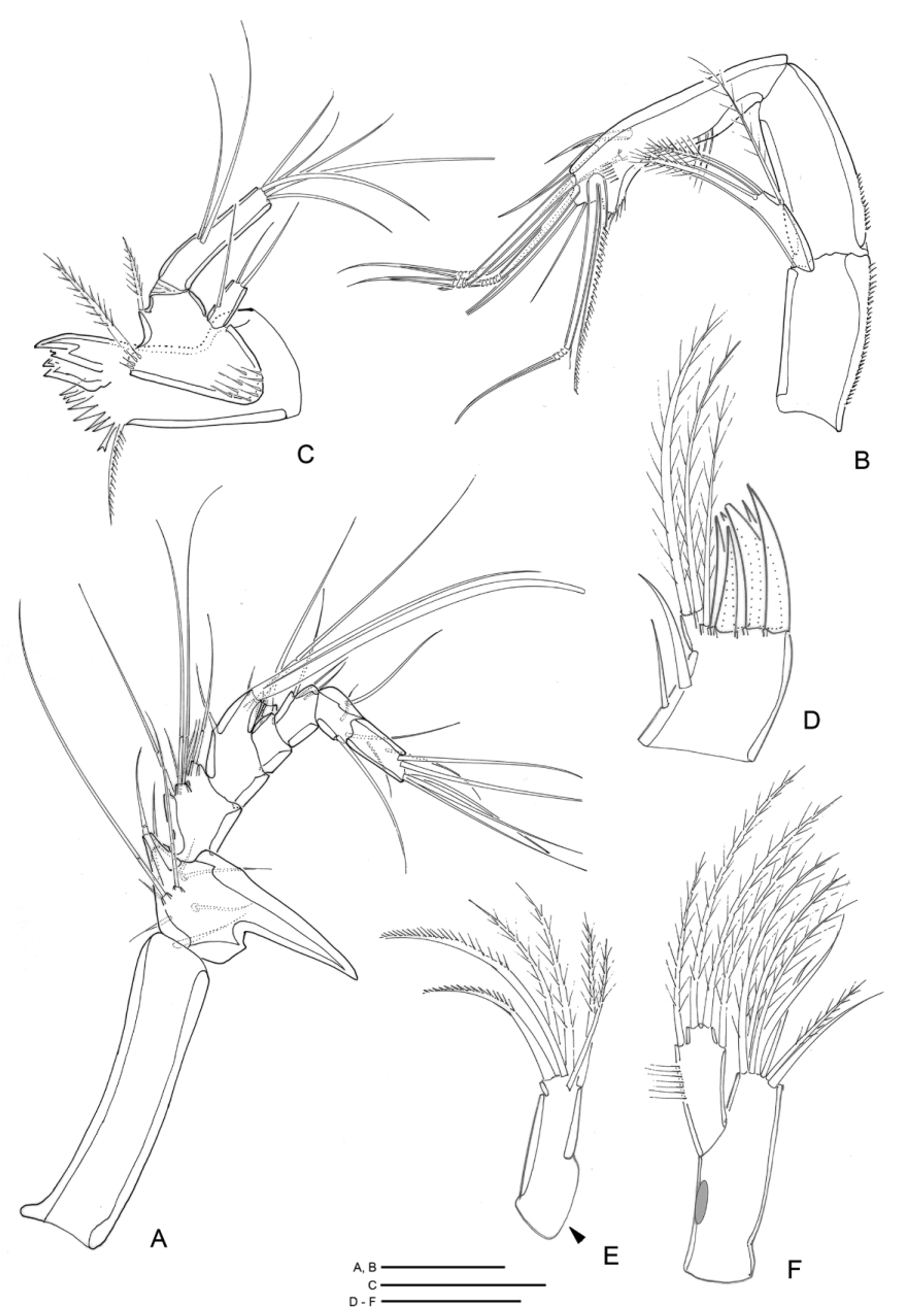
Maxilla ( Fig. 14 View FIGURE 14 A) with 4 syncoxal endites bearing 2, 1, 3, 3 spines proximal to distal segment, respectively. Basis armed with 1 spine and 2 setae. Endopod 2 -segmented, proximal segment with 3, distal segment with 4 setae.
Maxilliped ( Fig. 14 View FIGURE 14 B) prehensile. Syncoxa with 3 bipinnate setae. Basis with 1 seta. Endopod 1 -segmented, with terminal claw and 2 accompanying setae.
Leg 1 ( Fig. 14 View FIGURE 14 C) biramous. Coxa ornamented with rows of small spinules. Basis with rows of small spinules, bearing 1 bipinnate seta near proximal inner corner and 1 missing element on outer margin (arrowed). Exopodal segments with small spinules along outer margin and exp- 2 with setules along inner margin. Enp- 1 4.5 times longer than enp- 2, with spinules marginally; enp- 2 with small spinules along outer margin.
Legs 2–3 biramous ( Figs 14 View FIGURE 14 D, 15 A). Coxa ornamented with rows of spinules. Basis bearing 1 seta on outer corner, and spiniform projection on distal rim between exopod and endopod and on distal inner corner. Exopod 3 - segmented. Endopod 2 -segmented. Inner and outer margins of exopods and endopods ornamented with setules and spinules, spiniform projections on distal outer corner of exopods.
Leg 4 biramous ( Fig. 15 View FIGURE 15 B). Coxa ornamented with rows of spinules. Basis with rows of spinules, bearing 1 seta on outer corner. Exopod 3 -segmented; elongated segments with setules along outer margin. Endopod 2 - segmented; enp- 2 with setules marginally.
Legs 1–4 ( Figs 14 View FIGURE 14 C–D, 15 A–B) armature formula as follows:
Leg 5 ( Fig. 6 View FIGURE 6 D) large and foliaceous. Outer margin from proximal to distal with row of setules, 1 proximal slender seta (remnant of outer setophore), setules along margin, 1 seta inserted medially directed to inwards, 1 spine, 1 bippinate seta, 3 long spinules, 1 pointed process and 2 setae. Distal area with fine setules. Outer margin with 4 bipinnate setae.
Genital field as in Fig. 12 View FIGURE 12 B.
Leg 6 ( Fig. 12 View FIGURE 12 B) with 3 bipinnate setae on genital segment.
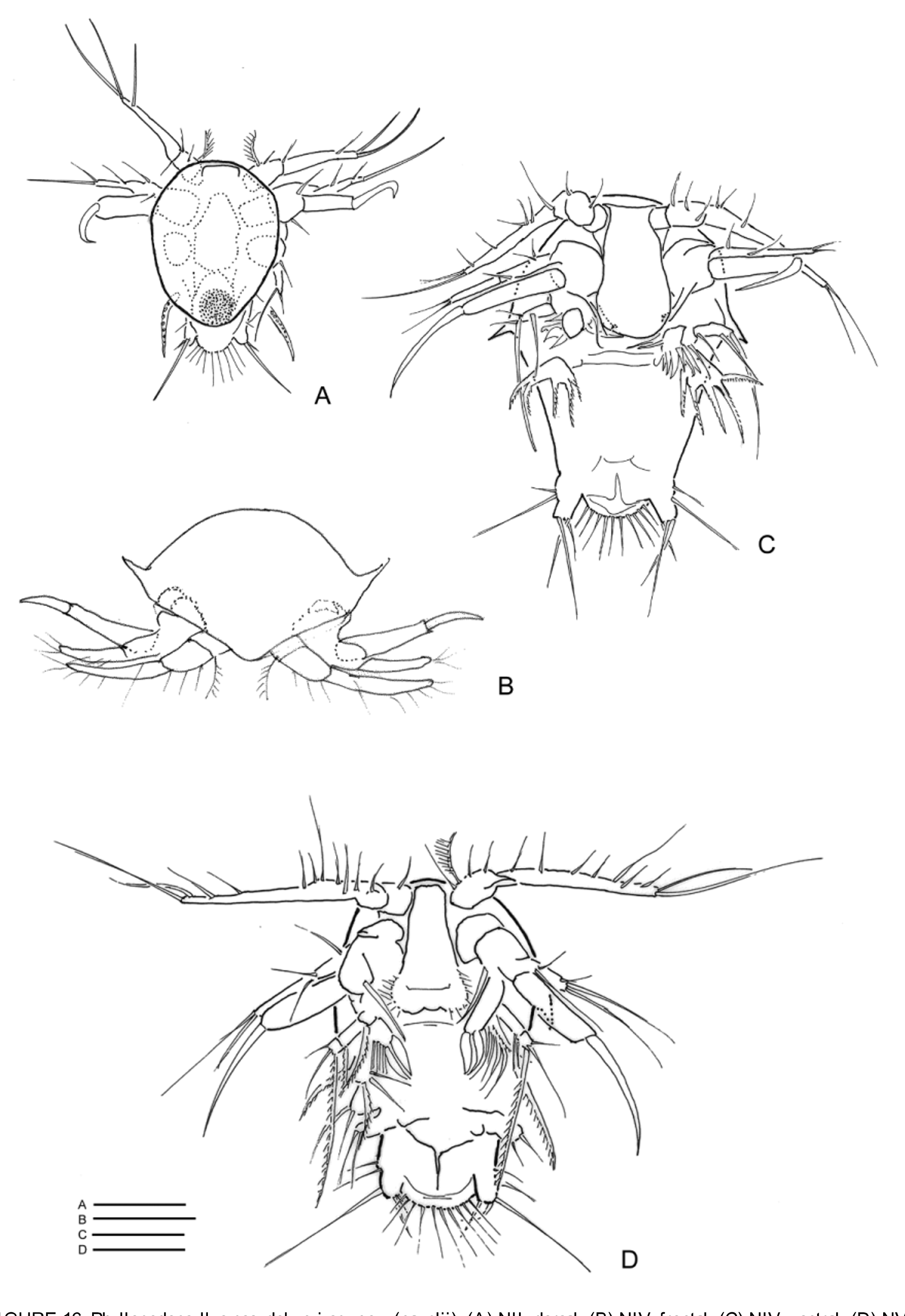
Description of naupliar stages. This tetragonicipitid nauplius ( Figs 16 View FIGURE 16 A–D), collected in the region, is the probable larva of this species because the females, sampled at the time, were ovigerous and the eggs in the broodpouches were ripe (transparent), so we expected to find nauplii. Those figured here were the only tetragonicipitid nauplii not yet identified which occurred in the region. The naupliar stages collected were NII, IV and VI.
NII ( Fig. 16 View FIGURE 16 A) 70 µm long. Dorsal shield oval, longer than wide, posterior region of body not covered by shield. Labrum straight. Antennule 3 -segmented, second segment bearing a pinnate seta and a simple seta, third segment bearing 4 setae. Antenna biramous: exopod with 2 lateral and one terminal seta, near to endopod insertion point an anterior seta; endopod one segmented with long hook-like terminal spine, below insertion point of endopod a posterior seta. Mandible with one-segmented exopod bearing 2 terminal setae and at insertion point another outer seta. Anlage of maxillule present in form of a thick spiny seta. Posterior region of body rounded, ornamented with a row of long setules. Two lateral ventral processes, one on each side of posterior region, bear a longer inner and a shorter outer seta. Another outer lateral seta inserted pre-terminally.
NIV ( Figs 16 View FIGURE 16 B–C) differs from NII as follows: 155 µm long, one lateral pointed process on each side of the first third of dorsal shield. Body longer, posteriorly wider and rounded, with strong indentation ventrally towards anal opening. On each side of the anal indentation a rectangular process bearing two lateral outer setae and three terminal setae of different lengths. Labrum is long, anteriorly straighter and posteriorly wider. Antennal exopod two-segmented: three setae on first segment and two terminal setae on second, long basal process directed medially partly covered by labrum is visible. Mandible: coxa with two seta-like processes directed medially towards labrum; exopod one-segmented bearing two terminal setae and a proximal seta; endopod bearing two thick pointed setae and two or more simple setae. Maxillule anlage hand-like, a more or less triangular structure bearing five thick pointed setae.
NVI ( Fig. 16 View FIGURE 16 D) differs from NIV as follows: Body 155 µm long; processes lateral to anal region shorter, anal region wider, outermost terminal seta on lateral process longer. Two lateral processes appear on each side of ventral body wall, bearing two setae each representing the anlage or primordium of future appendages. Antennule with increased number of setae (12–13) Labrum longer and straighter finely setulose along posterior margin. Antenna: coxa bears a masticatory process, and two pinnate setae turned medially towards labrum. Basis bears two pinnate setae turned posteriorly. Exopod: long, 4 or 5 segmented. Each segment bears one seta, except the last segment with two terminal setae. Mandible: endopod with 2 foliaceous or spatulate setae and four or five setae; exopod two or three segmented with a seta proximally, a lateral and distal seta on second segment and two terminally. Maxillule with 5 strongly setulose spines in last stages. Anlagen of maxilla and of maxillipeds: lateral processes of ventral body bearing a pair of setae each. Posterior region with 2 latertal processes bearing 3 lateral setules and, terminally, 1 longer and 1 shorter seta.
Remarks. The nauplii separated from the sample were very damaged and covered with debris, collected after a period of very ruff sea.
Etymology. The name species is derived from Greek pseudo (meaning false—used in science to denote a false resemblance to something). “Pseudo” and “ kunzi ” meaning “apparently like kunzi ”.

No known copyright restrictions apply. See Agosti, D., Egloff, W., 2009. Taxonomic information exchange and copyright: the Plazi approach. BMC Research Notes 2009, 2:53 for further explanation.
|
Kingdom |
|
|
Phylum |
|
|
Class |
|
|
Order |
|
|
Family |
|
|
Genus |