|
 Austropezia samuelsii Austropezia samuelsii
SynonymsEriopezia samuelsii
BiostatusPresent in region - Indigenous. Endemic
Images (click to enlarge)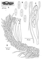
Caption: Figure 71. Austropezia samuelsii, P.D.D. 41744. A. Apothecia. B. Ascospores. C. Ascus
and paraphyses. D. Vertical section. | 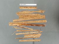
Caption: Dried type specimen
Owner: Herb PDD | 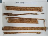
Caption: Dried type specimen
Owner: Herb PDD | 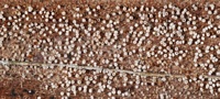
Caption: On dead frond nikau palm, Piha, June 2008.
Owner: Herb PDD | 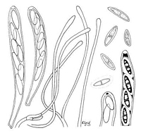
Caption: FIG. 2. Eriopezia samuelsii. Two asci mounted in lactic acid-cotton blue; four paraphyses in
Melzer's Reagent; five free ascospores, lowermost showing gel sheath, in cotton blue; ascus
apex showing J+ pore after 2% KOH pretreatment, in Melzer' | 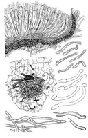
Caption: FIG. 3. Eriopezia samuelsii. Apothecial cross section and six marginal hairs in optical
section, in lactic acid-cotton blue; four hairs from base of apothecium, mounted in KOH-phloxine; portions of subicular hyphae, at left in optical section a |
Article: Spooner, B.M. (1987). Helotiales of Australasia: Geoglossaceae, Orbiliaceae, Sclerotiniaceae, Hyaloscyphaceae. Bibliotheca Mycologica 116: 711 p.
Description: APOTHECIA 0.4-0.6 mm diam., gregarious, black throughout when dry, basally immersed in
a white subiculum, very short-stipitate. DISC plano-convex, brownish with an olive tinge
when rehydrated, smooth. RECEPTACLE patellate, blackish, clothed to the margin with
white hairs and basally immersed in white subicular hyphae. ASCI 8-gpored, clavate-cylindric,
90-108(-115) x 11-13 µm, tapered below and expanded into a small foot 4-6 µm diam., apex
narrowed, rounded or obtusely conical, the pore blue in Melzer's reagent. ASCOSPORES 15-19 x (3.0-) 3.5-4.5 µm, hyaline, ellipso-fusoid, slightly inequilateral or curved, 1(-2)-septate,
surrounded by a gelatinous sheath 2-3 µm thick, biseriate. PARAPHYSES 1.5-2.0 µm diam.,
subcylindric, obtuse, hyaline or pale brown near the base, unbranched, surrounded by a
gelatinous sheath, coherent, exceeding the asci. SUBHYMENIUM not differentiated.
MEDULLARY EXCIPULUM hyaline or pale brown, composed or interwoven, thin-walled
hyphae and small cells 2-3 µm diam., forming a layer 40-50 µm thick at the centre of the
receptacle narrowed upwards and not developed in the parathecium. ECTAL EXCIPULUM
20-25 µm thick at the base of the receptacle, narrowed to 10-12 µm at the margin, brown,
composed in the lower part of small, subangular or subglobose, thin-walled cells 4-7 µm
diam., lying in irregular rows at an angle of about 30 degrees to the surface; towards the
margin becoming narrower and more elongated, forming parallel, septate hyphae 2.0-2.5 µm
diam., lying parallel to the surface. HAIRS arising from superficial cells, hyaline, obtuse,
cylindric or tapered, straight or flexuous, unbranched, septate, 20-35(-55) x 2.5-3.0 µm.
SUBICULAR HYPHAE 2.5-3.0 µm diam., hyaline, much branched, with thickened walls
roughened with superficial granules which are soluble in Melzer's reagent
Habitat: On dead leaves of Gahnia sp. (Cyperaceae). New
Zealand.
Notes: The apothecia are gregarious, nestled amongst conspicuous, white subicular hyphae. They
appear sessile but, when examined in longitudinal section, are seen to possess a very short,
central stipe by which they are at first loosely attached to the substrate, as figured by Korf
(1978). However, apothecia frequently become detached and suspended free from the
substrate amongst the subicular hyphae.
The appearance of this species is similar to that of Eriopezia caesia, having dark
apothecia which contrast markedly with a white subiculum, though in A. samuelsii the
subiculum is far less dense. The excipular structure is also comparable in these species,
comprising brown cells towards the base which become narrower upwards and form parallel
hyphae towards the margin. Apothecia of E. caesia have a well-developed stipe and a thick,
well-developed excipular tissue which gives rise at the surface to hyaline, much-branched
subicular hyphae. These hyphae, unlike those In the figure published by Korf (1978), arise
over the entire surface of the receptacle, even to the extreme margin. Thus, the apothecia of
Eriopezia are not so much seated on a subiculum as immersed in it, as though in a loosely
constructed stroma. The subicular hyphae have finely granulate walls, and this granulation is
not dissolved in Melzer's reagent, though is partially so after prolonged immersion in Cotton
blue in lactophenol. In contrast, apothecia of A. samuelsii have a very short stipe and a
reduced excipular tissue which gives rise to branched subicular hyphae only towards the base
of the receptacle, these hyphae being encrusted with small particles which are readily soluble
in Melzer's reagent. Only simple, hyaline hairs are present over the upper part of the
receptacle, which stands free of the subiculum.
Further significant differences between these species are apparent. The paraphyses of E.
caesia are frequently forked or branched whereas those of A. samuelsii are simple, coherent
and sheathed with gel. The asci of the former are minute, usually less than 35 µm long, and
contain small, unicellular ascospores whereas those of A. samuelsii are frequently greater than
100 µm in length and contain ascospores which are large, septate and surrounded by a 0.5 mm
thick gelatinous sheath. These differences make me unwilling to follow Korf in referring the
two species to the same genus, and, as E. caesia is the type species of Eriopezia, I propose
here a new genus to accommodate E. samuelsii.
Korf (1978) distinguished two tribes, Polydesmieae and Arachnopezizeae, in subfamily
Arachnopezizoideae, based on the position of the apothecia in relation to the substrate and
subiculum. Apothecia of species of Polydesmieae are attached to the substrate, whereas those
of Arachnopezizeae arise directly from subicular hyphae. If this division is maintained,
Austropezia must fall, with Eriopezia, in the Polydesmieae. However, the tenuous attachment
to the substrate exhibited by apothecia of A. samuelsii may render such a division difficult to
uphold, and there is no other indication that the genus is more closely related to Polydesmia
than to Arachnopeziza. Apothecia of Arachnopeziza have a comparable, though hyaline, ectal
excipulum, but develop directly from the subicular hyphae and are never attached to the
substrate. They differ further from those of Austropezia in having well-developed hairs. The
inter-relationships of these genera are difficult to interpret and, as discussed in the introduction
to the Hyaloscyphaceae, the taxonomy of the genera currently assigned to subfamily
Arachnopezizoideae is not yet entirely satisfactory.
Article: Korf, R.P. (1978). Revisionary studies in the Arachnopezizoideae: a monograph of the Polydesmieae. Mycotaxon 7: 457-492.
Notes: ETYMOLOGY: From the name of the collector of the holotype specimen.
|