[1] JONES SP, BAQAI K, CLEGG A, et al. Stroke in India: a systematic review of the incidence, prevalence, and case fatality. Int J Stroke. 2022;17(2):132-140.
[2] ZHANG Z, SUN GY, DING S. Glial cell line-derived neurotrophic factor and focal ischemic stroke. Neurochem Res. 2021;46(10):2638-2650.
[3] TANG HL, LI Y, TANG WJ, et al. Endogenous neural stem cell-induced neurogenesis after ischemic stroke: processes for brain repair and perspectives. Transl Stroke Res. 2022. doi: 10.1007/s12975-022-01078-5.
[4] FAN Y, LI YK, YANG YK, et al. Chlorogenic acid promotes angiogenesis and attenuates apoptosis following cerebral ischaemia-reperfusion injury by regulating the PI3K-Akt signalling. Pharm Biol. 2022;60(1):1646-1655.
[5] GAO X, ZHANG XJ, CUI LL, et al. Ginsenoside Rb1 promotes motor functional recovery and axonal regeneration in post-stroke mice through cAMP/PKA/CREB signaling pathway. Brain Res Bull. 2020;154:51-60.
[6] YANG HJ, QI CX, SU F, et al. Cerebral ischemia/reperfusion injury and pharmacologic preconditioning as a means to reduce stroke-induced inflammation and damage. Neurochem Res. 2022;47(12):3598-3614.
[7] LI L, ZHOU J, HAN L, et al. The specific role of reactive astrocytes in stroke. Front Cell Neurosci. 2022;16:850866.
[8] WANG H, SONG G, CHUANG H, et al. Portrait of glial scar in neurological diseases. Int J Immunopathol Pharmacol. 2018;31:2058738418801406.
[9] KANG J, KIM BJ, HAN MK, et al. The changing effect of blood pressure on stroke outcomes through acute to subacute stage of ischemic stroke. J Stroke Cerebrovasc Dis. 2019;28(9):2563-2568.
[10] WASAN H, SINGH D, JOSHI B, et al. Dihydromyricetin alleviates cerebral ischemia-reperfusion injury by attenuating apoptosis and astrogliosis in peri-infarct corte. Neurol Res. 2022;44(5):403-414.
[11] YAWOOT N, SENGKING J, WICHA P, et al. Melatonin attenuates reactive astrogliosis and glial scar formation following cerebral ischemia and reperfusion injury mediated by GSK-3 beta and RIP1K. J Cell Physiol. 2022;237(3):1818-1832.
[12] SU HB, FAN SP, ZHANG LQ, et al. TMAO aggregates neurological damage following ischemic stroke by promoting reactive astrocytosis and glial scar formation via the Smurf2/ALK5 axis. Front Cell Neurosci. 2021;15:569424.
[13] HEITHOFF BP, GEORGE KK, PHARES AN, et al. Astrocytes are necessary for blood-brain barrier maintenance in the adult mouse brain. Glia. 2021;69(2):436-472.
[14] VERKHRATSKY A, NEDERGAADR M. The homeostatic astroglia emerges from evolutionary specialization of neural cells. Philos Trans R Soc Lond B Biol Sci. 2016;371(1700):20150428.
[15] HE T, YANG GY, ZHANG Z. Crosstalk of astrocytes and other cells during ischemic stroke. Life (Basel). 2022;12(6):910.
[16] ZAMANIAN JL, XU L, FOO LC, et al. Genomic analysis of reactive astrogliosis. J Neurosci. 2012;32(18):6391-6410.
[17] LIDDELOW SA, GUTTENPLAN KA, LARKE LEC, et al. Neurotoxic reactive astrocytes are induced by activated microglia. Nature. 2017;541(7638):481-487.
[18] DIMITROVA-SHUMKOVSKA J, KRSTANOSKI L, VEENMAN L. Diagnostic and therapeutic potential of TSPO studies regarding neurodegenerative diseases, psychiatric disorders, alcohol use disorders, traumatic brain injury, and stroke: an update. Cells. 2020;9(4):870.
[19] SHEN XY, GAO ZK, HAN Y, et al. Activation and role of astrocytes in ischemic stroke. Front Cell Neurosci. 2021;15:755955.
[20] WANNER IB, ANDERSON MA, SONG B, et al. Glial scar borders are formed by newly proliferated, elongated astrocytes that interact to corral inflammatory and fibrotic cells via STAT3-dependent mechanisms after spinal cord injury. J Neurosci. 2013;33(31):12870-12886.
[21] CHAN SJ, NIU W, HAYAKAWA K, et al. Promoting neuro-supportive properties of astrocytes with epidermal growth factor hydrogels. Stem Cells Transl Med. 2019;8(12):1242-1248.
[22] CHENG X, YEUNG PKK, ZHONG K, et al. Astrocytic endothelin-1 overexpression promotes neural progenitor cells proliferation and differentiation into astrocytes via the Jak2/Stat3 pathway after stroke. J Neuroinflammation. 2019;16(1):227.
[23] BANITALEBI S, SKAULI N, GEISELER S, et al. Disassembly and mislocalization of AQP4 in incipient scar formation after experimental stroke. Int J Mol Sci. 2022; 23(3):1117.
[24] SUN CF, LIN LY, YIN LK, et al. Acutely inhibiting AQP4 with TGN-020 improves functional outcome by attenuating edema and peri-infarct astrogliosis after cerebral ischemic. Front Immunol. 2022;13:870029.
[25] SABELSTROM H, STENUDD M, REU P, et al. Resident neural stem cells restrict tissue damage and neuronal loss after spinal cord injury in mice. Science. 2013; 342(6158):637-640.
[26] HUGHES EG, KANG SH, FUKAYA M, et al. Oligodendrocyte progenitors balance growth with self-repulsion to achieve homeostasis in the adult brain. Nat Neurosci. 2013;16(6):668-676.
[27] AHMED A, NAKAGAWA H, ISAKSEN TJ, et al. The effects of Bone Morphogenetic Protein 4 on adult neural stem cell proliferation, differentiation and survival in an in vitro model of ischemic stroke. Neurosci Res. 2022;183:17-29.
[28] GALINDO LT, MUNDIM M, PINTO AS, et al. Chondroitin sulfate impairs neural stem cell migration through ROCK activation. Mol Neurobiol. 2018;55(4):3185-3195.
[29] LORENZANA AO, LEE JK, MUI M, et al. A surviving intact branch stabilizes remaining axon architecture after injury as revealed by in vivo imaging in the mouse spinal cord. Neuron. 2015;86(4):947-954.
[30] MAHAR M, CAVALLI V. Intrinsic mechanisms of neuronal axon regeneration. Nat Rev Neurosci. 2018;19(6):323-337.
[31] ZBESKO JC, NGUYEN TV, YANG T, et al. Glial scars are permeable to the neurotoxic environment of chronic stroke infarcts. Neurobiol Dis. 2018;112:63-78.
[32] ZHANG R, WU Y, XIE F, et al. RGMa mediates reactive astrogliosis and glial scar formation through TGFβ1/Smad2/3 signaling after stroke. Cell Death Differ. 2018; 25(8):1503-1516.
[33] CAO H, SETO SW, BHUYAN DJ, et al. Effects of thrombin on the neurovascular unit in cerebral ischemia. Cell Mol Neurobiol. 2022;42(4):973-984.
[34] SCHACHTRUP C, RYU JK, HELMRICK MJ, et al. Fibrinogen triggers astrocyte scar formation by promoting the availability of active TGF-beta after vascular damage. J Neurosci. 2010;30(17):5843-5854.
[35] RENAULT-MIHARA F, MUKAINO M, SHINOZAKI M, et al. Regulation of RhoA by STAT3 coordinates glial scar formation. J Cell Biol. 2017;216(8):2533-2550.
[36] SUI Y, BIAN LG, AI QL, et al. Gastrodin inhibits inflammasome through the STAT3 signal pathways in TNA2 astrocytes and reactive astrocytes in experimentally induced cerebral ischemia in rats. Neuromolecular Med. 2019;21(3):275-286.
[37] LIU J, ZHU YM, GUO Y, et al. Inhibition of GSK3β and RIP1K attenuates glial scar formation induced by ischemic stroke via reduction of inflammatory cytokine productio. Front Pharmacol. 2020;11:812.
[38] ZHU ZM, LIN L, WEI C, et al. The key regulator of necroptosis, RIP1 kinase, contributes to the formation of astrogliosis and glial scar in ischemic strok. Transl Stroke Res. 2021;12(6):991-1017.
[39] LIU H, SUN S, LIU B. Smurf2 exerts neuroprotective effects on cerebral ischemic injury. J Biol Chem. 2021;297(2):100537.
[40] SCHWEDHELM E, VON LUCADOU M, PEINE S, et al. Trimethyllysine, vascular risk factors and outcome in acute ischemic stroke (MARK-STROKE). Amino Acids. 2021;53(4):555-561.
[41] ZHANG YF, MENG LB, HAO ML, et al. Identification of co-expressed genes between atrial fibrillation and strok. Front Neurol. 2020;11:184.
[42] DAI Q, LI S, LIU T, et al. Interleukin-17A-mediated alleviation of cortical astrocyte ischemic injuries affected the neurological outcome of mice with ischemic stroke. J Cell Biochem. 2019. doi: 10.1002/jcb.28429.
[43] LIU SP, HUANG L, FLORES J, et al. Secukinumab attenuates reactive astrogliosis via IL-17RA/(C/EBPβ)/SIRT1 pathway in a rat model of germinal matrix hemorrhage. CNS Neurosci Ther. 2019;25(10):1151-1161.
[44] 崔迈尹,付艳琼,李卓丽,等.达珀利奈抑制急性缺血性脑卒中小鼠皮层神经炎症、胶质细胞活化及SARM1表达[J].中国组织化学与细胞化学杂志, 2022,31(2):107-115.
[45] YE SY, APPLE JE, REN X, et al. Microglial VPS35 deficiency regulates microglial polarization and decreases ischemic stroke-induced damage in the cortex. J Neuroinflammation. 2019;16(1):235.
[46] CHEN WC, CHANG LH, HUANG SS, et al. Aryl hydrocarbon receptor modulates stroke-induced astrogliosis and neurogenesis in the adult mouse brain. J Neuroinflammation. 2019;16(1):187.
[47] ZHAO X, ZHOU KS, LI ZH, et al. Knockdown of Ski decreased the reactive astrocytes proliferation in vitro induced by oxygen-glucose deprivation/reoxygenatio. J Cell Biochem. 2018;119(6):4548-4558.
[48] YANG X, GENG KY, ZHANG JF, et al. Sirt3 mediates the inhibitory effect of adjudin on astrocyte activation and glial scar formation following ischemic stroke. Front Pharmacol. 2017;8:943.
[49] LI Z, SONG Y, HE T, et al. M2 microglial small extracellular vesicles reduce glial scar formation via the miR-124/STAT3 pathway after ischemic stroke in mice. Theranostics. 2021;11(3):1232-1248.
[50] YUAN YJ, LIU LJ, DU Y, et al. P-hydroxy benzaldehyde revitalizes the microenvironment of peri-infarct cortex in rats after cerebral ischemia-reperfusion. Phytomedicine. 2022;105:154379.
[51] LI W, LIU J, CHEN JR, et al. Neuroprotective effects of DTIO, a novel analog of Nec-1, in acute and chronic stages after ischemic stroke. Neuroscience. 2018; 390: 12-29.
[52] CHITIMUS DM, POPESCU MR, VOICULESCU SE, et al. Melatonin’s impact on antioxidative and anti-inflammatory reprogramming in homeostasis and disease. Biomolecule. 2020;10(9):1211.
[53] LU T, LI H, ZHOU Y, et al. Neuroprotective effects of alisol A 24-acetate on cerebral ischaemia-reperfusion injury are mediated by regulating the PI3K/AKT pathway. J Neuroinflammation. 2022;19(1):37.
[54] LUO D, ZHANG Y, YUAN X, et al. Oleoylethanolamide inhibits glial activation via moudulating PPARα and promotes motor function recovery after brain ischemi. Pharmacol Res. 2019;141:530-540.
[55] GAO X, ZEB S, HE YY, et al. Valproic acid inhibits glial scar formation after ischemic stroke. Pharmacology. 2022;107(5-6):263-280.
[56] YEW WP, DJUKIC ND, JAYASEELAN JSP, et al. Differential effects of the cell cycle inhibitor, olomoucine, on functional recovery and on responses of peri-infarct microglia and astrocytes following photothrombotic stroke in rats. J Neuroinflammation. 2021;18(1):168.
[57] ZHANG X, DU QM, YANG Y, et al. Salidroside alleviates ischemic brain injury in mice with ischemic stroke through regulating BDNK mediated PI3K/Akt pathway. Biochem Pharmacol. 2018;156:99-108.
[58] WEI YC, HONG HM, ZHANG XQ, et al. Salidroside inhibits inflammation through PI3K/Akt/HIF signaling after focal cerebral ischemia in rats. Inflammation. 2017; 40(4):1297-1309.
[59] DONG C, WEN S, ZHAO S, et al. Salidroside inhibits reactive astrogliosis and glial dcar formation in late cerebral ischemia via the Akt/GSK-3β pathway. Neurochem Res. 2021;46(4):755-769.
[60] OU-YANG L, LIU Y, WANG BY, et al. Carnosine suppresses oxygen-glucose deprivation/recovery-induced proliferation and migration of reactive astrocytes of rats in vitro. Acta Pharmacol Sin. 2018;39(1):24-34.
[61] ZHANG Y, LIU J, YAO M, et al. Sailuotong capsule prevents the cerebral ischaemia-induced neuroinflammation and impairment of recognition memory through inhibition of LCN2 expression. Oxid Med Cell Longev. 2019; 2019:8416105.
[62] GHAZAVI H, SHIRZAD S, FOROUZANFAR F, et al. The role of resveratrol as a natural modulator in glia activation in experimental models of stroke. Avicenna J Phytomed. 2020;10(6):557-573.
[63] DZYUBENKO E, MANRIQUE-CASTANO D, PILLATH-EILERS M, et al. Tenascin-C restricts reactive astrogliosis in the ischemic brain. Matrix Biol. 2022;110:1-15.
[64] HUANG L, LI S, DAI Q, et al. Astrocytic Yes-associated protein attenuates cerebral ischemia-induced brain injury by regulating signal transducer and activator of transcription 3 signaling. Exp Neurol. 2020;333:113431.
[65] CONFORTI P, MEZEY S, NATH S, et al. Fibrinogen regulates lesion border-forming reactive astrocyte properties after vascular damage. Glia. 2022;70(7):1251-1266.
[66] HETTIARATCHI MH, O’MEARA MJ, TEAL CJ, et al. Local delivery of stabilized chondroitinase ABC degrades chondroitin sulfate proteoglycans in stroke-injured rat brains. J Control Release. 2019;297:14-25.
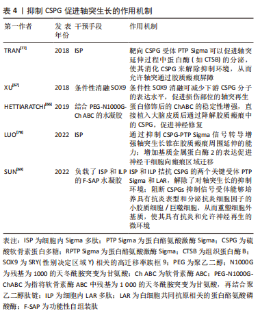
[67] XU X, BASS B, MCKILLOP WM, et al. Sox9 knockout mice have improved recovery following stroke. Exp Neurol. 2018;303:59-71.
[68] ZHU YM, GAO X, NI Y, et al. Sevoflurane postconditioning attenuates reactive astrogliosis and glial scar formation after ischemia-reperfusion brain injury. Neuroscience. 2017;356:125-141.
[69] SUN XM, Liu HQ, TAN Z, et al. Remodeling microenvironment for endogenous repair through precise modulation of chondroitin sulfate proteoglycans following spinal cord injury. Small. 2022. doi:1002/smll.202205012.
[70] GARCIA JH, YOSHIDA Y, CHEN H, et al. Progression from ischemic-injury to infarct following middle cerebral-artery occlusion in the rat. Am J Pathol. 1993; 142(2):623-635.
[71] YAMASHITA K, VOGEL P, FRITZE K, et al. Monitoring the temporal and spatial activation pattern of astrocytes in focal cerebral ischemia using in situ hybridization to GFAP mRNA: comparison with sgp-2 and hsp70 mRNA and the effect of glutamate receptor antagonist. Brain Res. 1996;735(2):285-297.
[72] BADAN I, BUCHHOLD B, HAMM A, et al. Accelerated glial reactivity with reduced to stroke in aged rats correlates functional recover. J Cereb Blood Flow Metab. 2003;23(7):845-854.
[73] ZHU Z, ZHANG Q, YU Z, et al. Inhibiting cell cycle progression reduces reactive astrogliosis initiated by scratch injury in vitro and by cerebral ischemia in vivo. Glia. 2007;55(5):546-558.
[74] HAYAKAWA K, NAKANO T, IRIE K, et al. Inhibition of reactive astrocytes with fluorocitrate retards neurovascular remodeling and recovery after focal cerebral ischemia in mice. J Cereb Blood Flow Metab. 2010;30(4):871-882.
[75] BAO Y, QIN LY, KIM E, et al. CD36 is involved in astrocyte activation and astroglial scar formation. J Cereb Blood Flow Metab. 2012;32(8):1567-1577.
[76] HUANG LJ, WU ZB, ZHUGE QC, et al. Glial scar formation occurs in the human brain after ischemic stroke. Int J Med Sci. 2014;11(4):344-348.
[77] TRAN AP, SUNDAR S, YU MG, et al. Modulation of receptor protein tyrosine phosphatase sigma increases chondroitin sulfate proteoglycan degradation through cathepsin B secretion to enhance axon outgrowt. J Neurosci. 2018; 38(23):5399-5414.
[78] LUO FC, WANG JP, ZHANG Z, et al. Inhibition of CSPG receptor PTPs promotes migration of newly born neuroblasts, axonal sprouting, and recovery from stroke. Cell Rep. 2022;40(4):111137. |