Quick Facts
Origin: Anterior tibial artery.
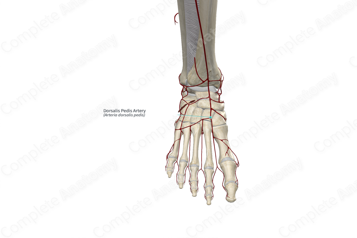
Course: Travels distally to the base of the first metatarsal.
Branches: Medial and lateral tarsal, arcuate, first dorsal metatarsal, and deep plantar arteries.
Supplied Structures: Dorsum of the foot.
Related parts of the anatomy
Origin
The dorsalis pedis artery arises from the anterior tibial artery. In up to 5% of individuals, it may arise from the perforating branch of the fibular artery (Tubbs, Shoja and Loukas, 2016).
The dorsalis pedis artery may be enlarged if the main plantar arterial supply is diminished. Conversely, it is absent in approximately 12% of individuals and may be replaced by an enlarged perforating branch from the fibular artery (Tubbs, Shoja and Loukas, 2016).
Course
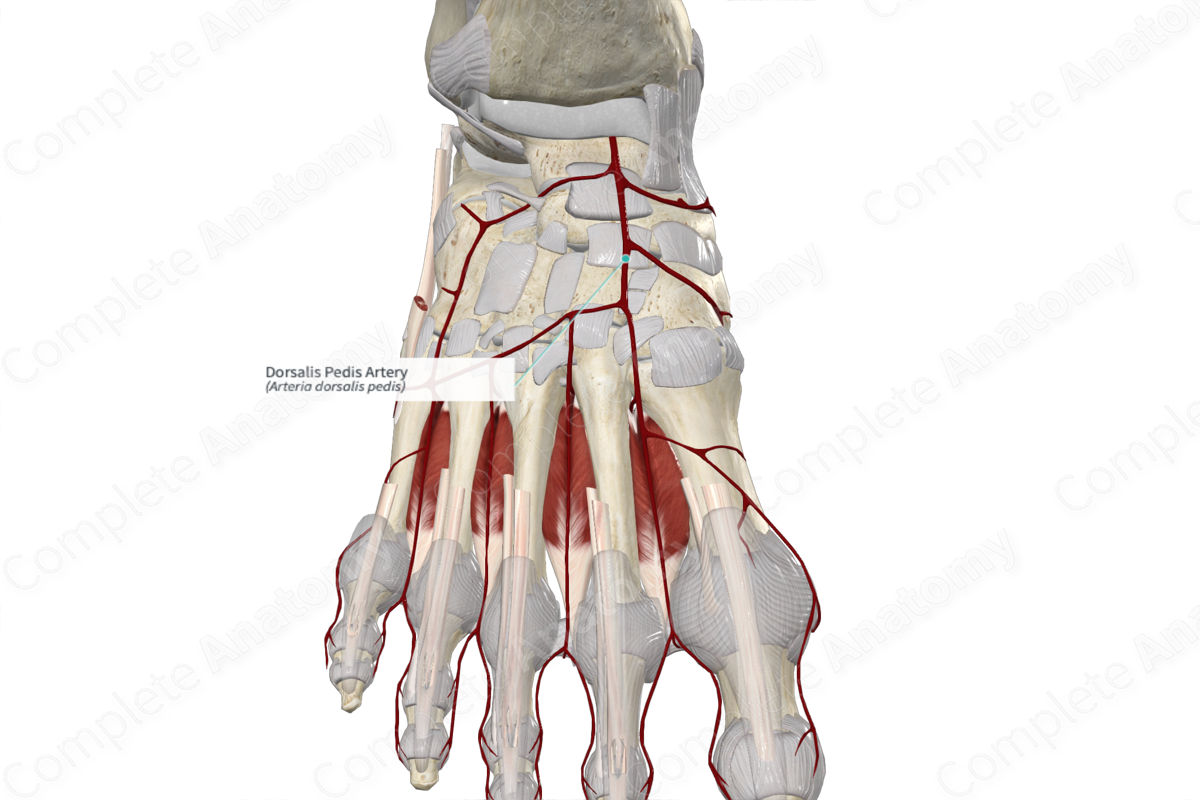
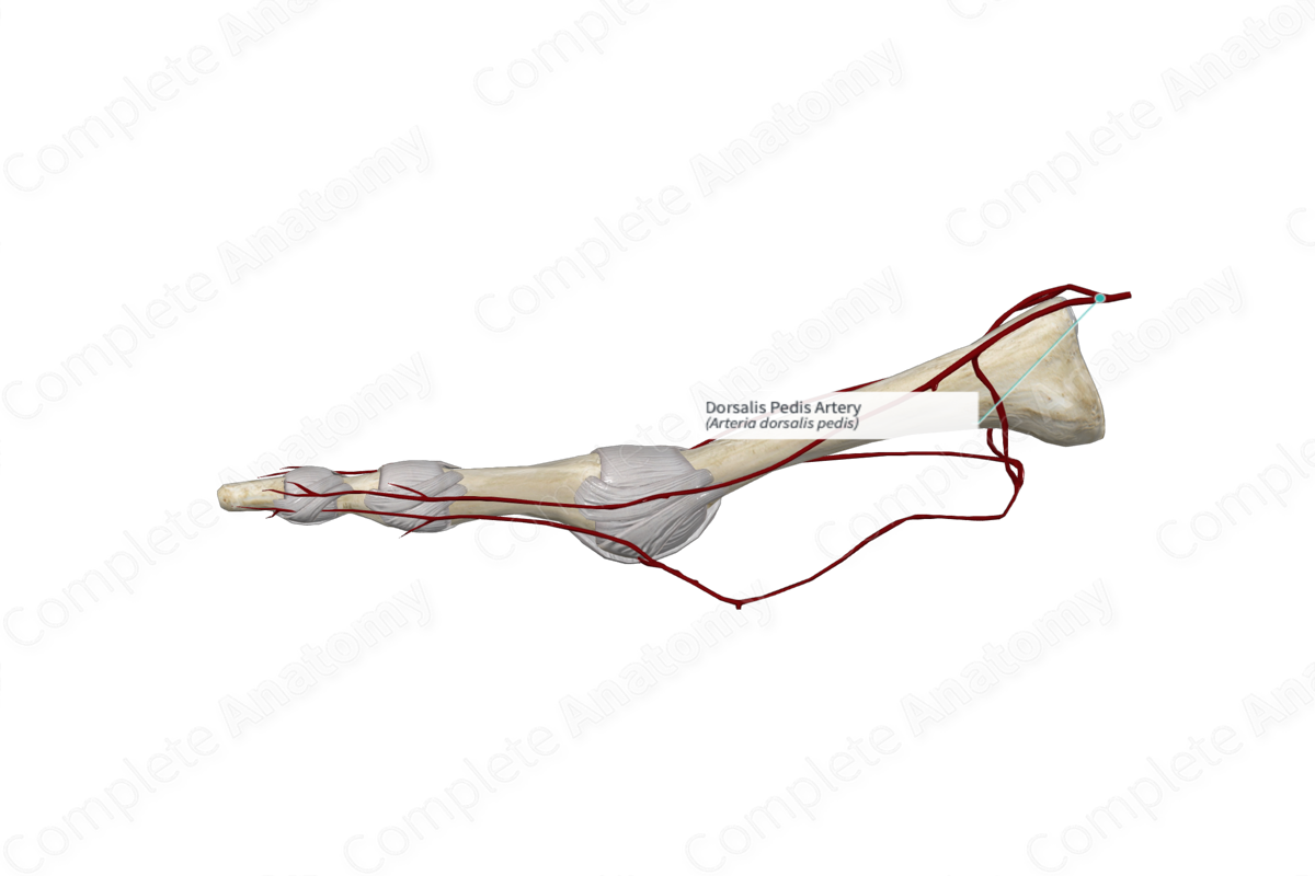
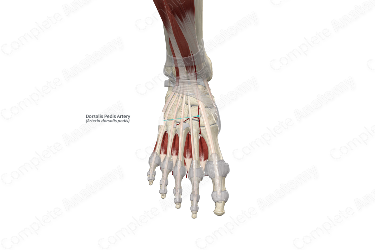
The dorsalis pedis artery runs distally along the talocrural joint and tarsal bones, deep to the extensor hallucis brevis muscle. The deep fibular nerve sits lateral to the dorsalis pedis artery.
The dorsalis pedis artery continues along the dorsal aspect of the foot, between the long tendons of the extensor hallucis longus muscle medially and the extensor digitorum longus muscle laterally. These tendons provide important anatomical landmarks in order to avoid the vasculature during surgery (Standring, 2016). They are also important in finding the dorsalis pedis pulse. In the region of the navicular bone, the vessel is superficial and can be palpated when the ankle is in slight dorsiflexion (Moore, Dalley and Agur, 2013). This pulse point is important clinically, whereby a weak pulse is indicative of vascular occlusion and arterial disease.
The dorsalis pedis artery reaches the first intermetatarsal space and passes between the two heads of the first dorsal interosseous muscle, into the plantar aspect of the foot. Here, where it gives rise to the deep plantar artery, which joins the deep plantar arch.
The dorsalis pedis often strays either medially or laterally from the route described above; however, lateral deviation is more common (Tubbs, Shoja and Loukas, 2016).
Branches
As the dorsalis pedis runs along the tarsal bones, it gives off the medial and lateral tarsal arteries. As it reaches the proximal head of the first metatarsal bone, it gives off the arcuate artery. It continues to the proximal end of the first intermetatarsal space where it gives off the first dorsal metatarsal artery, before traveling into the plantar aspect of the foot as the deep plantar artery.
Supplied Structures
The dorsalis pedis artery contributes to the supply of the extensor hallucis brevis and the extensor digitorum brevis muscles. It may also send some twigs to the long tendons of the dorsal aspect of the foot.
It supplies some of the surrounding bones, namely the navicular bone, and gives cutaneous supply to the dorsal aspect of the foot.
List of Clinical Correlates
- Dorsalis pedis pulse
- Vascular occlusive disorder of the lower limb
References
Moore, K. L., Dalley, A. F. and Agur, A. M. R. (2013) Clinically Oriented Anatomy. Clinically Oriented Anatomy 7th edn. Wolters Kluwer Health/Lippincott Williams & Wilkins.
Standring, S. (2016) Gray's Anatomy: The Anatomical Basis of Clinical Practice. Gray's Anatomy Series 41st edn.: Elsevier Limited.
Tubbs, R. S., Shoja, M. M. and Loukas, M. (2016) Bergman's Comprehensive Encyclopedia of Human Anatomic Variation. Wiley.
Learn more about this topic from other Elsevier products
Dorsalis Pedis Artery

The dorsalis pedis artery also gives cutaneous branches that supply the dorsal foot skin.
