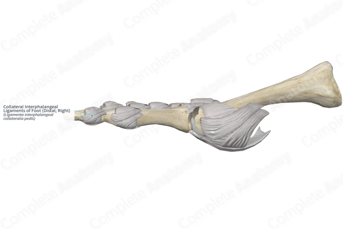
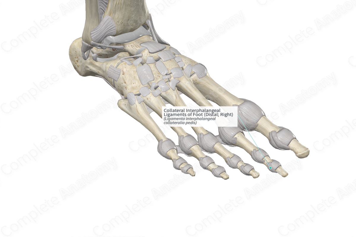
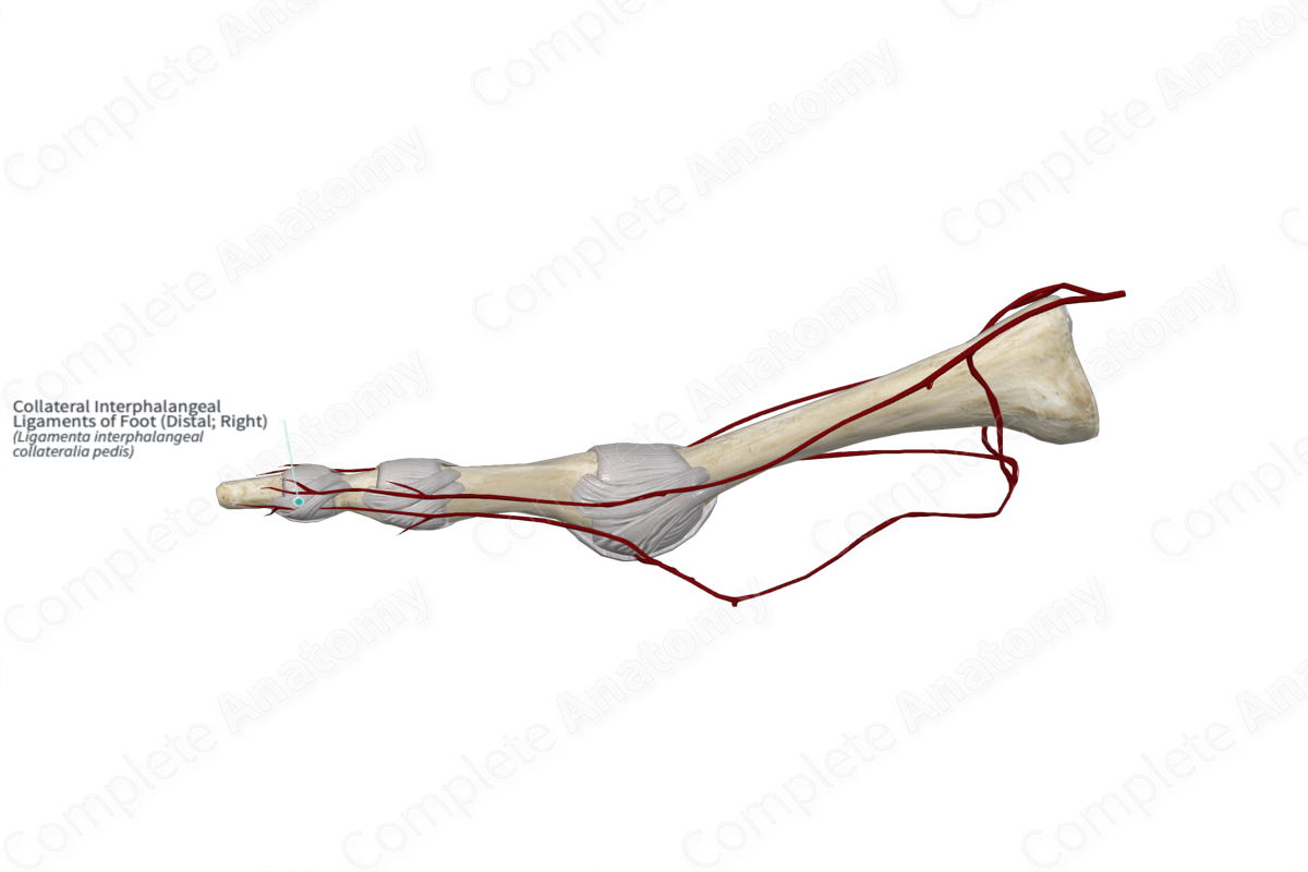
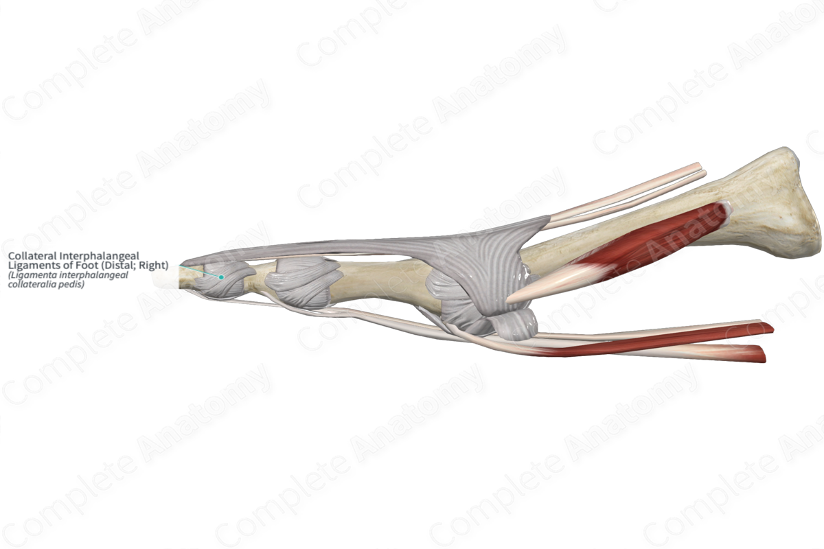
Collateral Interphalangeal Ligaments of Foot (Distal; Right)
Ligamenta interphalangeal collateralia pedis
Read moreStructure
The collateral interphalangeal ligaments of the distal interphalangeal joints are strong fibers that extend from the lateral and medial sides of the heads of the middle phalanges to the bases of the distal phalanges. Additional fibers, also called the accessory collateral ligaments, insert into the plantar ligaments.
Related parts of the anatomy
Function
The collateral interphalangeal ligaments help reinforce the articular capsules surrounding the joints and stabilize the joints during plantar and dorsiflexion.
Learn more about this topic from other Elsevier products
Interphalangeal Joint

To inject the interphalangeal joints under ultrasound guidance, the patient is placed in the sitting position with the elbow flexed to approximately 90 degrees with the forearm and the palm of the hand resting comfortably on a pillow.
