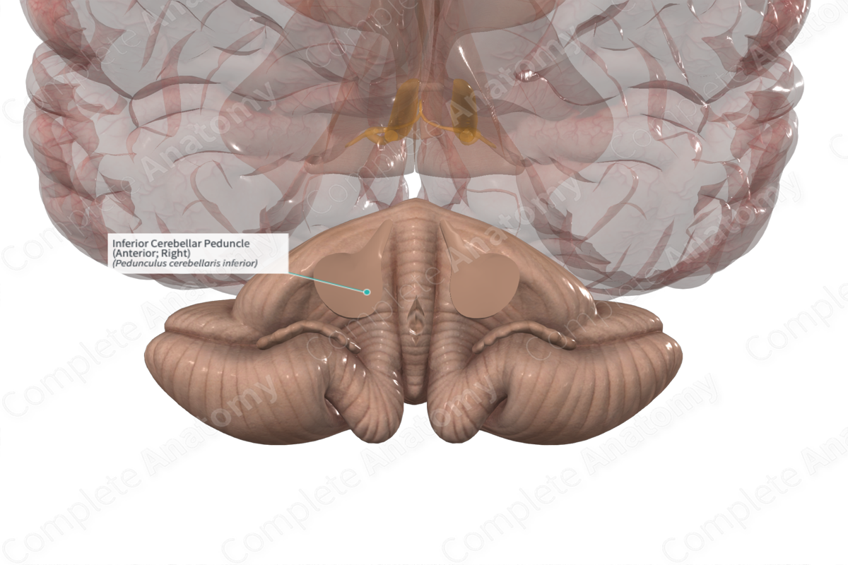
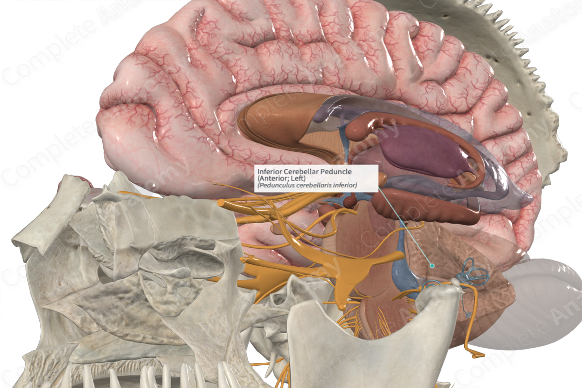
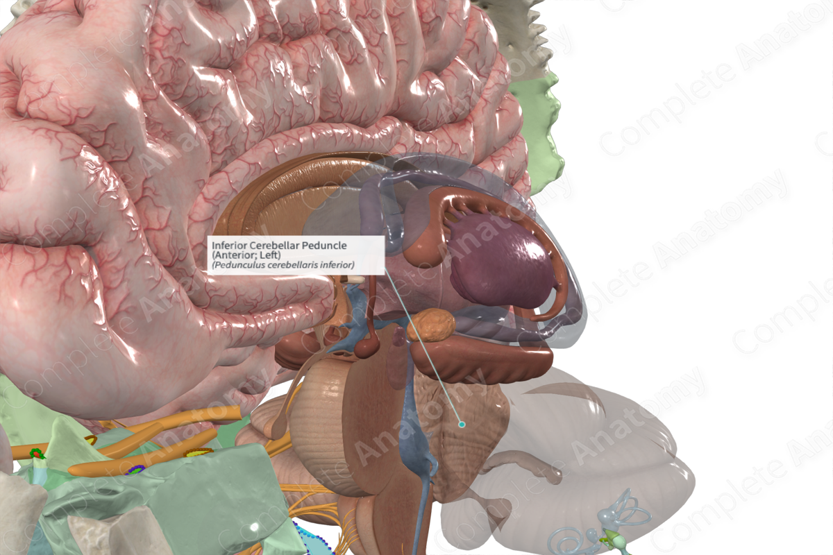
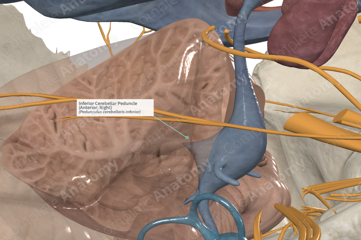
Inferior Cerebellar Peduncle (Anterior; Right)
Pedunculus cerebellaris inferior
Read moreQuick Facts
The inferior cerebellar peduncle (aka caudal cerebellar peduncle) is a thick, large bundle of nerve fibres that emerge from the posterolateral surface of the upper medulla. It extends under the lateral recesses of the rhomboid fossa and curves superiorly into the cerebellum, caudomedial to the middle cerebellar peduncle.
The fibres originate from spinal neurons and medullary relay nuclei. Each peduncle connects the spinal cord and medulla oblongata with the cerebellum and comprises the small, medial juxtarestiform body and large, lateral restiform body.
The peduncles carry many types of input and output fibers which integrate proprioceptive sensory input with motor vestibular functions such as balance and postural maintenance.
Related parts of the anatomy
Learn more about this topic from other Elsevier products
Inferior Cerebellar Peduncle

These are the inferior cerebellar peduncle, the middle cerebellar peduncle (or brachium pontis), and the superior cerebellar peduncle (or brachium conjunctivum), connecting the cerebellum to the medulla oblongata, basilar pons, and midbrain, respectively.
