Quick Facts
The cranium is the large round superior part of the skull; it encloses the brain, forms the skeleton of the midface, and is made up of the cranial bones held together by sutures, synchondroses, and bony unions (Dorland, 2011).
Structure
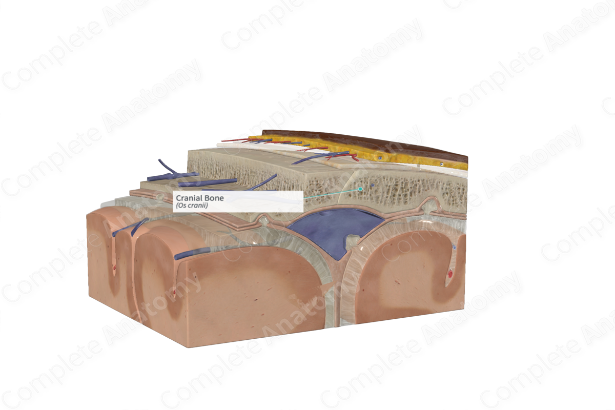
The bones that make up the neurocranium, the portion of the cranium that houses the brain, are the frontal, parietal, temporal, occipital, ethmoid, and sphenoid bones. The cranial bones are considered flat bones derived from the mesenchyme and develop by intramembranous and endochondral ossification. The bones contain a thin inner and outer layer of compact bone, separated by cancellous bone, known as diploë. The inner layer of compact bone is thinner and more brittle than its external layer (Moore, Dalley and Agur, 2013; Snell, 2011).
The frontal, parietal, temporal, and occipital bones make up the calvaria, which is covered by the scalp. The scalp attaches to the calvaria via the pericranium. The outer surface of the pericranium is loosely fused with the epicranial aponeurosis of the scalp via a loose connective tissue. The inner surface of the pericranium firmly attaches to the calvaria. Due to its elasticity, the pericranium can be easily removed from the calvaria, except at the location of the suture lines where it is firmly attached.
Anatomical Relations
The cranial bone is located deep to the scalp and superficial to the meninges and brain parenchyma.
Function
The cranial bones protect the brain within the cranial vault.
List of Clinical Correlates
—Skull fracture
References
Dorland, W. (2011) Dorland's Illustrated Medical Dictionary. 32nd edn. Philadelphia, USA: Elsevier Saunders.
Moore, K. L., Dalley, A. F. and Agur, A. M. R. (2013) Clinically Oriented Anatomy. Clinically Oriented Anatomy 7th edn.: Wolters Kluwer Health/Lippincott Williams & Wilkins.
Snell, R. S. (2011) Clinical Anatomy by Regions. Lippincott Williams & Wilkins.
