Quick Facts
Location: Hand.
Bone Type: Long bone.
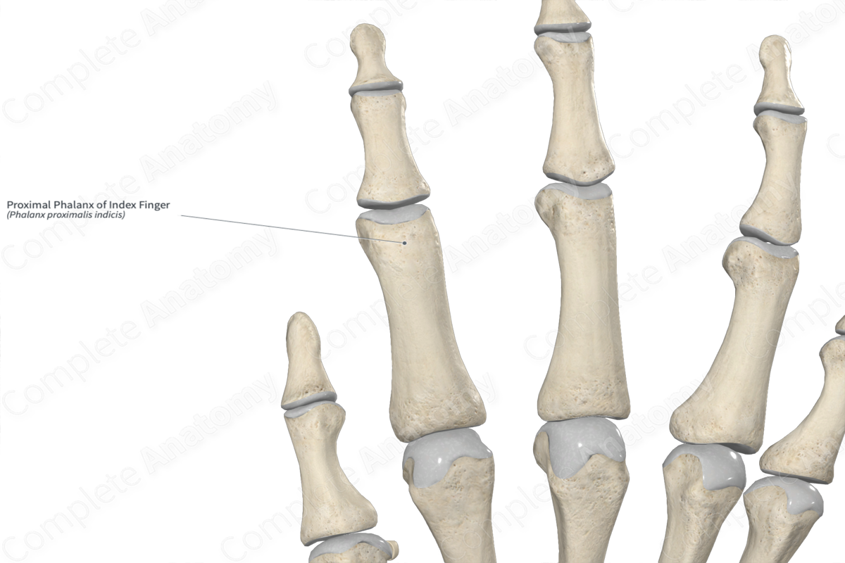
Key Features: Head, body, base, and proximal and distal articular facets.
Articulates With: Second metacarpal bone and middle phalanx of index finger.
Arterial Supply: Proper palmar digital arteries.
Key Features & Anatomical Relations
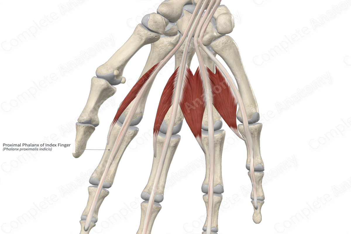
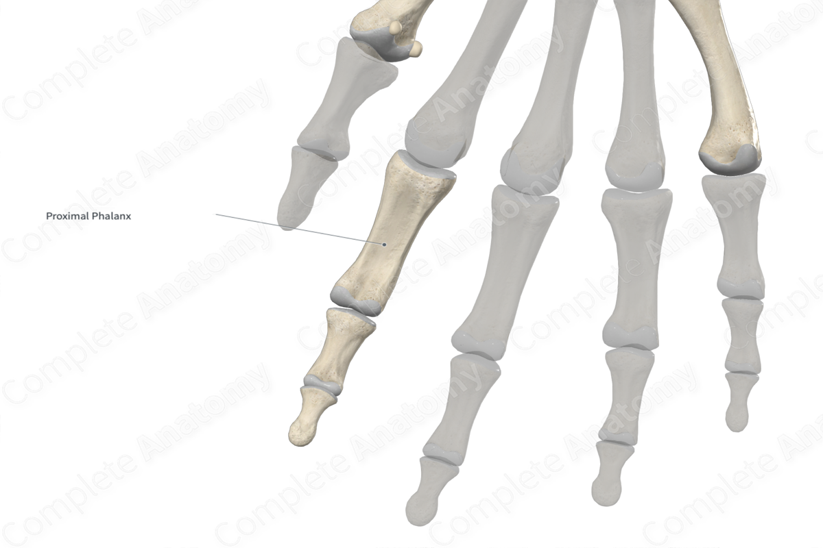
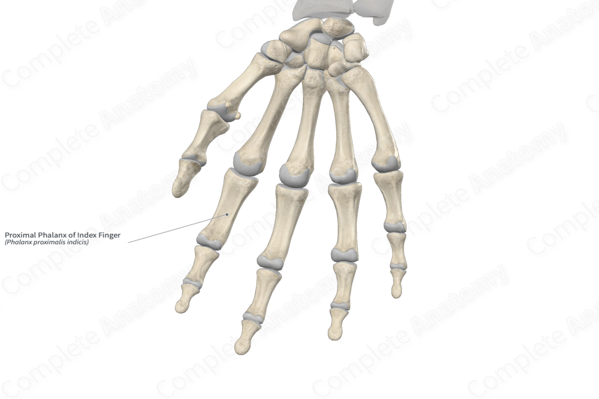
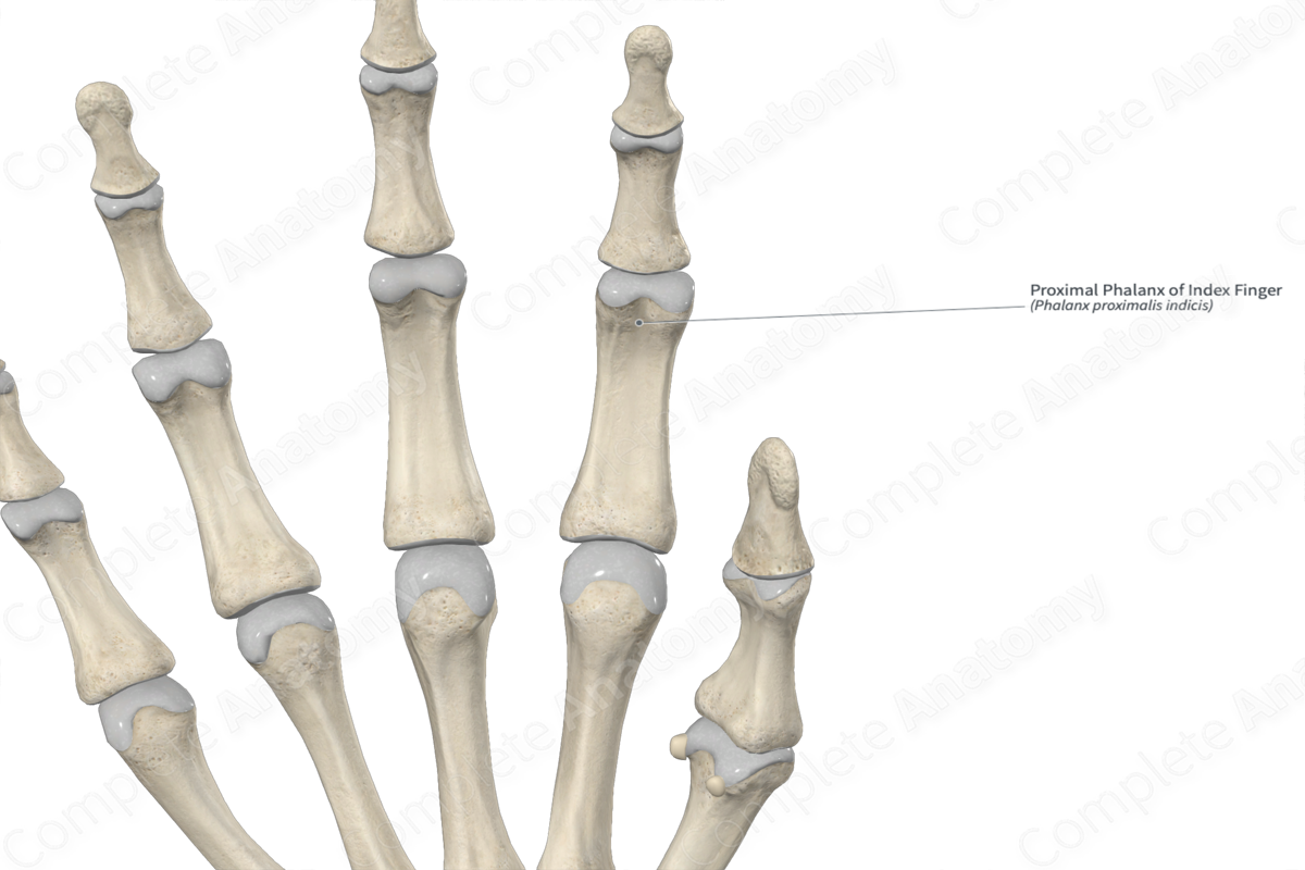
The proximal phalanx of the index finger is one of the fourteen phalangeal bones of the hand. It’s classified as a long bone and includes the following bony features:
- parts: head, body, and base;
- landmarks: proximal and distal articular facets.
More information regarding these bony features can be found in the Parts and Landmarks tabs for this bone.
The proximal phalanx is located:
- proximal to the middle phalanx of index finger;
- distal to the second metacarpal bone;
It articulates with the:
- middle phalanx at the proximal interphalangeal joint;
- metacarpal bone at the metacarpophalangeal joint.
Ossification
Ossification of the proximal phalanx occurs at two ossification centers, these are found in the:
- body, which appears in utero during the third month;
- base, which appears during the second year.
These ossification centers fuse with each other during the fifteenth to eighteenth years (Standring, 2016).
Surface Anatomy
The head, body, and base of the proximal phalanx can be easily palpated.
List of Clinical Correlates
- Fracture
- Brachyphalangia
- Symphalangia
- Thiemann’s disease
References
Standring, S. (2016) Gray's Anatomy: The Anatomical Basis of Clinical Practice. Gray's Anatomy Series 41st edn.: Elsevier Limited.
Learn more about this topic from other Elsevier products
Proximal Phalanx

Based on the second proximal phalanx, it was possible to make a further comparison between the antemortem radiograph and the postmortem material, specifically an X-ray and a photograph of the bone.
