Quick Facts
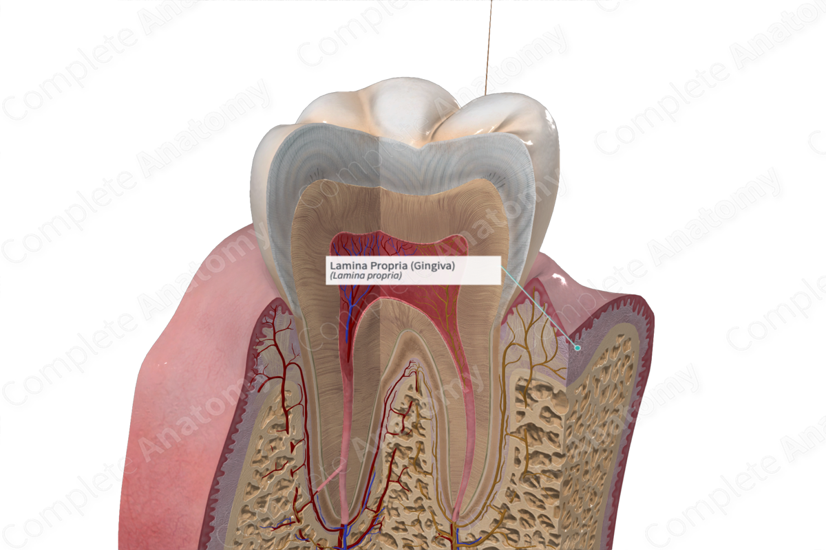
The lamina propria is the connective tissue coat of a mucous membrane just deep to the epithelium and basement membrane (Dorland, 2011)
Structure and/or Key Feature(s)
Lamina propria is a thin layer of connective tissue that comprises part of the oral mucosa. Lying subjacent to the epithelium, it primarily consists of collagen fibers, fibroblasts, vessels and nerves. Mast cells, macrophages and inflammatory cells are also found in the lamina propria.
Fibroblasts of the lamina propria produce many connective tissue fibers including collagen, reticulin, oxytalan, and elastic fibers (Fehrenbach and Popowics, 2015).
The lamina propria of the attached gingiva has tall connective tissue papillae and rete pegs which gives the overall gingiva tissue a stippled surface effect. The lamina propria is directly attached to the underlying mandible and maxilla (jaw bones), making the attached gingiva firm and immobile. The epithelium and lamina propria attached to the underlying periosteum of the jaw bone is collectively known as a mucoperiosteum.
The lamina propria of the marginal gingiva is continuous with the lamina propria of the gingival sulcus. Unlike attached gingiva, marginal gingiva is not directly attached to the underlying mandible and maxilla (jaw bones), making it firm but mobile. The lamina propria of the marginal gingiva is also continuous with the lamina propria of the attached gingiva and the periodontal ligament.
Function
Lamina propria plays an important role in supporting the superjacent mucosal epithelial layer. It facilitates anchoring of the epithelium to the underlying jaw bones and plays a further role in immune defense due to the presence of immune cells (Rajendran and Sivapathasundharam, 2014).
References
Dorland, W. (2011) Dorland's Illustrated Medical Dictionary. 32nd edn. Philadelphia, USA: Elsevier Saunders.
Fehrenbach, M. J. and Popowics, T. (2015) Illustrated Dental Embryology, Histology, and Anatomy - E-Book. Elsevier Health Sciences.
Rajendran, A. and Sivapathasundharam, B. (2014) Shafer's Textbook of Oral Pathology. Elsevier Health Sciences.
