Quick Facts
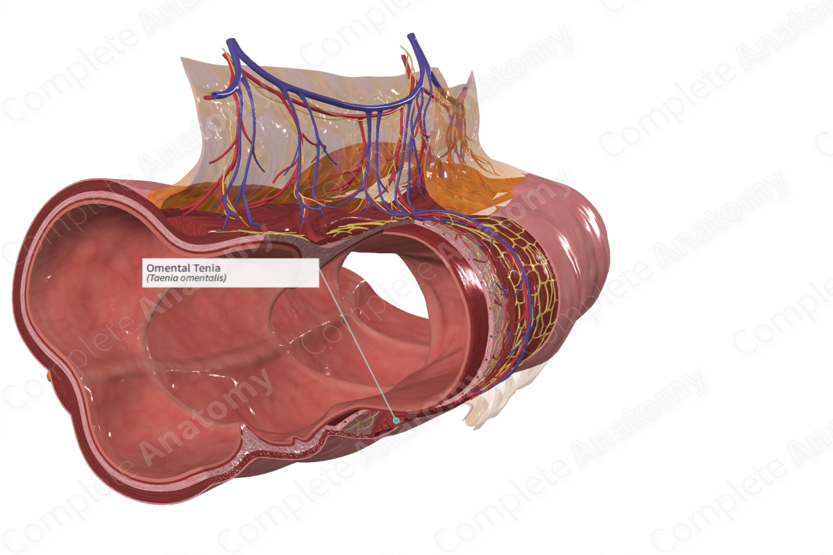
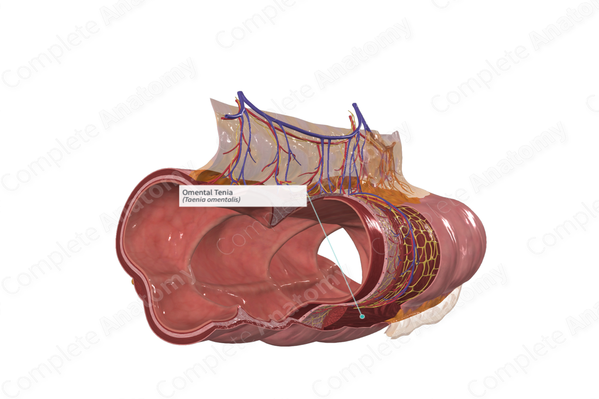
The omental tenia is the band of longitudinal muscle fibers of the large intestine along the site of attachment of the gastrocolic ligament of the greater omentum to the transverse colon (Dorland, 2011).
Related parts of the anatomy
Structure/Morphology
The omental tenia is one of three distinct bands of longitudinal smooth muscle that are collectively termed the teniae coli. These strips of longitudinal smooth muscle cells run along the large intestine and reside beneath its outer serosal surface.
Anatomical Relations
The three teniae coli are termed the mesocolic tenia, the free tenia, and the omental tenia. The free tenia is located opposite to the mesentery, the omental tenia is located on the posterolateral aspect of the colon, and the mesocolic tenia is located on the posteromedial aspect of the colon.
Due to the hepatic flexure at the junction of the transverse colon the teniae coli have a rotated position. Thus, the anterior free tenia becomes inferior, the posteromedial mesocolic tenia becomes posterior, and the posterolateral omental tenia becomes superior.
Once they reach the sigmoid colon, the teniae expand to take up a greater portion of the circumference of the colon where they form distinct bands located anteriorly and posteriorly. Both of these bands eventually converge and unite to form a complete longitudinal muscle layer, a feature characteristic of the rectum.
Function
The longitudinal and circular layers of the muscular layer of the colon are responsible for producing peristaltic contractions of the gut, thus propelling the ingested substance through the lumen of the gut. Additionally, contraction of the teniae coli results in bulging of the colonic haustra, giving rise to the sacculation, which give the colon its segmented appearance.
References:
Dorland, W. (2011) Dorland's Illustrated Medical Dictionary. 32nd edn. Philadelphia, USA: Elsevier Saunders.
