Congenital Heart Disease: Cyanotic Defects Made Easy
Congenital heart diseases made easy! Learn these cyanotic congenital heart defects with a simple counting trick. Tetralogy of Fallot, transposition of great arteries, truncus arteriosus, tricuspid atresia, and more!
Save Time with a Video!
Save time by watching the video first, then supplement it with the lecture below!
Click below to view the EZmed video library. Subscribe to stay in the loop!
Download the Study Guide & Lecture!
Click below to instantly download and enjoy :)

Become a Member!
Instant Access to All PDF Notes and Study Guides!
Instant access to a members-only page of ALL the flashcards, study guides, and PDF lectures. Cancel anytime.
Congenital Heart Diseases/Defects
Learning the different types of congenital heart diseases can be a challenge.
Wouldn’t it be nice if you had a simple trick to remember them all?
Fortunately, you have come to the right spot!
We are going to walk through a simple trick that will help you remember the main cyanotic congenital heart defects.
And the best part is…..the trick only takes a few minutes to learn!
By the end of this post, you will know the names and descriptions of:
Truncus Arteriosus
Transposition of Great Arteries
Tricuspid Atresia
Tetralogy of Fallot
Total Anomalous Pulmonary Venous Return
What Are Congenital Heart Defects?
Let’s quickly define congenital heart defects, and then we will go over the trick to remember them all!
Congenital heart defects are structural abnormalities of the heart and/or great vessels occurring during fetal development.
They are also referred to as congenital heart diseases, or CHD.
CHD can be subdivided into 2 main types: Cyanotic and Acyanotic.
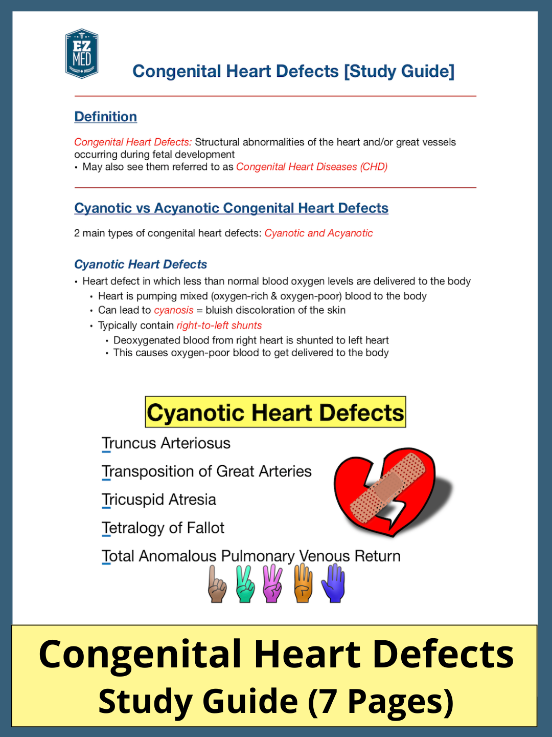
Cyanotic Congenital Heart Defects
Cyanotic heart defects are cardiac defects in which the blood pumped to the rest of the body contains less than normal amounts of oxygen.
In other words, the heart pumps mixed oxygen-poor and oxygen-rich blood to the body.
This can lead to cyanosis which is a bluish discoloration of the skin.
Cyanotic heart defects typically contain right-to-left shunts, meaning deoxygenated blood from the right heart is shunted to the left heart.
As a result, oxygen-poor blood is delivered to the body and can cause cyanosis.
Acyanotic Congenital Heart Defects
Acyanotic heart defects are cardiac defects that can affect the normal flow of blood, but blood oxygen levels delivered to the body typically remain normal.
Acyanotic heart defects often contain left-to-right shunts in which oxygenated blood from the left heart is shunted to the right heart.
The blood in the left heart still remains oxygen-rich, and that is why there is no cyanosis as oxygen-rich blood is delivered to the rest of the body.
We will focus on the main cyanotic congenital heart defects in this post.
Let’s now walk through the main defects and the tricks to remember them all!
Cyanotic vs Acyanotic Congenital Heart Diseases (CHD): Heart defects can be cyanotic or acyanotic depending on the amount of oxygen delivered to the rest of the body.
Cyanotic Heart Defects - Trick!
The 5 main cyanotic congenital heart defects discussed in this post include:
Truncus Arteriosus
Transposition of Great Arteries
Tricuspid Atresia
Tetralogy of Fallot
Total Anomalous Pulmonary Venous Return (TAPVR)
You can see they all start with the letter “T”.
So for the students reading this post - whenever you see a congenital heart defect on an exam that starts with a “T”, there is a good chance it is a cyanotic heart defect.
Now for the trick you’ve been waiting for!
All of the defects above can be remembered by counting them off on your fingers.
What do we mean by that?
Let’s go through each one below!
Cyanotic Congenital Heart Defect List: Truncus Arteriosus, Transposition of Great Arteries, Tricuspid Atresia, Tetralogy of Fallot, Total Anomalous Pulmonary Venous Return
1. Truncus Arteriosus
Trick: Hold up 1 finger
Truncus Arteriosus: One great vessel leaving the heart, instead of 2
The first cyanotic congenital heart defect is truncus arteriosus.
You can hold up 1 finger to remember this.
Truncus arteriosus is when one blood vessel leaves the heart instead of 2.
You might remember from the anatomy of the heart lecture that normally there are 2 main arteries leaving the heart.
The main pulmonary artery leaves the right side of the heart and delivers deoxygenated blood to the lungs.
The aorta leaves the left side of the heart and delivers oxygenated blood to the rest of the body.
In the case of truncus arteriosus, the great vessel coming out of the heart fails to divide during development.
This leaves a connection between the aorta and pulmonary artery.
A ventricular septal defect (VSD) is typically present, and the blood from the right and left ventricle combine and exit the heart through one great vessel.
A VSD is a hole in the wall between the right and left ventricle.
As a result, oxygen-poor blood from the right heart and oxygen-rich blood from the left heart are delivered to the rest of the body.
This can lead to potential cyanosis.
So again, use 1 finger to remember truncus arteriosus and one great vessel leaving the heart.
For more information on the normal blood flow through the heart, check out this simple 12 step guide!
Truncus Arteriosus: Hold up 1 finger to remember one great vessel leaves the heart, instead of two (normally pulmonary artery [1] and aorta [2] leave the heart).
2. Transposition of Great Arteries
Trick: Hold up 2 fingers and cross them
Transposition of Great Arteries: Two great arteries leaving the heart are transposed
The second cyanotic heart defect is transposition of great arteries.
You can remember this by holding up 2 fingers and crossing them (to represent the transposition).
Transposition of great arteries is when the 2 main arteries leaving the heart (main pulmonary artery and aorta) are transposed or reversed.
Remember we said the main pulmonary artery normally leaves the right heart and goes to the lungs, and the aorta leaves the left heart and goes to the rest of the body.
In transposition of great arteries, the pulmonary artery and aorta are reversed.
Therefore, the main pulmonary artery arises from the left ventricle instead of the right, and the aorta arises from the right ventricle instead of the left.
Transposition of Great Arteries: Hold up 2 fingers and cross them to remember the pulmonary artery (1) and aorta (2) are transposed (reversed) as shown by the arrows.
The transposition of the aorta and pulmonary artery creates 2 separate circuits.
Circuit 1: Deoxygenated blood from the right heart flows to the rest of the body (via the aorta) and back to the right side of the heart again.
Normally deoxygenated blood would flow from the right heart to the lungs via the pulmonary artery.
Circuit 2: Oxygenated blood from the left heart flows to the lungs (via the pulmonary artery) and back to the left side of the heart again.
Normally oxygenated blood would flow from the left heart to the rest of the body via the aorta.
In order to be compatible with life, there needs to be a connection between the 2 circuits to allow for mixing of oxygen-rich and poor blood.
So there is typically a patent ductus arteriosus or a ventricular septal defect present.
As you can imagine, this will lead to the delivery of oxygen-poor blood to the body and subsequent cyanosis.
So again, use 2 fingers and cross them to remember transposition of great arteries and how the 2 great arteries are reversed or transposed.
Transposition of Great Arteries: The reversal of the aorta and pulmonary artery creates 2 separate circuits (blue and red). A connection must exist to be compatible with life.
3. Tricuspid Atresia
Trick: Hold up 3 fingers
Tricuspid Atresia: Tricuspid valve fails to form (Tri = 3)
The third cyanotic heart defect is tricuspid atresia.
Hold up 3 fingers to remember this defect.
Tricuspid atresia is a congenital heart defect in which the tricuspid valve fails to form.
Remember in our medical terminology lecture, we learned the prefix “tri-” means 3.
So holding up 3 fingers will help you remember tricuspid atresia.
You might remember from the anatomy of the heart lecture that the tricuspid valve is located between the right atrium and right ventricle.
In the case of tricuspid atresia, the tricuspid valve fails to form.
As a result, blood from the right atrium cannot enter the right ventricle.
Instead, an atrial septal defect is present (a hole in the wall between the right and left atrium).
This allows for deoxygenated blood in the right atrium to flow into the left atrium.
As a result, oxygen-poor blood from the right heart mixes with the oxygen-rich blood in the left heart.
This can lead to decreased oxygen levels in the blood delivered to the rest of the body, which can cause cyanosis.
There are different types of tricuspid atresia, but the right ventricle is typically underdeveloped and the presence of a ventricular septal defect allows blood from the left ventricle to enter the right ventricle.
Remember the right ventricle is not receiving blood from the right atrium in this case, so it receives blood from the left ventricle instead.
So again, use 3 fingers to remember tricuspid atresia and how the tricuspid valve fails to form.
Tricuspid Atresia: Hold up 3 fingers to remember the tricuspid valve (star) fails to form and blood is unable to flow from the right atrium to the right ventricle (red X)
4. Tetralogy of Fallot
Trick: Hold up 4 fingers
Tetralogy of Fallot: Tetrad of 4 cardiac defects (Tetra = 4)
The fourth cyanotic heart defect is tetralogy of Fallot.
Hold up 4 fingers to remember this, as tetralogy of Fallot is a tetrad of 4 cardiac defects.
Remember in our medical terminology lecture, the prefix “tetra-” means 4.
So holding up 4 fingers will help you remember tetralogy of Fallot is a tetrad.
The tetrad includes:
Pulmonary Stenosis
Right Ventricular Hypertrophy (RVH)
Overriding Aorta
Ventricular Septal Defect (VSD)
Pulmonary stenosis is narrowing of the pulmonary valve and main pulmonary artery.
Right ventricular hypertrophy is thickening of the right ventricular wall.
Overriding aorta refers to the enlarged aortic valve that seems to open from both ventricles and sits on top of the ventricular septal defect.
Finally, the ventricular septal defect is a hole in the wall between the right and left ventricle.
The pulmonary stenosis, RVH, and VSD can alter pressure gradients and create a right-to-left shunt, allowing oxygen-poor blood in the right heart to flow to the left heart.
This can lead to cyanosis.
So again, use 4 fingers to remember tetralogy of Fallot and how there is a tetrad of 4 cardiac defects.
Tetralogy of Fallot: Hold up 4 fingers to remember the tetrad of cardiac defects including pulmonary stenosis (1), right ventricular hypertrophy (2), overriding aorta (3), and ventricular septal defect (4)
5. Total Anomalous Pulmonary Venous Return (TAPVR)
Trick: Hold up 5 fingers
Total Anomalous Pulmonary Venous Return (5 words): Pulmonary veins connect to systemic venous system rather than the left atrium
The fifth cyanotic heart defect is total anomalous pulmonary venous return (TAPVR).
Hold up 5 fingers to remember this because there are 5 words that make up the defect.
TAPVR is when the pulmonary veins connect to the systemic venous system rather than the left atrium.
Normally the 4 pulmonary veins deliver oxygenated blood from the lungs to the left atrium.
In the case of TAPVR, the pulmonary veins do not connect to the left atrium.
They connect to the systemic venous system instead.
As a result, the oxygenated blood from the lungs mixes with the deoxygenated venous blood from the body, and the mixed blood flows back to the right atrium.
Since the pulmonary veins are not delivering blood to the left atrium, there is usually an atrial septal defect present to allow blood to travel from the right atrium to the left atrium.
Remember the right atrial blood in this case is mixed oxygen-rich and oxygen-poor blood coming from the rest of the body.
So the left side of the heart is receiving blood with less than normal amounts of oxygen (compared to the oxygenated blood it would normally receive from the pulmonary veins).
This can cause cyanosis.
So again, use 5 fingers to remember total anomalous pulmonary venous return as the defect contains 5 words.
This is when the pulmonary veins connect to the systemic venous system rather than the left atrium.
Total Anomalous Pulmonary Venous Return (TAPVR): Hold up 5 fingers to remember the 5 words in TAPVR. The pulmonary veins do not connect to the left atrium (X) like they normally should (star), instead they connect to the systemic venous system.
Cyanotic Heart Defects - Recap!
So these are the 5 main cyanotic congenital heart defects.
They all start with the letter “T” and you can count them off on your fingers.
Use 1 finger to remember truncus arteriosus, which is 1 great vessel leaving the heart instead of 2.
Use 2 fingers and cross them to remember transposition of great arteries, which is when the pulmonary artery and aorta are transposed or reversed.
Use 3 fingers to remember tricuspid atresia, which is when the tricuspid valve fails to form.
Use 4 fingers to remember tetralogy of Fallot, which is a tetrad of cardiac defects (pulmonary stenosis, RVH, overriding aorta, VSD).
Use 5 fingers to remember total anomalous pulmonary venous return (5 words), which is when the pulmonary veins connect to the systemic venous system rather than the left atrium.
Cyanotic Congenital Heart Diseases: Use the counting method to remember the 5 main cardiac defects that can cause cyanosis
Before You Go….
Save time studying and reviewing!
Make sure to sign up for FREE to the EZmed newsletter below, and never miss out on future medical and science topics made easy!
A weekly notification is sent right to your inbox filled with new lectures, videos, and exam prep!
Did you enjoy this lecture? Leave a comment down below!
Feedback or suggestions for future topics? Reach out using the contact button above!
Thank you for using EZmed!
Make Your Learning Experience Even Easier!
Perform well in class, ace your exams, and keep up with your medical knowledge throughout your career using the following EZmed platforms:
YouTube Channel: EZmed - Animations and videos that simplify medicine and science
Instagram: @ezmedlearning - High yield exam content
Pinterest: ezmedlearning - Easy illustrations and flashcards
Referenceshttps://www.ncbi.nlm.nih.gov/books/NBK500001/https://www.ncbi.nlm.nih.gov/books/NBK538434/https://www.ncbi.nlm.nih.gov/books/NBK554495/https://www.mottchildren.org/health-library/tx4010https://www.cincinnatichildrens.org/health/t/tricuspidhttps://stanfordhealthcare.org/medical-conditions/blood-heart-circulation/congenital-heart-defects/types/cyanotic-congenital-heart-defects.html