Transposition of the great arteries
Transposition of the great arteries is a congenital heart anomaly that usually requires surgery within the first few weeks of life.
Overview
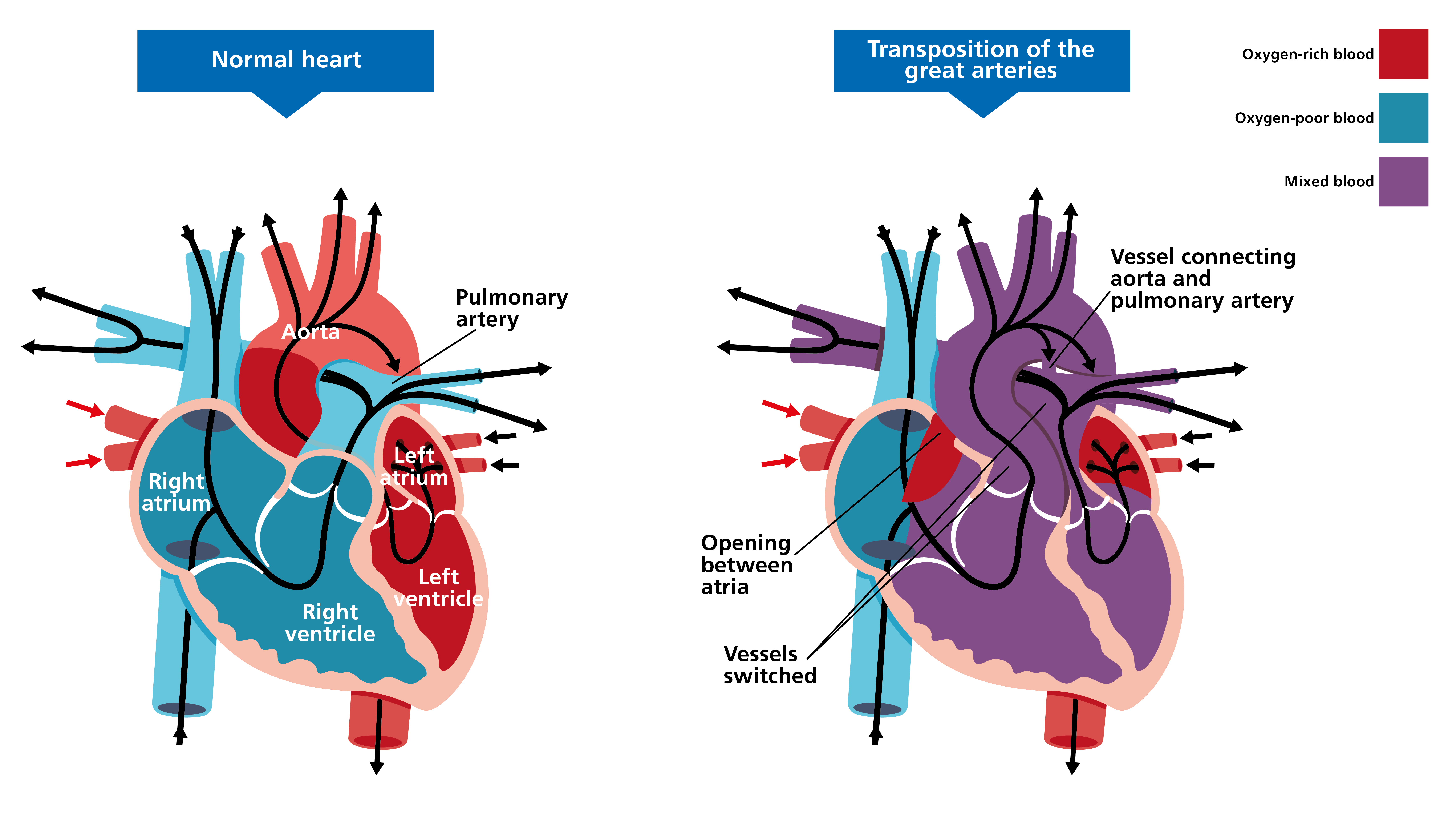
In transposition of the great arteries, the position of the two main arteries carrying blood out of the heart are switched: the pulmonary artery is joined to the left ventricle, and the aorta to the right ventricle. The condition can be diagnosed during ultrasound in both the antenatal and postnatal period.
Clinical features
Clinical features of transposition of the great arteries (TGA) are:
- rapid breathing or problems with breathing;
- weak pulse;
- blue or purple tint to lips and/or skin (cyanosis);
- difficulty feeding;
- poor appetite; and
- inadequate weight gain.
For comparison with a normal heart, see figure 1.
Figure 1. Transposition of the great arteries
View this video, shared with kind permission from Leeds Congenital Hearts, to see how TGA works.
Potential genetic causes
Most of the time, TGA occurs on its own; however, it can occur with other cardiac differences, such as a ventricular septal anomaly.
The cause of TGA is often unknown and there are therefore no commonly associated genetic changes considered to be responsible for it. TGA, in association with other anomalies detected during ultrasound or postnatally, may warrant a genomic test. The type of test would be determined following full assessment and consultation.
Inheritance and genomic counselling
TGA is considered to be associated only rarely with genetic syndromes and to have a low risk of recurrence among relatives of affected patients.
Management
Most babies will be given prostaglandin quickly after birth to allow blood to circulate more effectively. A balloon septostomy will be performed in most cases to ensure adequate oxygenation while awaiting surgery. Surgery, during which the major arteries are switched to the normal position, is usually performed within the first three weeks of life.
Resources
For clinicians
- American Heart Association: Transposition of the Great Arteries
- Great Ormond Street Hospital for Children NHS Foundation Trust: Transposition of the great arteries
- NHS England: National Genomic Test Directory
For patients
- British Heart Foundation. Understanding your child’s heart: Transposition of the great arteries
- Tiny Tickers