Inter J Stomatol ›› 2019, Vol. 46 ›› Issue (2): 171-176.doi: 10.7518/gjkq.2019016
• Periodontal Diseases • Previous Articles Next Articles
Manying Huang1,Yun Fu2( )
)
CLC Number:
| [1] |
Shah R, Sowmya NK, Mehta DS . Prevalence of gin-gival biotype and its relationship to clinical parame-ters[J]. Contemp Clin Dent, 2015,6(Suppl 1):S167-S171.
doi: 10.4103/0976-237X.166824 pmid: 26604569 |
| [2] |
Frost NA, Mealey BL, Jones AA , et al. Periodontal biotype: gingival thickness as it relates to probe visi-bility and buccal plate thickness[J]. J Periodontol, 2015,86(10):1141-1149.
doi: 10.1902/jop.2015.140394 pmid: 26110452 |
| [3] |
Cook DR, Mealey BL, Verrett RG , et al. Relationship between clinical periodontal biotype and labial plate thickness: an in vivo study[J]. Int J Periodontics Res-torative Dent, 2011,31(4):345-354.
doi: 10.4012/dmj.2011-028 pmid: 21837300 |
| [4] |
Younes F, Eghbali A, Raes M , et al. Relationship be- tween buccal bone and gingival thickness revisited using non-invasive registration methods[J]. Clin Oral Implants Res, 2016,27(5):523-528.
doi: 10.1111/clr.12618 pmid: 26010518 |
| [5] |
Peixoto A, Marques TM, Correia A . Gingival biotype characterization: a study in a Portuguese sample[J]. Int J Esthet Dent, 2015,10(4):534-546.
pmid: 26794050 |
| [6] |
Sin YW, Chang HY, Yun WH , et al. Association of gingival biotype with the results of scaling and root planing[J]. J Periodontal Implant Sci, 2013,43(6):283-290.
doi: 10.5051/jpis.2013.43.6.283 pmid: 3891860 |
| [7] |
Arora R, Narula SC, Sharma RK , et al. Evaluation of supracrestal gingival tissue after surgical crown leng-thening: a 6-month clinical study[J]. J Periodontol, 2013,84(7):934-940.
doi: 10.1902/jop.2012.120162 pmid: 23088528 |
| [8] |
Tao JX, Wu Y, Chen JR , et al. A follow-up study of up to 5 years of metal-ceramic crowns in maxillary central incisors for different gingival biotypes[J]. Int J Periodontics Restorative Dent, 2014,34(5):e85-e92.
doi: 10.11607/prd.2024 pmid: 25171044 |
| [9] |
Rasperini G, Acunzo R, Cannalire P , et al. Influence of periodontal biotype on root surface exposure during orthodontic treatment: a preliminary study[J]. Int J Periodontics Restorative Dent, 2015,35(5):665-675.
doi: 10.11607/prd.2239 pmid: 26357696 |
| [10] |
Kim DM, Neiva R . Periodontal soft tissue non-root coverage procedures: a systematic review from the AAP Regeneration Workshop[J]. J Periodontol, 2015,86(2 Suppl):S56-S72.
doi: 10.1902/jop.2015.130684 |
| [11] |
Chen ST, Darby IB, Reynolds EC , et al. Immediate implant placement postextraction without flap eleva-tion[J]. J Periodontol, 2009,80(1):163-172.
doi: 10.1902/jop.2009.080243 pmid: 19228102 |
| [12] |
Evans CD, Chen ST . Esthetic outcomes of immediate implant placements[J]. Clin Oral Implants Res, 2008,19(1):73-80.
doi: 10.1111/j.1600-0501.2007.01413.x pmid: 17956569 |
| [13] |
De Rouck T, Eghbali R, Collys K , et al. The gingival biotype revisited: transparency of the periodontal probe through the gingival margin as a method to discriminate thin from thick gingiva[J]. J Clin Perio-dontol, 2009,36(5):428-433.
doi: 10.1111/j.1600-051X.2009.01398.x pmid: 19419444 |
| [14] |
Eghbali A, De Rouck T, De Bruyn H , et al. The gingival biotype assessed by experienced and inexperienced clinicians[J]. J Clin Periodontol, 2009,36(11):958-963.
doi: 10.1111/j.1600-051X.2009.01479.x pmid: 19811580 |
| [15] |
Cuny-Houchmand M, Renaudin S, Leroul M , et al. Gingival biotype assessement: visual inspection re-levance and maxillary versus mandibular comparison[J]. Open Dent J, 2013,7:1-6.
doi: 10.2174/1874210601307010001 pmid: 23400554 |
| [16] |
乐迪, 张豪, 胡文杰 , 等. 牙周探诊法判断牙龈生物型的初步研究[J]. 中华口腔医学杂志, 2012,47(2):81-84.
doi: 10.3760/cma.j.issn.1002-0098.2012.02.003 |
|
Le D, Zhang H, Hu WJ , et al. Preliminary study on gingival biotype by periodontal probing[J]. Chin J Stomatol, 2012,47(2):81-84.
doi: 10.3760/cma.j.issn.1002-0098.2012.02.003 |
|
| [17] |
Kaya Y, Alkan ö, Keskin S . An evaluation of the gingival biotype and the width of keratinized gingiva in the mandibular anterior region of individuals with different dental malocclusion groups and levels of crowding[J]. Korean J Orthod, 2017,47(3):176-185.
doi: 10.4041/kjod.2017.47.3.176 pmid: 5432439 |
| [18] |
Sharma S, Thakur SL, Joshi SK , et al. Measurement of gingival thickness using digital vernier caliper and ultrasonographic method: a comparative study[J]. J Investig Clin Dent, 2014,5(2):138-143.
doi: 10.1111/jicd.12026 pmid: 23355379 |
| [19] |
Baldi C, Pini-Prato G, Pagliaro U , et al. Coronally advanced flap procedure for root coverage. Is flap thickness a relevant predictor to achieve root coverage? A 19-case series[J]. J Periodontol, 1999,70(9):1077-1084.
doi: 10.1902/jop.1999.70.9.1077 pmid: 10505811 |
| [20] |
Rathee M, Rao PL, Bhoria M . Prevalence of gingival biotypes among young dentate north indian popula-tion: a biometric approach[J]. Int J Clin Pediatr Dent, 2016,9(2):104-108.
doi: 10.5005/jp-journals-10005-1343 pmid: 27365928 |
| [21] |
Memon S, Patel JR, Sethuraman R , et al. A com-parative evaluation of the reliability of three methods of assessing gingival biotype in dentate subjects in different age groups: an in vivo study[J]. J Indian Prosthodont Soc, 2015,15(4):313-317.
doi: 10.4103/0972-4052.171830 pmid: 26929533 |
| [22] |
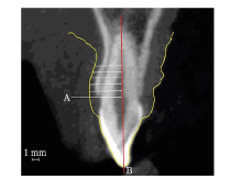
Alpiste-Illueca F . Dimensions of the dentogingival unit in maxillary anterior teeth: a new exploration technique (parallel profile radiograph)[J]. Int J Periodontics Restorative Dent, 2004,24(4):386-396.
doi: 10.1111/j.1365-2591.2004.00849.x pmid: 15446409 |
| [23] |
Galgali SR, Gontiya G . Evaluation of an innovative radiographic technique—parallel profile radiography—to determine the dimensions of dentogingival unit[J]. Indian J Dent Res, 2011,22(2):237-241.
doi: 10.4103/0970-9290.84294 pmid: 21891892 |
| [24] |
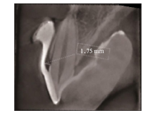
Kim YJ, Park JM, Kim S , et al. New method of asse-ssing the relationship between buccal bone thickness and gingival thickness[J]. J Periodontal Implant Sci, 2016,46(6):372-381.
doi: 10.5051/jpis.2016.46.6.372 pmid: 28050315 |
| [25] |
Esfahanizadeh N, Daneshparvar N, Askarpour F , et al. Correlation between bone and soft tissue thickness in maxillary anterior teeth[J]. J Dent (Tehran), 2016,13(5):302-308.
pmid: 5250627 |
| [26] |
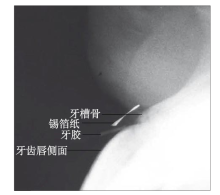
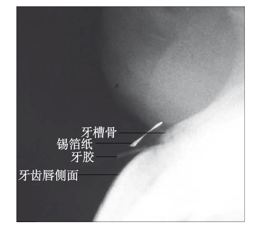
曹洁, 胡文杰, 张豪 , 等. 基于锥形束计算机体层摄影术测量牙龈厚度[J]. 北京大学学报(医学版), 2013,45(1):135-139.
doi: 10.3969/j.issn.1671-167X.2013.01.028 |
|
Cao J, Hu WJ, Zhang H , et al. Method and its app-lication of gingival thickness measurement based on cone-beam computed tomography[J]. J Peking Univ (Health Sci), 2013,45(1):135-139.
doi: 10.3969/j.issn.1671-167X.2013.01.028 |
|
| [27] |
Lee SP, Kim TI, Kim HK , et al. Discriminant ana-lysis for the thin periodontal biotype based on the data acquired from three-dimensional virtual models of Korean young adults[J]. J Periodontol, 2013,84(11):1638-1645.
doi: 10.1902/jop.2013.120594 pmid: 23305168 |
| [28] |
Malhotra R, Grover V, Bhardwaj A , et al. Analysis of the gingival biotype based on the measurement of the dentopapillary complex[J]. J Indian Soc Perio-dontol, 2014,18(1):43-47.
doi: 10.4103/0972-124X.128199 pmid: 3988642 |
| [1] | Fan Shengzi, Xie Zhigang.. Research progress on the effect of gingival biotype to implant aesthetic problems [J]. Inter J Stomatol, 2017, 44(5): 580-582. |
| [2] | Wang Tong, Wan Qianbing. Research progress on root surface area measurement [J]. Inter J Stomatol, 2016, 43(4): 490-494. |