Lamina Propria Pictures, Images and Stock Photos
Browse 70+ lamina propria stock photos and images available, or start a new search to explore more stock photos and images.
Most popular
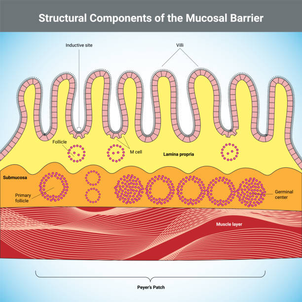
Structural Components of the Mucosal Barrier vector medical illustration
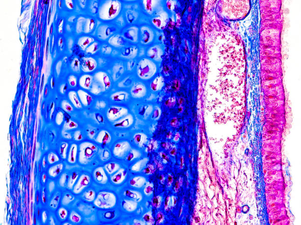
Human tongue cross section with taste buds or gustatory cells. Optical microscope, magnification X40.
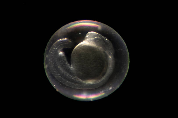
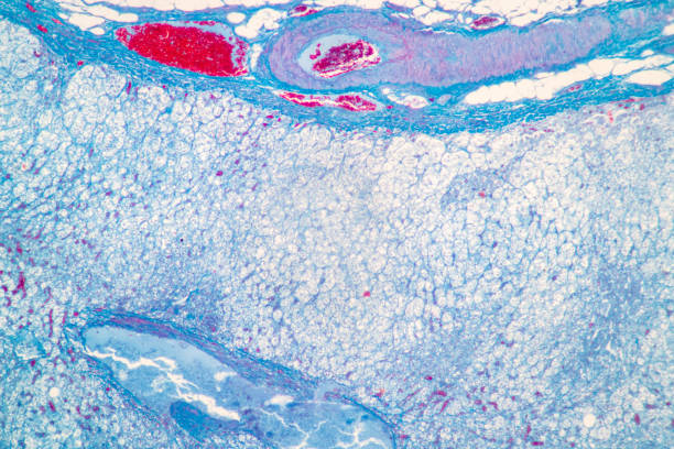
Zebrafish (Danio rerio) embryo about 24 hours after fertilization. Visible are the chorion surrounding the embryo, yolk, somites, chorda, and brain and eye vesicles.
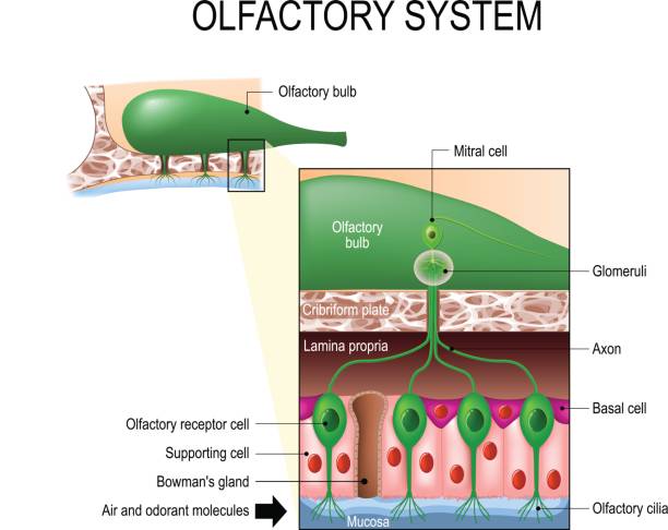
olfactory system inside the human head. Sense of smell. the olfactory bulb at the top which connects to scent cells at the bottom to identify odors
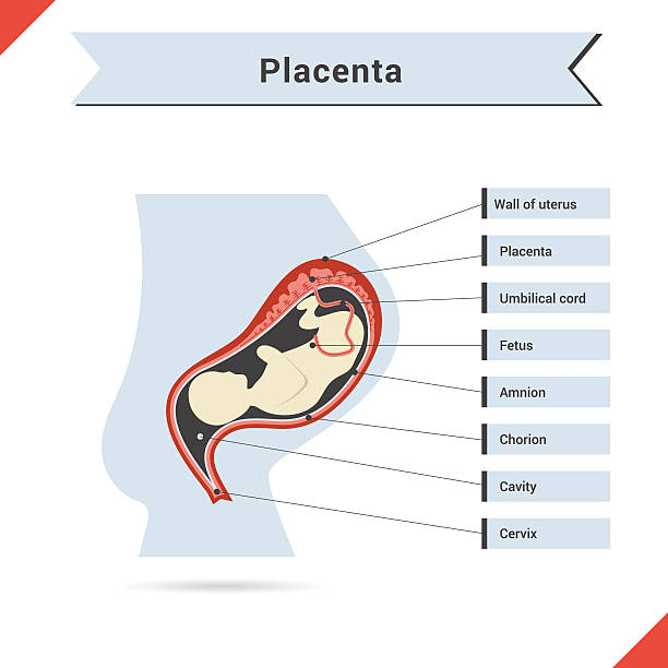
Vector infographic with fetus of womb and placenta.
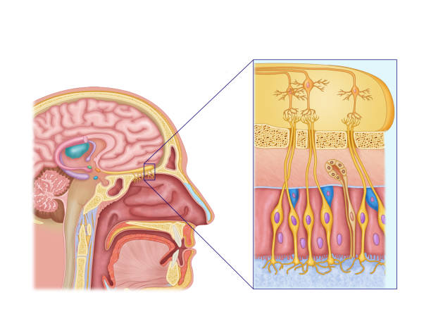
Illustration of the olfactory system, or sense of smell, is the sensory system used for smelling, also known as “olfaction”.
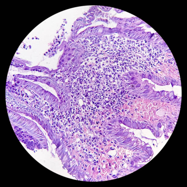
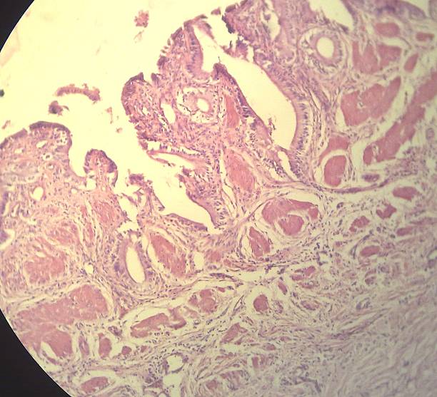
Urinary bladder(biopsy): Chronic cystitis, microscopic show bladder mucosa, infiltration of inflammatory cells in the lamina propria, interstitial cystitis, no malignancy seen.
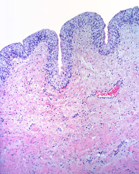
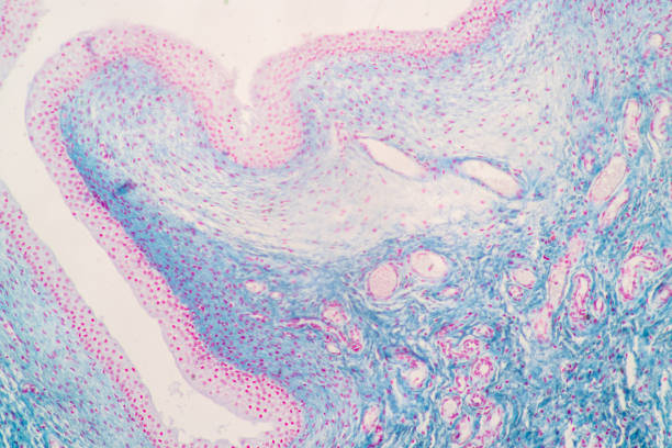
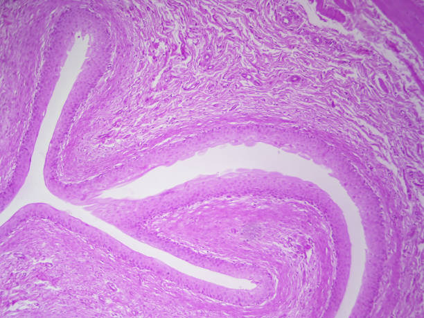
Mucosa of the urinary bladder formed by a transitional epithelium and a lamina propria of connective tissue. Below, the muscular layer formed by smooth muscle fibers begins
Tongue cross section with taste buds or gustatory cells. Optical microscope, magnification X40.
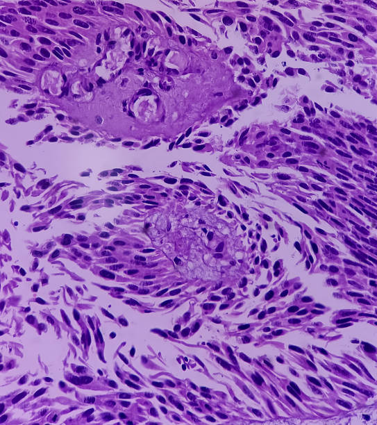
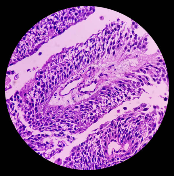
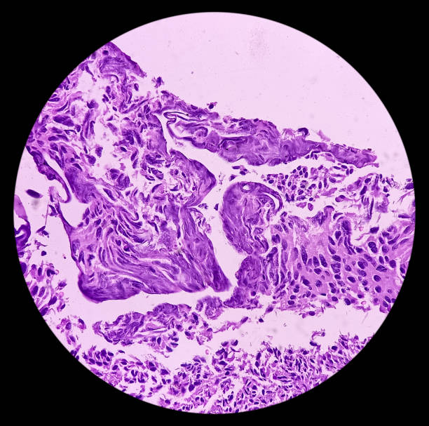
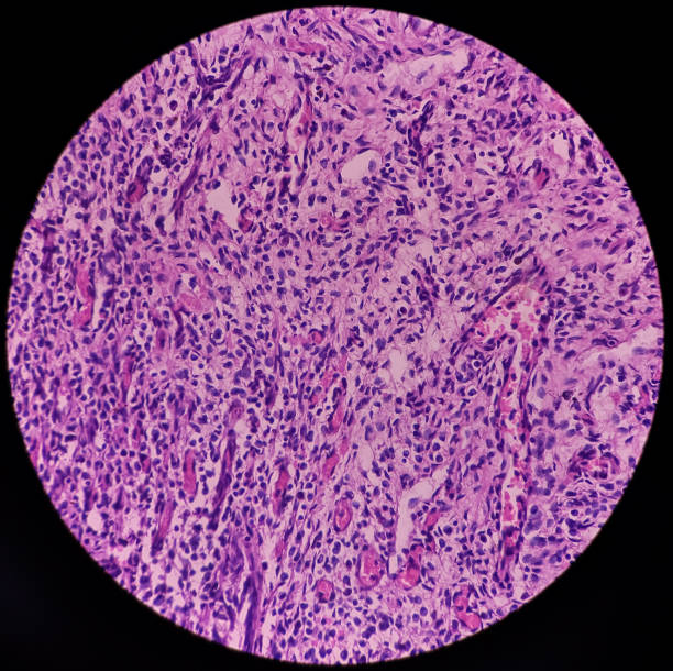
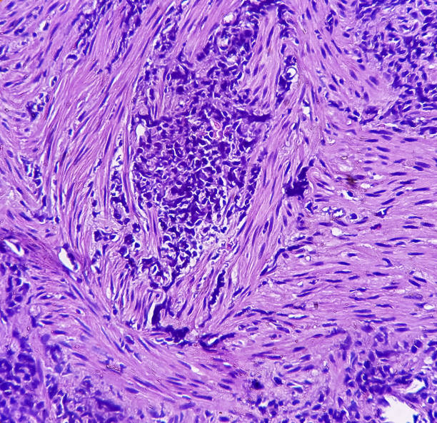
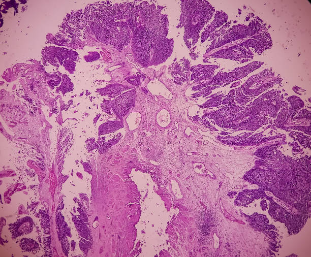
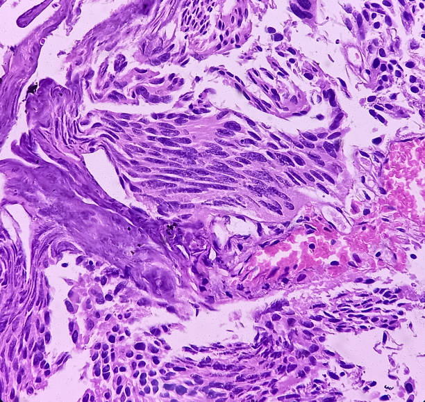
Urinary bladder cancer. Transitional cell carcinoma. show malignant neoplasm, 40x view.
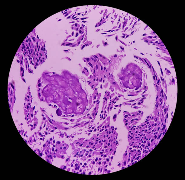
Urinary bladder cancer. Transitional cell carcinoma. show malignant neoplasm, 40x view.
Urinary bladder cancer. Transitional cell carcinoma. show malignant neoplasm, 40x view.
Urinary bladder cancer. Transitional cell carcinoma. show malignant neoplasm, 40x view.
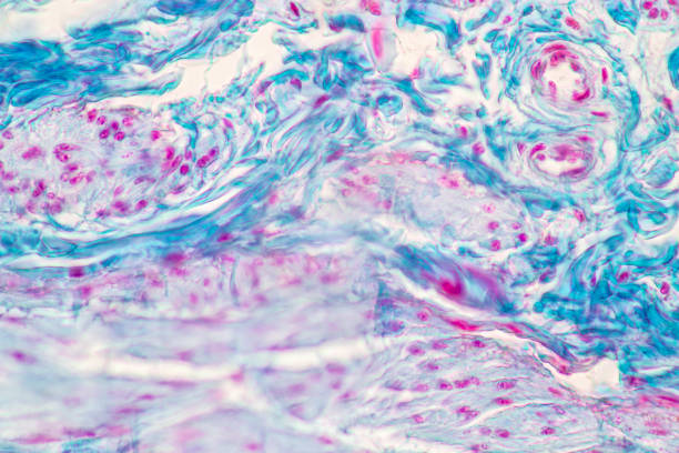
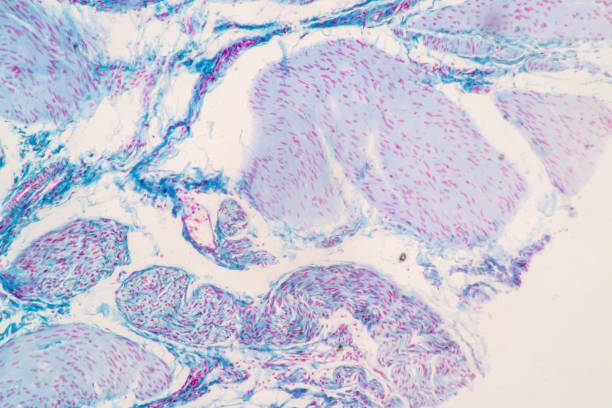
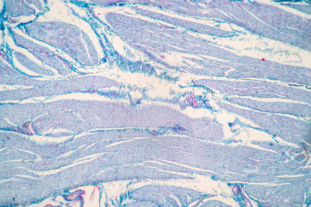
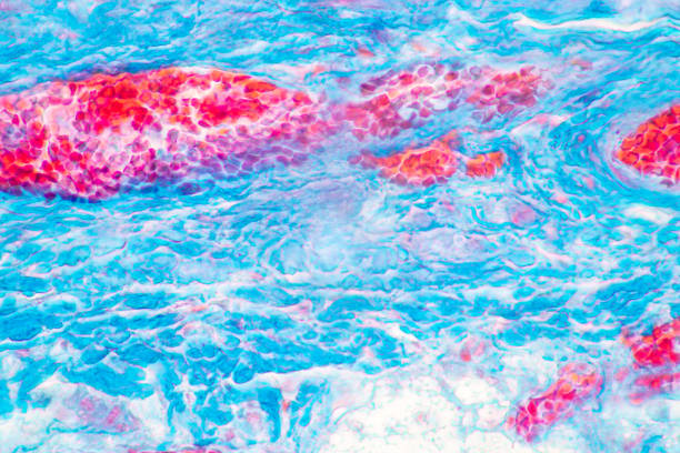
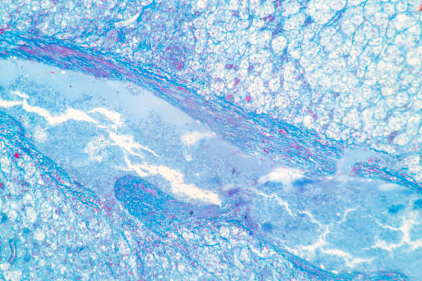
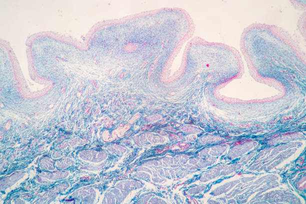
Showing Light micrograph of the Urinary bladder human under the microscope for education in the laboratory.
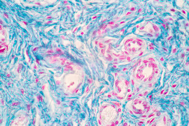
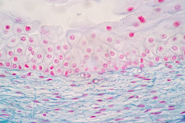
Showing Light micrograph of the Urinary bladder human under the microscope for education in the laboratory.
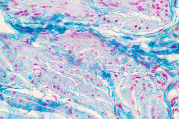
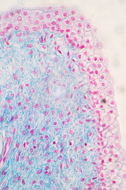
Showing Light micrograph of the Urinary bladder human under the microscope for education in the laboratory.
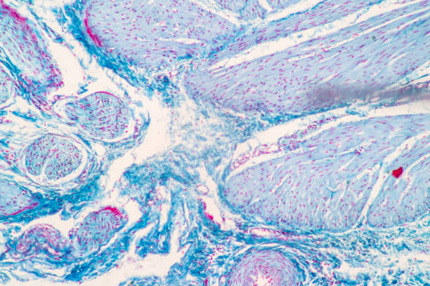
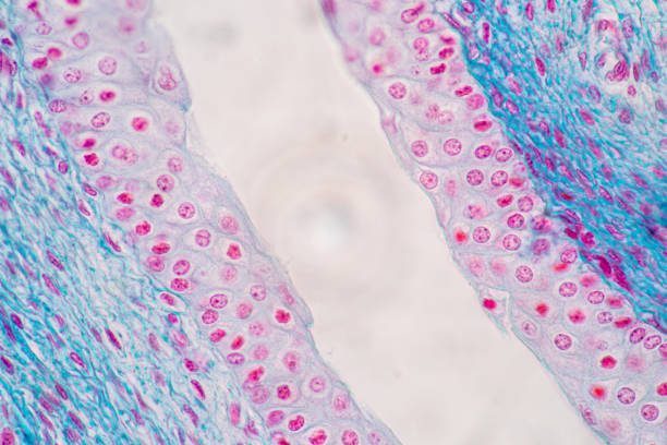
Showing Light micrograph of the Urinary bladder human under the microscope for education in the laboratory.
Showing Light micrograph of the Urinary bladder human under the microscope for education in the laboratory.
Showing Light micrograph of the Urinary bladder human under the microscope for education in the laboratory.
Showing Light micrograph of the Urinary bladder human under the microscope for education in the laboratory.
Showing Light micrograph of the Urinary bladder human under the microscope for education in the laboratory.
Urinary bladder cancer. Transitional cell carcinoma. show malignant neoplasm, 40x view.
Showing Light micrograph of the Urinary bladder human under the microscope for education in the laboratory.
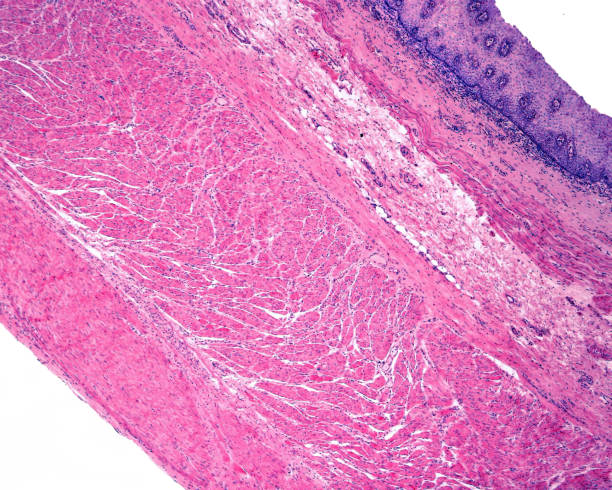
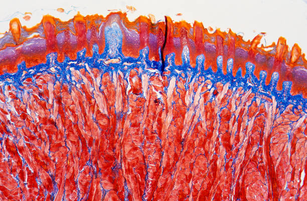
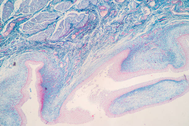
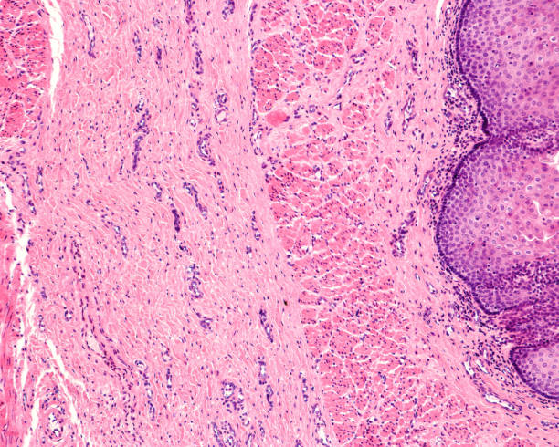
Layers of the wall of a human esophagus: mucosa (with epithelium, lamina propria, and muscularis mucosae), submucosa, muscular (inner and outer layers) and a very thin adventitia. "n"n

Urinary bladder(biopsy): Chronic cystitis, microscopic show bladder mucosa, infiltration of inflammatory cells in the lamina propria, interstitial cystitis, no malignancy seen.
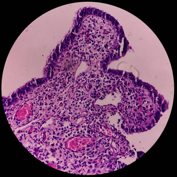
Urinary bladder(biopsy): Chronic cystitis, microscopic show bladder mucosa, infiltration of inflammatory cells in the lamina propria, interstitial cystitis, no malignancy seen.
Urinary bladder(biopsy): Chronic cystitis, microscopic show bladder mucosa, infiltration of inflammatory cells in the lamina propria, interstitial cystitis, no malignancy seen.
This plant is also known as Crataegus bibas or Mespilus japonica.
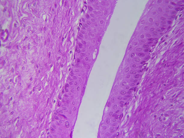
Esophagus. From right to left: stratified epithelium, lamina propria, a well-developed muscularis mucosa (with longitudinally-oriented smooth muscle fibers), and submucosa.
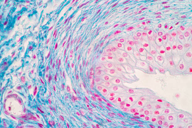
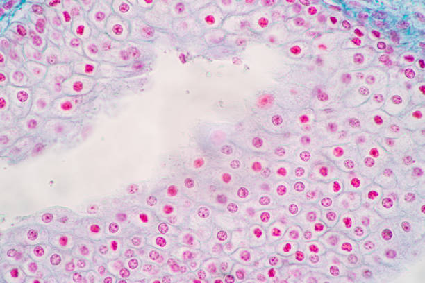
Microscope histology image of transitional epithelial of urinary bladder (400x)
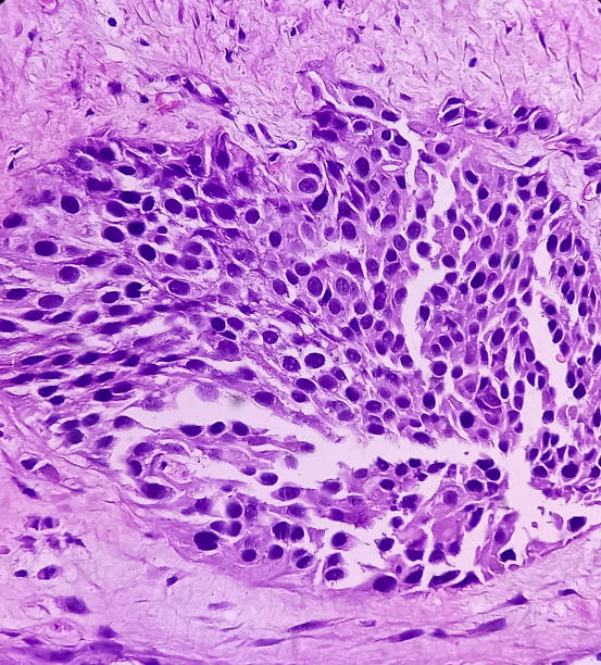
Pathology slide. The surface epithelium has lost its normal delicate papillary appearance with an increase in fibrous tissue and mild chronic inflammation in the lamina propria

Fashion patch badges in 80s-90s style. Vector illustration, set of vector illustrations in retro stereo tape style,Stylish people with objects90s,
Urinary bladder cancer. Transitional cell carcinoma. show malignant neoplasm, 40x view.
Urinary bladder cancer. Transitional cell carcinoma. show malignant neoplasm, 40x view.
Showing Light micrograph of the Urinary bladder human under the microscope for education in the laboratory.
Showing Light micrograph of the Urinary bladder human under the microscope for education in the laboratory.
Showing Light micrograph of the Urinary bladder human under the microscope for education in the laboratory.
Showing Light micrograph of the Urinary bladder human under the microscope for education in the laboratory.
Showing Light micrograph of the Urinary bladder human under the microscope for education in the laboratory.
Urinary bladder cancer. Transitional cell carcinoma. show malignant neoplasm, 40x view.
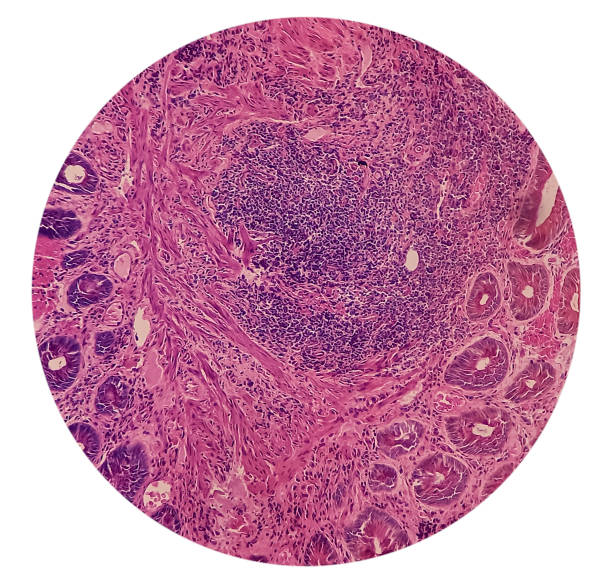
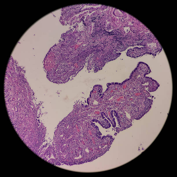
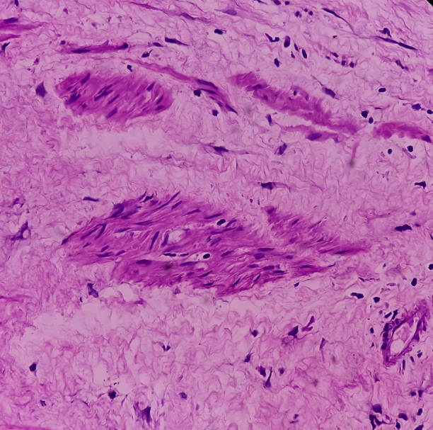
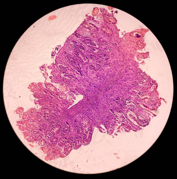
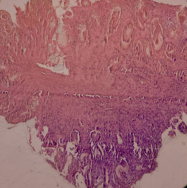
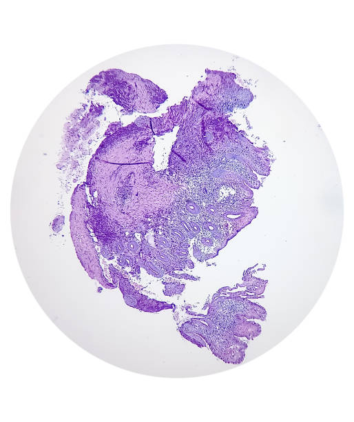
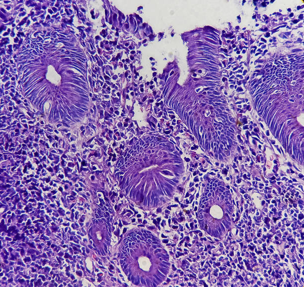
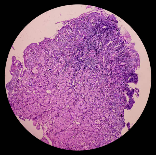
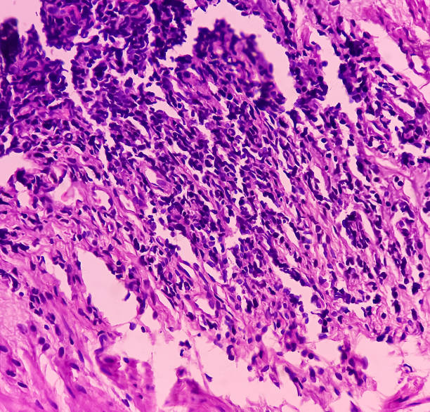
Anal canal ulcer biopsy: Chronic nonspecific proctitis, show anal mucosa, dense infiltration of lymphocytes, histiocytes and plasma cell in lamina propria with area of ulceration.
Anal canal ulcer biopsy: Chronic nonspecific proctitis, show anal mucosa, dense infiltration of lymphocytes, histiocytes and plasma cell in lamina propria with area of ulceration.
Microscope histology image of transitional epithelial of urinary bladder (100x)
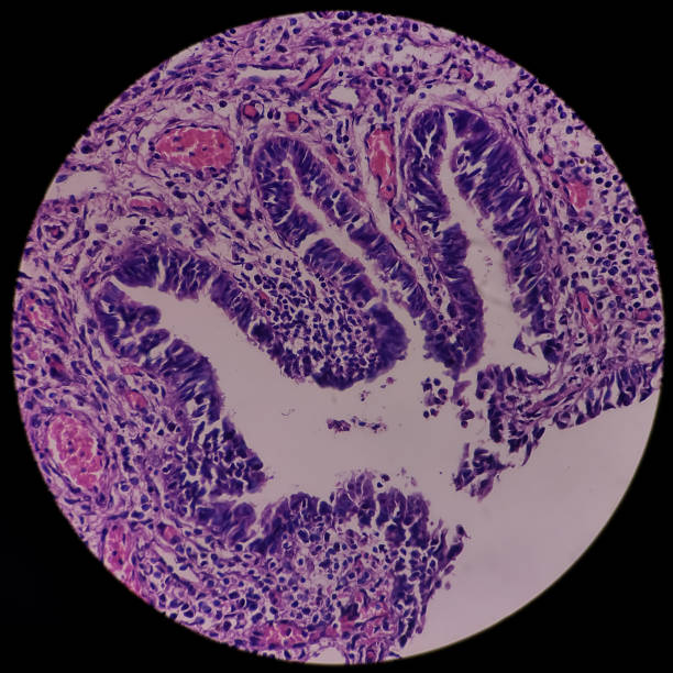
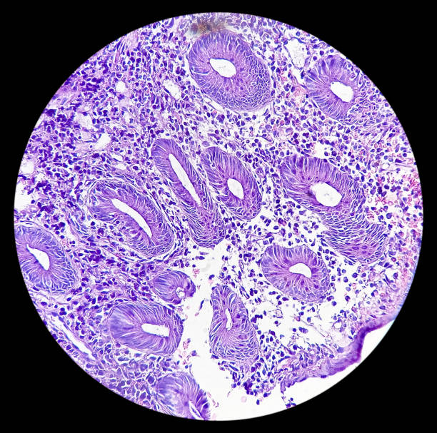
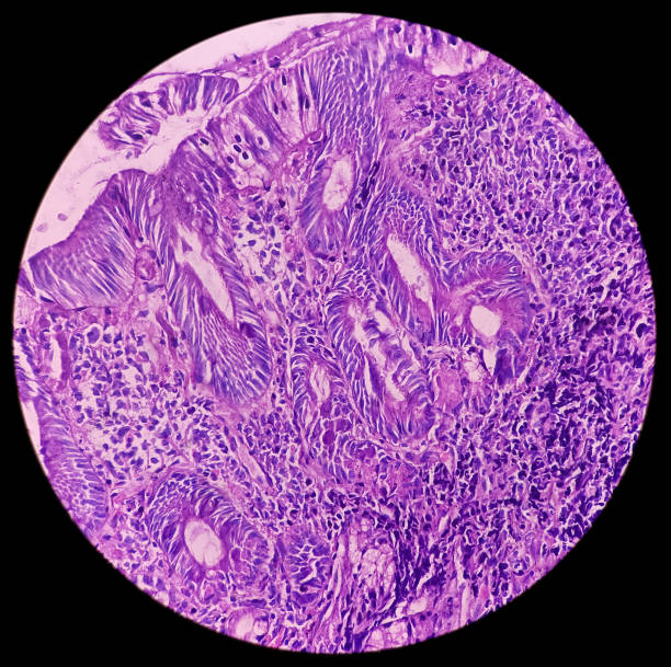
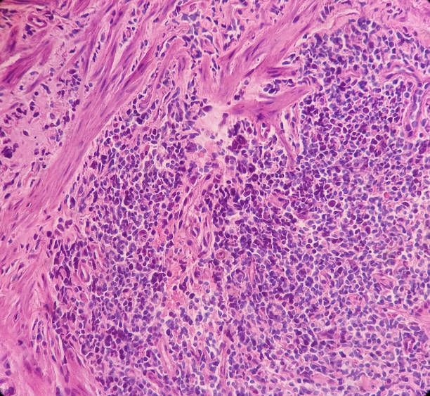
Tissue from rectum(colonoscopic biopsy): Chronic nonspecific proctitis. Show rectal mucosa, dense infiltration of lymphocytes, histiocytes and plasma cell in lamina propria. IBD.