Positron Emission Tomography - Computed Tomography (PET/CT)
Positron emission tomography (PET) uses small amounts of radioactive materials called radiotracers, a special camera and a computer to help evaluate your organ and tissue functions. By identifying body changes at the cellular level, PET may detect the early onset of disease before it is evident on other imaging tests.
Tell your doctor if there’s a possibility you are pregnant or if you are breastfeeding. Your doctor will instruct you based on the type of exam to be performed. Discuss any recent illnesses, medical conditions, medications you’re taking and allergies – especially to contrast material. You will likely be told not to eat anything and to drink only water several hours before your scan. Leave jewelry at home and wear loose, comfortable clothing. You may be asked to wear a gown.
What is Positron Emission Tomography – Computed Tomography (PET/CT) Scanning?
Positron emission tomography, also called PET imaging or a PET scan, is a type of nuclear medicine imaging.
Nuclear medicine is a branch of medical imaging that uses small amounts of radioactive material to diagnose and determine the severity of or treat a variety of diseases, including many types of cancers, heart disease, gastrointestinal, endocrine, neurological disorders and other abnormalities within the body. Because nuclear medicine procedures are able to pinpoint molecular activity within the body, they offer the potential to identify disease in its earliest stages as well as a patient’s immediate response to therapeutic interventions.
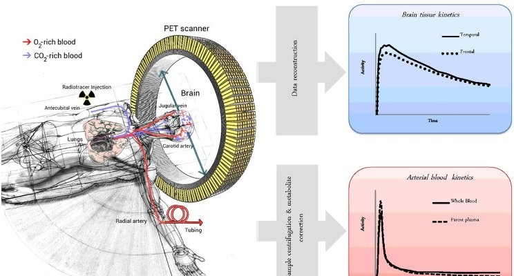
Nuclear medicine imaging procedures are noninvasive and, with the exception of intravenous injections, are usually painless medical tests that help physicians diagnose and evaluate medical conditions. These imaging scans use radioactive materials calledradiopharmaceuticals or radiotracers.
Depending on the type of nuclear medicine exam, the radiotracer is either injected into the body, swallowed or inhaled as a gas and eventually accumulates in the organ or area of the body being examined. Radioactive emissions from the radiotracer are detected by a special camera or imaging device that produces pictures and provides molecular information.

In many centers, nuclear medicine images can be superimposed with computed tomography (CT) or magnetic resonance imaging(MRI) to produce special views, a practice known as image fusion or co-registration. These views allow the information from two different exams to be correlated and interpreted on one image, leading to more precise information and accurate diagnoses. In addition, manufacturers are now making single photon emission computed tomography/computed tomography (SPECT/CT) and positron emission tomography/computed tomography (PET/CT) units that are able to perform both imaging exams at the same time. An emerging imaging technology, but not readily available at this time is PET/MRI.
A PET scan measures important body functions, such as blood flow, oxygen use, and sugar (glucose) metabolism, to help doctors evaluate how well organs and tissues are functioning.
CT imaging uses special x-ray equipment, and in some cases a contrast material, to produce multiple images or pictures of the inside of the body. These images can then be interpreted by a radiologist on a computer monitor. CT imaging provides excellent anatomic information.
Today, almost all PET scans are performed on instruments that are combined PET and CT scanners. The combined PET/CT scans provide images that pinpoint the anatomic location of abnormal metabolic activity within the body. The combined scans have been shown to provide more accurate diagnoses than the two scans performed separately.
What are some common uses of the procedure?
PET and PET/CT scans are performed to:
- detect cancer.
- determine whether a cancer has spread in the body.
- assess the effectiveness of a treatment plan, such as cancer therapy.
- determine if a cancer has returned after treatment.
- determine blood flow to the heart muscle.
- determine the effects of a heart attack, or myocardial infarction, on areas of the heart.
- identify areas of the heart muscle that would benefit from a procedure such as angioplasty or coronary artery bypass surgery (in combination with a myocardial perfusion scan).
- evaluate brain abnormalities, such as tumors, memory disorders, seizures and other central nervous system disorders.
- map normal human brain and heart function.