Clinical Images Journal Indexed in PubMed
  • contact@literaturepublishers.org
  • Whitley Bay, NE26 2HU, England, UK
  • Submit Manuscript
Article Details

Clinical Image

Volume 2, Issue 4 (November Issue)

Mondor Disease: A Rare Diagnosis

Ines B. Ferreira*, Ana O. Monteiro and Jorge Almeida

Internal Medicine Department, Centro Hospitalar Universitario Sao Joao, Porto, Portugal

*Corresponding author: Ines Branco Ferreira, Internal Medicine Department, Centro Hospitalar Universitario Sao Joao, Alameda Prof. Hernani Monteiro, 4200-319 Porto, Portugal, Tel: +351918335210; E-mail: inessalomebf@gmail.com

Received: August 17, 2020; Accepted: August 24, 2020; Published: September 15, 2020

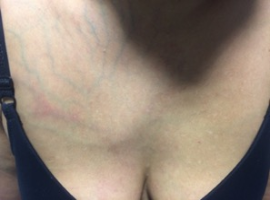
Citation: Ines B. Ferreira, Ana O. Monteiro, Jorge Almeida, et al. Mondor’s Disease: A Rare Diagnosis. Clin Image Case Rep J. 2020; 2(4): 122.

Mondor Disease: A Rare Diagnosis
Abstract

A 38-year-old woman presented to the emergency department with a tender and red mass in the right side of the sternum, alongside with collateral circulation on the right hemithorax. She had no history of trauma or risk factors.
Blood labs, including full blood count, erythrocyte sedimentation rate, renal and liver profile and coagulation, were unremarkable. Color Doppler sonography revealed a superficial vein thrombosis. Based on the history and clinical examination, a clinical diagnosis of Mondor’s disease was made.

The patient was reassured and treated with a course of ibuprofen.
She was reassessed in the outpatient clinic three weeks later. A mammography and a thrombophilic workup (protein C and S deficiency and factor V Leiden mutation) were performed and became normal. Her symptoms resolved completely at five weeks.
Mondor’s disease is a rare, benign and self-limited disease, more prevalent in women, generally between the second and fifth decade of life. It is characterized by superficial thrombophlebitis of the subcutaneous veins of the thoracoabdominal wall and presents with a palpable subcutaneous cord-like induration. Most cases are unilateral and the most affected vessel is the thoracoepigastral vein.

It is more often a primary disease, but it can also be secondary to local trauma, inflammatory conditions, breast surgery (breast augmentation or reconstruction) and rarely there are some cases associated with breast cancer.
The treatment is based on anti-inflammatory drugs to relieve symptoms. It usually resolves in four to eight weeks. The use of pf prophylactic or intermediate doses of low-molecular-weight heparin remains controversial.
This disease is often underdiagnosed due to lack of awareness and it is a differential diagnosis for chest pain.

Keywords: Mondor’s disease