

Viens (Human Anatomy): Image, Functions, Diseases and Treatments
Last Updated: Feb 15, 2023
Viens Image
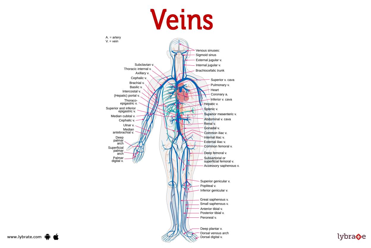
Veins collect oxygen-poor blood to return to the heart. The circulatory system includes veins. They help your heart and other blood vessels move blood. Veins contain most of your blood. 75% of blood is in veins.Veins transport deoxygenated blood to the heart. Gravity forces your leg veins to send blood to your heart. Chronic venous insufficiency, DVT, and varicose veins are all examples of vein problems.
What type of blood do veins carry?
The kind of blood in arteries and veins differs. Unlike arteries, veins contain oxygen-poor blood. Pulmonary veins are an exception.
What are venules?
- Venules are tiny blood channels that connect your capillaries to your veins all over your body.
- Your venules play a crucial role in transporting waste-laden blood from your capillaries to your veins. From there, your blood can return to your heart.
- Venules are larger than capillaries but narrower than veins. Venules vary in size, however even the largest venule is around 16 times smaller than a typical vein.
What do veins look like?
Your veins form a network of blood channels throughout your body. Veins and blood vessels are important parts of your circulatory system. Veins connect to venules and capillaries.
.jpg)
What colour are veins?
Many individuals believe that veins are blue because they seem blue through our skin. But it's merely a deception our eyes play on us. Your veins contain dark red blood, which is darker than the cherry red blood in your arteries.
The blood in one's veins is redder because of the lack of oxygen. Your veins appear blue due to the way light rays penetrate your skin. The blood in one's veins and arteries is always crimson.
What are veins made of?
Each vein has three layers of tissues and fibres:
- The tunica adventitia (outer layer) gives your vein structure and shape.
- Smooth muscle cells in the tunica medium (middle layer) allow your vein to widen or shrink as blood flows through it.
- The tunica intima (inner layer) of your vein is coated with smooth endothelial cells, allowing blood to flow freely.
- The general form of arteries and veins is shown here. Veins, on either hand, are distinguished from arteries by the presence about one valves that keep blood pumping in the proper direction.
- These valves are especially crucial in the legs, where they help blood flow upward into the heart. If these valves become broken, blood can leak backward, causing varicose veins and other issues.
- When it comes to the thickness of their walls, veins differ from arteries. Vein walls are thinner and less muscular. This is due to veins having lower pressure than arteries. As a result, their walls do not need to be as thick to withstand the strain.
What are the various types of veins?
You have three types of veins that help your circulatory system function.
- Deep veins
These veins can be detected in the muscles as well as along the bones. Your deep veins are responsible for transporting oxygen-depleted blood back into your heart. Deep veins in your legs store around 90% of the circulation that returns to your heart. One-way valves in your deep veins keep your blood flowing in the appropriate direction. - Superficial veins
In general, your superficial veins are smaller than your deep veins. They, like deep veins, have valves. They are not surrounded by muscle, unlike deep veins. Your superficial veins, on the other hand, can be discovered just beneath your skin. As a result, they are plainly seen.Ones superficial veins carry blood from the skin's outermost tissues to ones deep veins (via the perforating veins). However, because it is not immediately pushed into action by surrounding muscles, the blood moves more slowly.The great saphenous vein is the largest superficial vein in your body. It extends from your ankle to your thigh in each leg. - Perforating veins
They are superficial veins that transport blood to deeper veins. Perforating veins have valves that close when the calf muscles contract, preventing blood from flowing from deep veins to superficial veins in reverse.
Veins Functions
- Veins provide two functions. One function is to collect oxygen-depleted blood from your body and return it to your heart. Another function is to transport oxygen-rich blood from your lungs to your heart. Veins only convey oxygen-rich blood at this time.
- The function of each vein is determined by its location throughout your body. Veins form a complicated network known as the venous system.
- The venous system is the network of veins in your body and how it communicates to certain other organs and blood vessels. One venous system is separated into two major circuits or sections. These are the pulmonary and systemic circuits. Each circuit uses arteries (veins, arteries, and small blood vessels) to keep the blood circulating.Your veins have two primary circuits. Systemic and pulmonary circuits. Each circuit uses veins, arteries, and capillaries to move blood.
- Blood oxygenates cells and removes waste such as carbon dioxide once it enters capillaries. At that point, blood loses oxygen as well as continues to gain waste. So, recharge it. Blood enters venules before joining veins. Veins transport blood back into the heart to charge up it. Through your inferior and superior vena cava, oxygen-depleted blood reaches your heart.
- While blood comes back to the heart, the systemic circuit comes to an end. It is necessary to complete the pulmonary circuit. In this circuit, blood enters the lungs. During exercise, your veins just carry oxygen-rich blood. To reboot the systemic circuit, your heart must pump oxygen-rich blood.
Viens Conditions and Disorders
There are numerous venous diseases which prevent veins from functioning properly. Some typical issues include:
- Varicose Veins: Twisted, bulging veins close to the skin's surface are called varicose veins, and they develop when blood pools in the veins due to faulty valves.
- Superficial Thrombophlebitis: Inflammation of a vein due to a blood clot is called thrombophlebitis, and it most commonly affects the legs.
- Deep-vein Thrombophlebitis: Thrombophlebitis that affects the deeper veins (deep vein thrombosis) is more dangerous.
- Blood clot: A blood clot is a mass of coagulated blood that has the consistency of a gel. When formed in response to a wound, they stop bleeding by occluding the damaged blood vessel.
- Thrombophlebitis: Inflammation of a vein (often an extremity vein, notably a leg vein) due to a thrombus. A mild condition characterised by a red, sore vein is called superficial thrombophlebitis when it affects a vein close to the skin's surface.
- Deep vein thrombosis: DVT occurs when a blood clot (embolus) forms within a deep vein, typically in the legs (DVT). Edema and leg pain may be symptomatic of deep vein thrombosis. In some instances, no symptoms will be present.
- Phlebitis: A blood clot is not always present in cases of phlebitis. The superficial or deep veins may be affected. This condition is referred to as thrombophlebitis when a blood clot is to blame. The cause may be vein trauma, like that induced by an intravenous ( iv ) line.
- Spider veins: The legs are a common place for spider veins, which are enlarged, twisted veins, to manifest. Women are more likely to have spider veins. Spider veins are more likely to appear as you get older, gain weight, or become pregnant.
- Ulcers: Venous stasis ulcers, often known as ulcers, result from stagnant blood flow. Ulcers are open sores or wounds that refuse to heal or that continually coming back. Ulcers caused by venous insufficiency typically appear on the inner aspect of the lower leg, slightly above the ankle, and below the knee.
- Chronic venous obstruction: The condition characterised by vein occlusion is known as chronic venous obstruction.
- Femoral Vein Thrombosis: There's a clot in the femoral vein in the thigh. Symptoms are uncommon, but in rare cases, you can experience leg edema, redness, and pain.
- Paget-Schroetter Syndrome (PSS): It's a rare kind of DVT that tends to strike young, healthy athletes who engage in sports that place a premium on using the arms, such as baseball and swimming. Muscles in the area can put pressure on the vein.
- Superior Vena Cava Thrombosis: Blood flows from your upper body to your heart via this large vein in your chest. A central line (used to administer medication) or a catheter inserted into a vein is typically the cause of this complication.
- Jugular Vein Thrombosis: Blood from the head and neck is drained back to the heart via the two sets of jugular veins in the neck.
What are the common signs and symptoms of vein problems?
The indications and symptoms you experience are determined by your specific disease. They typically comprise:
- Swelling (edema) in your legs, ankles, or feet, especially after standing for a long period of time.
- Tenderness or pain Legs that are achy, weary, or throbbing Your legs have leathery skin.Itchy, flaking skin on the legs or feet. experience any of these symptoms, or if previously absent purple or bulging veins appear.
- Many venous disorders can be treated if detected early. And it's extremely critical to catch DVT early on before it develops into a pulmonary embolism.
Veins Tests
- Venous Doppler ultrasound: Ultrasound in the veins (venous Doppler) ,is a specialised method for assessing blood flow in the body's major arteries and veins, such as those in the chest, back, legs, and neck.
- Venogram: Medical professionals can examine your veins, particularly those in your legs, with a venogram. Injecting a particular dye visible on X-rays.
- CTV: Using contrast dye, high-tech x-ray equipment, and computer imaging, CT venography (CTV) generates maps of the veins, most frequently those in the legs. First, a technologist inserts an intravenous (IV) line in the patient and injects a contrast dye containing iodine into one of the patient's veins.
- Magnetic resonance imaging: MRI is a diagnostic technique that creates high-resolution pictures of the inside of a human body by using a powerful magnet, radio waves, and a computer. The veins can be seen in an MRV because of the combination of magnetic resonance imaging and IV contrast dye.
Veins Treatments
- Sclerotherapy: Superficial varicose veins may be treated with sclerotherapy. To do this operation, the doctor will visit the patient at home.
- Injection: injects a solution into smaller and medium-sized varicose veins to cause scarring. The procedure seals off the veins, diverting blood to more efficient routes.
- Laser therapy: A laser fibre or electrode is threaded to the targeted vein. Veins are then heated using a laser. This will cause the vein to close off and disappear.
- Surgical ligation: In severe cases, the only effective treatment is surgical varicose vein ligation (tying off) and removal (stripping). Others step forward to fill the void left by the absence of certain veins.
- Ambulatory phlebectomy: Ambulatory phlebectomy that can be performed in an outpatient setting are called 'ambulatory' . Smaller varicose veins can be removed by a medical professional through a series of very small punctures in the skin. This outpatient technique involves numbing the leg only where needles will be inserted.
- Catheter-based procedures using radiofrequency: Small varicose veins can be removed by a medical professional through a series of punctures in the skin. This outpatient technique involves numbing the leg only where needles will be inserted.
Table of content


Find Vascular Surgeon near me

Ask a free question
Get FREE multiple opinions from Doctors