The family physician made a diagnosis of neuropathic ulceration based upon the clinical findings. Neuropathy causes approximately 50% of diabetic foot ulcers. Having diabetic neuropathy alone makes a patient 1.7 times more likely to develop foot ulceration. Patients with diabetic neuropathy combined with pedal deformity are 12.1 times more likely to develop ulceration. The mechanism of injury is commonly described as moderate pressure with repetitive trauma.
Overall 15% of people with diabetes will experience a foot ulcer during their lifetime, and 15% of these will have osteomyelitis. Foot ulcers can develop in any location of the foot but are more common under the metatarsal heads, hallux, heel, or other weight-bearing areas. Foot ulcers may also result from poorly fitting footwear, which creates excessive pressure, friction, or irritation.
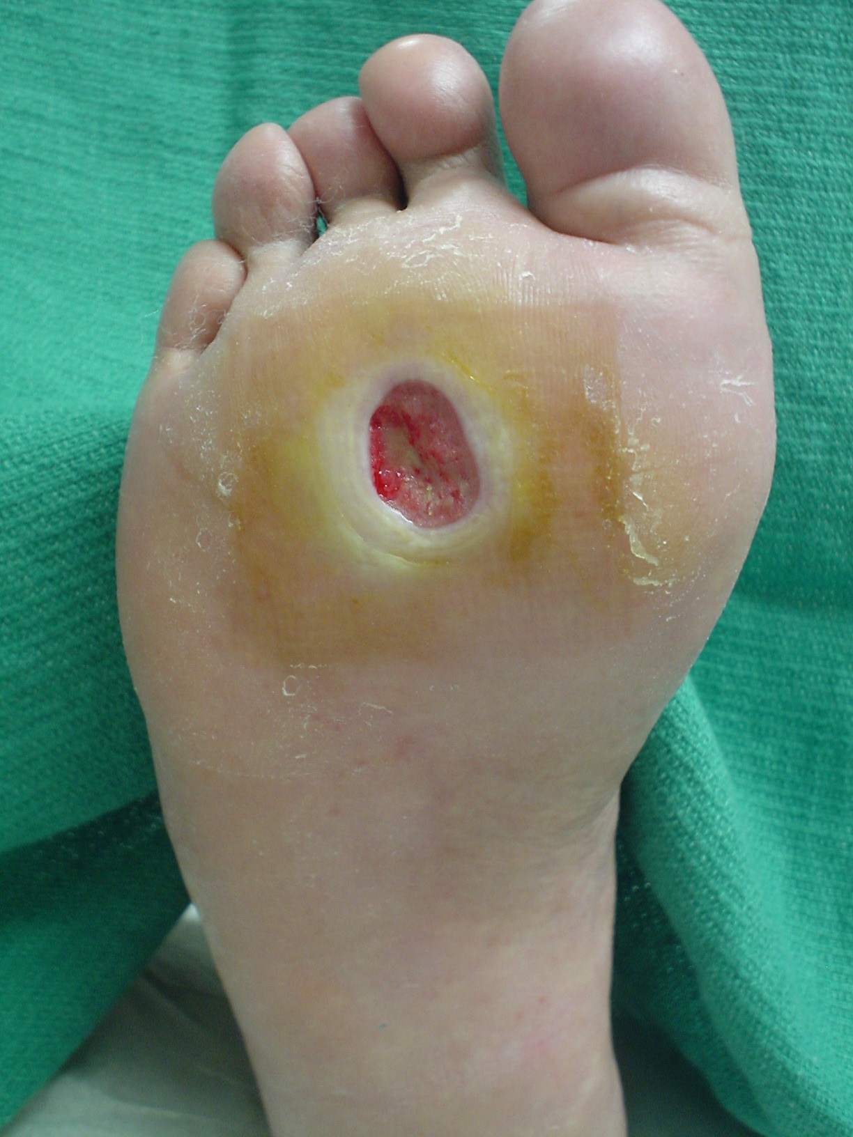
Neuropathic ulcers often have a red, granular base with surrounding hyperkeratosis with white, macerated margins. The ulcer is most commonly found under the metatarsal heads, but it can also be present in the distal and plantar aspects of the toes. Cultures are only indicated if infection is suspected. Swab cultures are not reliable. Curettage of the base of the wound may be more reliable. Radiographs may identify a foreign body, or underlying osteomyelitis. A biopsy may be necessary to rule out a suspected malignancy.
Off-loading pressure from the foot is the standard of care. Multiple devices (eg, removable cast boot, surgical shoes, and wedge shoes) are used for off-loading; however, a total contact cast is the gold standard. Serial tissue debridement should be performed weekly to biweekly to maintain minimal bacterial load, low pressure surrounding the ulcer, and a metabolically active wound base.
If no improvement in the ulcer is noticed in 4 weeks, the ulcer should be considered a chronic wound, and adjunctive therapy (eg, growth factors) must be considered.
In this case, the primary physician referred the patient to a podiatrist, who immediately off-loaded the foot with a total contact cast. The patient’s ulcer healed in one month and he was subsequently fitted with orthopedic shoes.
Text for Photo Rounds Friday courtesy of Richard P. Usatine, MD. Photo courtesy of Javier La Fontaine, DPM. This case was adapted from: La Fontaine J, Shibuya, N. Neuropathic ulcer. In: Usatine R, Smith M, Mayeaux EJ, et al, eds. The Color Atlas of Family Medicine. New York, NY: McGraw-Hill; 2009:907-908.
To learn more about The Color Atlas of Family Medicine, see:
• http://www.amazon.com/Color-Atlas-Family-Medicine/dp/0071474641
You can now get The Color Atlas of Family Medicine as an app for mobile devices including the iPhone and iPad by clicking this link: