
Phytochemical Investigation of New Algerian Lichen Species: Physcia Mediterranea Nimis

 , , ,
, , ,  ,
,
Abstract
:1. Introduction
2. Results and Discussion
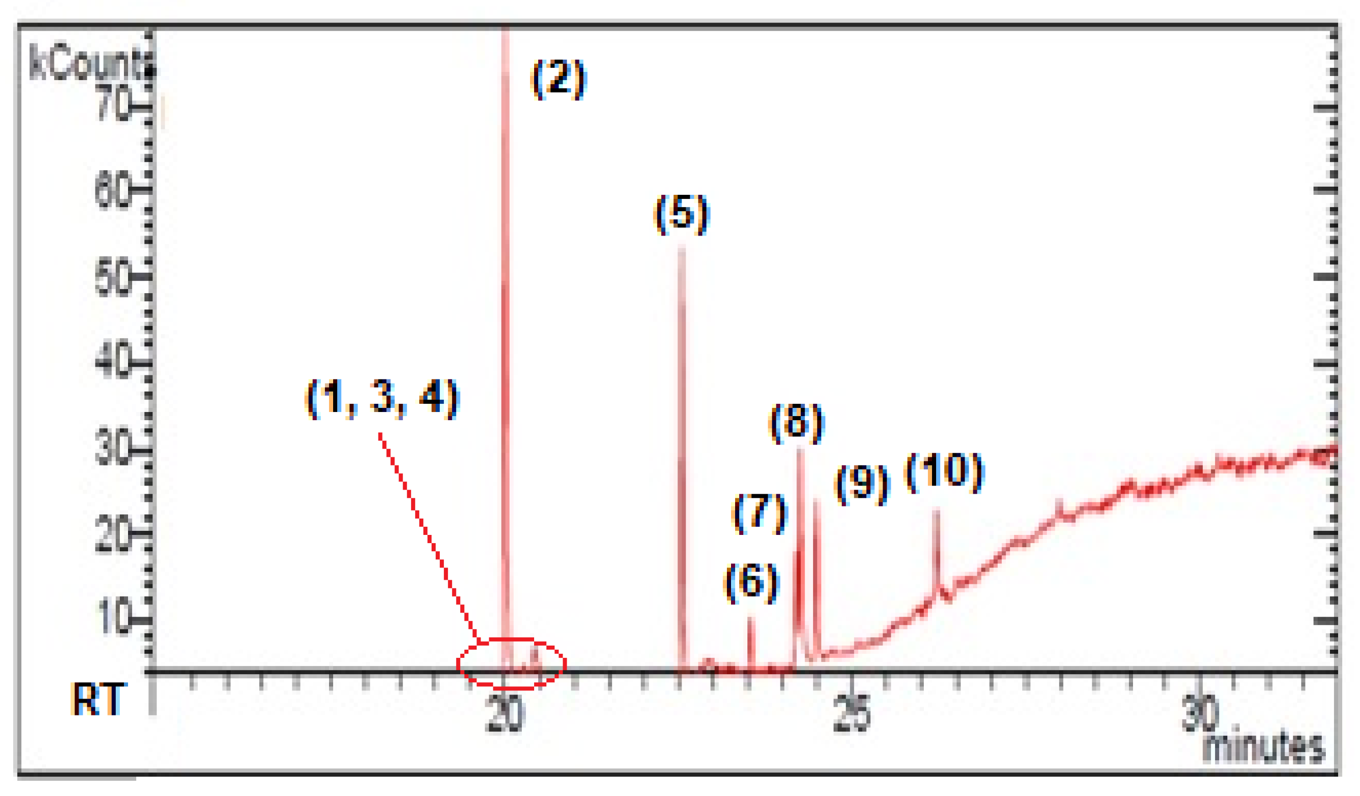
2.1. GC-MS Analysis
2.2. HPLC-ESI-MS-MS Analysis
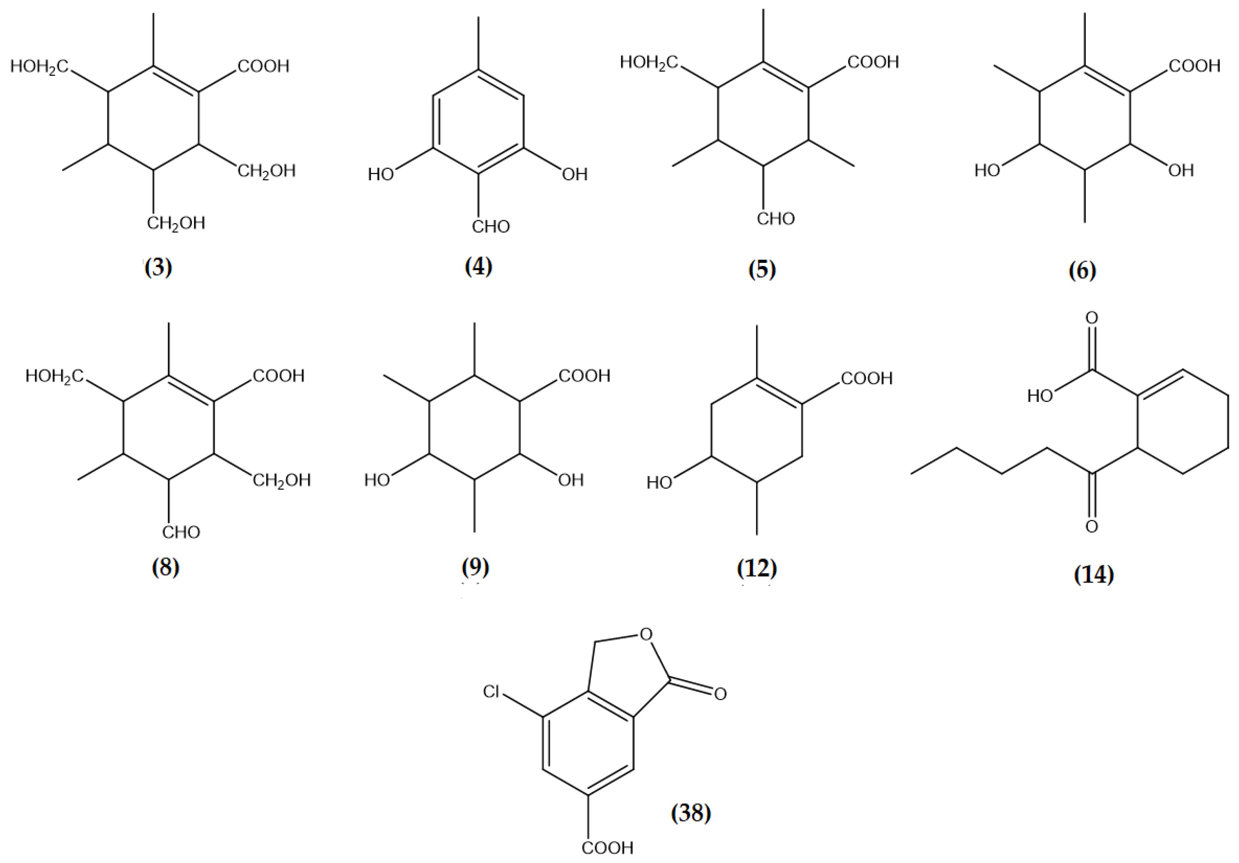
2.2.1. WA-hex Extract Analysis of Physcia mediterranea
2.2.2. ME-hex Extract Analysis of Physcia mediterranea
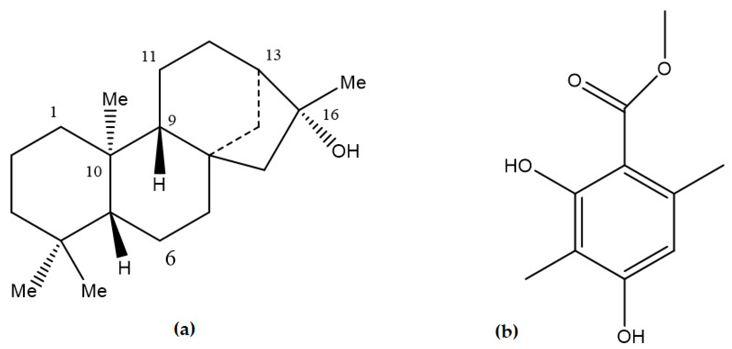
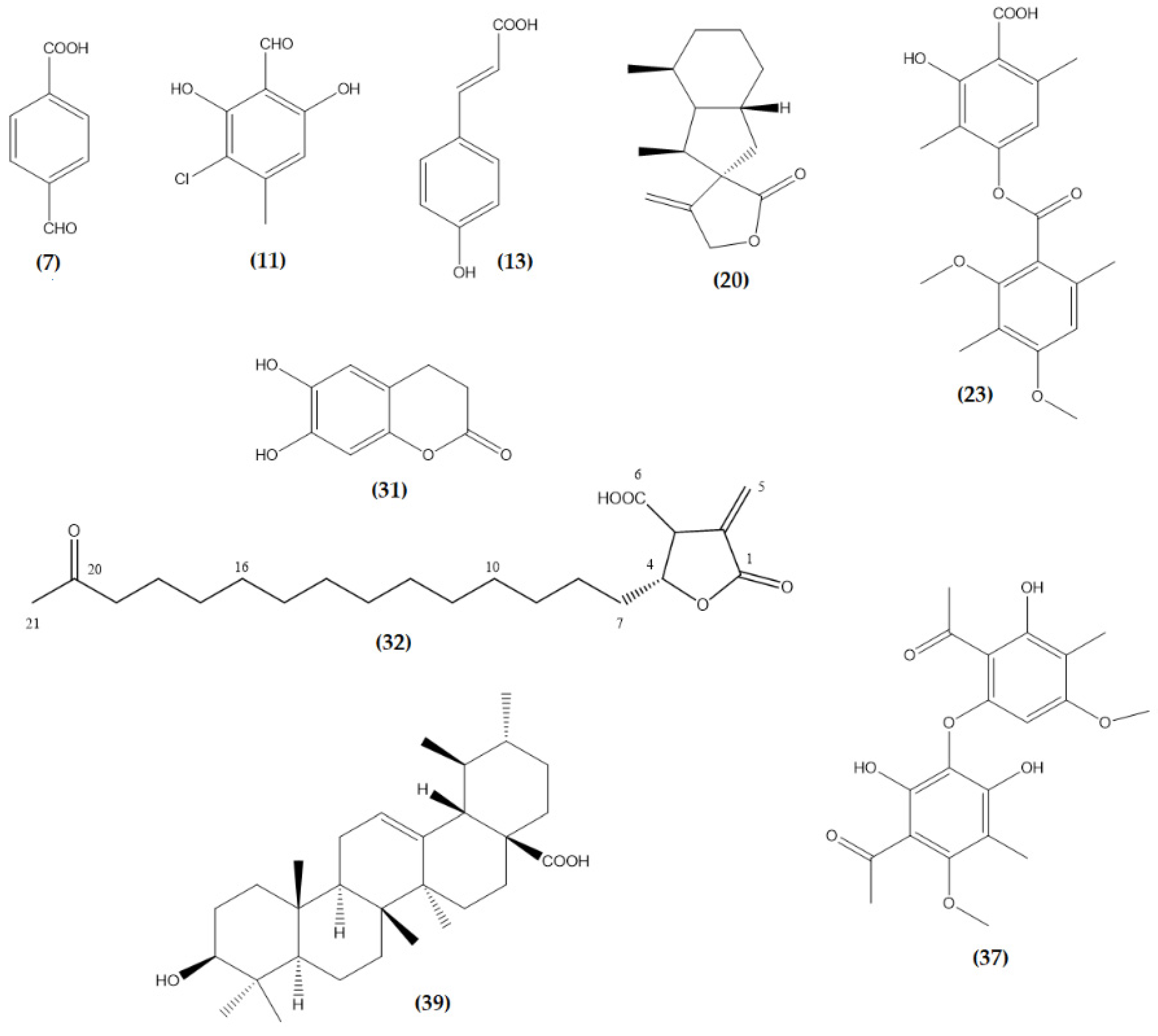
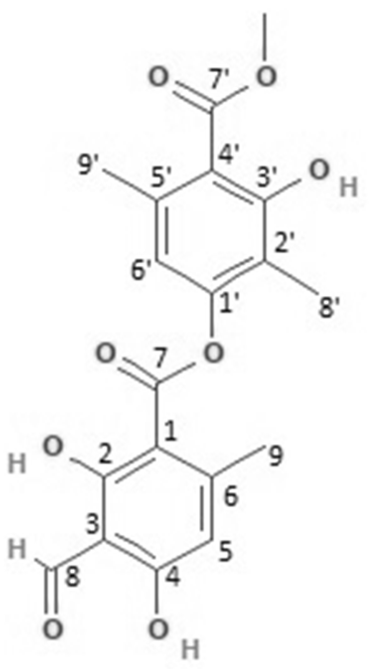
2.3. NMR Analysis
3. Materials and Methods
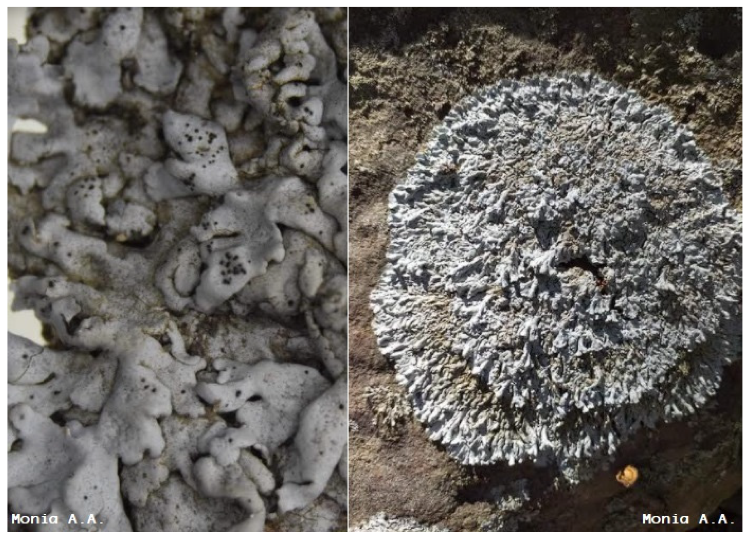
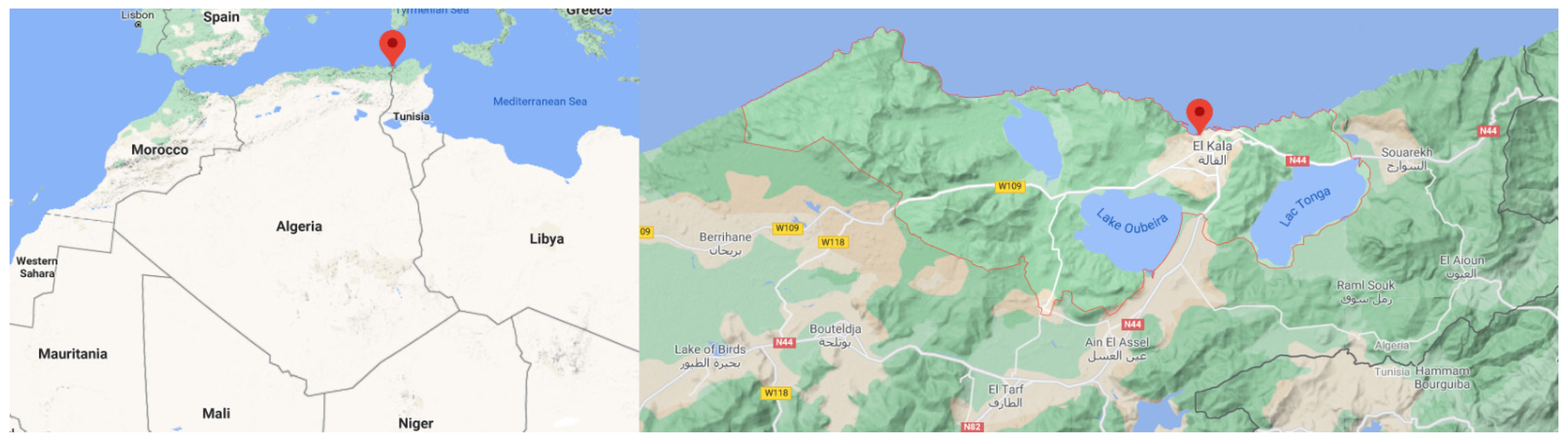
3.1. Lichen Material
3.2. Sample Preparation
3.3. Preparation of Physcia mediterranea Extracts
3.4. Instrumentation and Analysis Parameters
3.4.1. GC-MS Analysis
3.4.2. NMR Analysis
3.4.3. HPLC-ESI-MS-MS
4. Conclusions
Supplementary Materials
Author Contributions
Funding
Institutional Review Board Statement
Informed Consent Statement
Data Availability Statement
Acknowledgments
Conflicts of Interest
Sample Availability
References
- Boullard, B. Guerre et Paix Dans le Règne Végétal; Ellipses: Paris, France, 1990. [Google Scholar]
- Armaleo, D.; Müller, O.; Lutzoni, F.; Andrésson, Ó.S.; Blanc, G.; Bode, H.B.; Collart, F.R.; Dal Grande, F.; Dietrich, F.; Grigoriev, I.V.; et al. The lichen symbiosis re-viewed through the genomes of Cladonia grayi and its algal partner Asterochloris glomerata. BMC Genom. 2019, 20, 1–33. [Google Scholar] [CrossRef] [Green Version]
- Weber, B.; Büdel, B. Fungi and lichens. In Encyclopedia of Earth Sciences Series; Springer: Amsterdam, The Netherlands, 2011; pp. 401–409. [Google Scholar]
- Taylor, T.N.; Krings, M. Fossil Plants: Fungi and Lichens. In Encyclopedia of Geology; Elsevier: Amsterdam, The Netherlands, 2004; pp. 436–443. ISBN 9780123693969. [Google Scholar]
- Adams, D.G.; Bergman, B.; Nierzwicki-Bauer, S.A.; Duggan, P.S.; Rai, A.N.; Schüßler, A. Cyanobacterial-plant symbioses. In The Prokaryotes: Prokaryotic Biology and Symbiotic Associations; Springer: Berlin/Heidelberg, Germany, 2013; pp. 359–400. ISBN 9783642301940. [Google Scholar]
- Werth, S.; Sork, V.L. Identity and genetic structure of the photobiont of the epiphytic lichen Ramalina menziesii on three oak species in southern California. Am. J. Bot. 2010, 97, 821–830. [Google Scholar] [CrossRef] [PubMed] [Green Version]
- Van-Haluwyn, C.; Asta, J.; Gaveriaux, J.P. Guide des Lichens de France: Lichens Des Arbres; Belin, Ed.; Belin: Paris, France, 2009; ISBN 978-2701147000. [Google Scholar]
- Meeßen, J.; Sánchez, F.J.; Brandt, A.; Balzer, E.M.; de la Torre, R.; Sancho, L.G.; de Vera, J.P.; Ott, S. Extremotolerance and Resistance of Lichens: Comparative Studies on Five Species Used in Astrobiological Research I. Morphological and Anatomical Characteristics. Orig. Life Evol. Biosph. 2013, 43, 283–303. [Google Scholar] [CrossRef] [PubMed]
- Kranner, I.; Cram, W.J.; Zorn, M.; Wornik, S.; Yoshimura, I.; Stabentheiner, E.; Pfeifhofer, H.W. Antioxidants and photoprotection in a lichen as compared with its isolated symbiotic partners. Proc. Natl. Acad. Sci. USA 2005, 102, 3141–3146. [Google Scholar] [CrossRef] [PubMed] [Green Version]
- Reyes, A.; Lopez-Bilbao, M.G.; Del, M.; Molina, C. Relationship between Isozymic Variability and Envir-onmental Conditions in the Lichen Xanthoria parietina. Phyton 1996, 36, 265–275. [Google Scholar]
- Goga, M.; Elečko, J.; Marcinčinová, M.; Ručová, D.; Bačkorová, M.; Bačkor, M. Lichen Metabolites: An Overview of Some Secondary Metabolites and Their Biological Potential. In Co-Evolution of Secondary Metabolites; Springer: Cham, Germany, 2018; pp. 1–36. [Google Scholar]
- Kaushik, A.; Giri, P.P. Lichens. In Lichen-Derived Products; Wiley: Hoboken, NJ, USA, 2020; pp. 179–187. [Google Scholar]
- Upreti, D.K.; Divakar, P.K.; Shukla, V.; Bajpai, R. Recent Advances in Lichenology: Modern Methods and Approaches in Lichen Systematics and Culture Techniques; Springer: New Delhi, India, 2015; Volume 2, ISBN 9788132222354. [Google Scholar]
- Yamamoto, Y.; Kinoshita, Y.; Kinoshita, K.; Koyama, K.; Takahashi, K. A zearalenone derivative from the liquid culture of the lichen, Baeomyces placophyllus. J. Hattori Bot. Lab. 2002, 92, 285–289. [Google Scholar]
- Chicita, F.C. Chemical and Botanical Guide to Lichen Products; UNC Press: Chapel Hill, NC, USA, 1969; ISBN 978-0-8078-6859-1. [Google Scholar]
- Moreira, A.S.N.; Braz-Filho, R.; Mussi-Dias, V.; Vieira, I.J.C. Chemistry and biological activity of Ramalina lichenised fungi. Molecules 2015, 20, 8952–8987. [Google Scholar] [CrossRef] [PubMed] [Green Version]
- Oksanen, I. Ecological and biotechnological aspects of lichens. Appl. Microbiol. Biotechnol. 2006, 73, 723–734. [Google Scholar] [CrossRef]
- Huneck, S.; Yoshimura, I.; Huneck, S.; Yoshimura, I. Identification of Lichen Substances. In Identification of Lichen Substances; Springer: Berlin/Heidelberg, Germany, 1996; pp. 11–123. [Google Scholar]
- Calcott, M.J.; Ackerley, D.F.; Knight, A.; Keyzers, R.A.; Owen, J.G. Secondary metabolism in the lichen symbiosis. Chem. Soc. Rev. 2018, 47, 1730. [Google Scholar] [CrossRef]
- Elshobary, M.E.; Osman, M.E.; Abo-Shady, A.M.; Komatsu, E.; Perreault, H.; Sorensen, J.; Piercey-Normore, M.D. Algal carbohydrates affect polyketide synthesis of the lichen-forming fungus Cladonia rangiferina. Mycologia 2016, 108, 646–656. [Google Scholar] [CrossRef] [Green Version]
- Elkhateeb, W.; Daba, G. Occurrence of terpenes, polyketides, and tannins in some Japanese lichens and green mosses. Egypt. Pharm. J. 2020, 19, 216. [Google Scholar] [CrossRef]
- Solárová, Z.; Liskova, A.; Samec, M.; Kubatka, P.; Büsselberg, D.; Solár, P. Anticancer potential of lichens’ secondary metabolites. Biomolecules 2020, 10, 87. [Google Scholar] [CrossRef] [Green Version]
- Jha, B.N.; Shrestha, M.; Pandey, D.P.; Bhattarai, T.; Bhattarai, H.D.; Paudel, B. Investigation of antioxidant, antimicrobial and toxicity activities of lichens from high altitude regions of Nepal. BMC Complement. Altern. Med. 2017, 17, 1–8. [Google Scholar] [CrossRef] [PubMed] [Green Version]
- Thadhani, V.M.; Karunaratne, V. Potential of Lichen Compounds as Antidiabetic Agents with Antioxidative Properties: A Review. Oxid. Med. Cell. Longev. 2017, 2017. [Google Scholar] [CrossRef] [PubMed] [Green Version]
- Rodriguez, C.M.; Bennett, J.P.; Johnson, C.J. Lichens: Unexpected anti-prion agents? Prion 2012, 6, 11–16. [Google Scholar] [CrossRef] [Green Version]
- Millot, M.; Girardot, M.; Dutreix, L.; Mambu, L.; Imbert, C. Antifungal and anti-biofilm activities of acetone lichen extracts against Candida albicans. Molecules 2017, 22, 651. [Google Scholar] [CrossRef] [PubMed] [Green Version]
- Zambare, V.P.; Christopher, L.P. Biopharmaceutical potential of lichens. Pharm. Biol. 2012, 50, 778–798. [Google Scholar] [CrossRef] [PubMed]
- White, P.A.S.; Oliveira, R.C.M.; Oliveira, A.P.; Serafini, M.R.; Araújo, A.A.S.; Gelain, D.P.; Moreira, J.C.F.; Almeida, J.R.G.S.; Quintans, J.S.S.; Quintans-Junior, L.J.; et al. Antioxidant activity and mechanisms of action of natural compounds isolated from lichens: A systematic review. Molecules 2014, 19, 14496–14527. [Google Scholar] [CrossRef]
- Shukla, V.; Joshi, G.P.; Rawat, M.S.M. Lichens as a potential natural source of bioactive compounds: A review. Phytochem. Rev. 2010, 9, 303–314. [Google Scholar] [CrossRef]
- Behera, B.C.; Mahadik, N.; Morey, M. Antioxidative and cardiovascular-protective activities of metabolite usnic acid and psoromic acid produced by lichen species Usnea complanata under submerged fermentation. Pharm. Biol. 2012, 50, 968–979. [Google Scholar] [CrossRef] [Green Version]
- Crawford, S.D. Lichens used in traditional medicine. In Lichen Secondary Metabolites: Bioactive Properties and Pharmaceutical Potential; Springer International Publishing: Berlin/Heidelberg, Germany, 2015; pp. 27–80. ISBN 9783319133744. [Google Scholar]
- Crawford, S.D. Lichens Used in Traditional Medicine. In Lichen Secondary Metabolites; Springer International Publishing: Berlin/Heidelberg, Germany, 2019; pp. 31–97. [Google Scholar]
- Komaty, S.; Letertre, M.; Dang, D.; Jungnickel, H.; Laux, P. Sample preparation for an optimised extraction of localised metabolites in lichens: Application to Pseudevernia furfuracea. Talanta 2015. [Google Scholar] [CrossRef]
- Rafika, B.; Monia, A. Antibacterial Activity of the Chloroform, Acetone, Methanol and queous Extracts of Algerian Lichens. Jordan J. Pharm. Sci. 2018, 11, 55–67. [Google Scholar]
- Monia, A.A.; Rafika, B.; Tarek, H. Lichen diversity in the edough peninsula, North East of Algeria. Bot. Complut. 2018, 42, 9–18. [Google Scholar] [CrossRef] [Green Version]
- Brakni, R.; Serradj, M.; Fernandez, X.; Michel, T. Chemical and biological investigation of Algerian lichens. Planta Med. 2016, 82, P657. [Google Scholar] [CrossRef]
- Aljabeili, H.S.; Barakat, H.; Abdel-Rahman, H.A. Chemical Composition, Antibacterial and Antioxidant Activities of Thyme Essential Oil (Thymus vulgaris). Food Nutr. Sci. 2018, 9, 433–446. [Google Scholar] [CrossRef] [Green Version]
- Serradj, M.; El Oualidi, J.; Slimani, A.; Boumedris, Z. Contribution to the lichens;inventory from the Oubeira Lake (NE Algeria). Bull. Inst. Sci. Rabat Sect. Sci. Vie 2013, 35, 15–17. [Google Scholar]
- Amrani, S.; Nacer, A.; Noureddine, N.E.; Seaward, M.R. Lichenological exploration of Algeria: Historical overview and annotated bibliography, 1799–2013. Willdenowia 2015, 45, 15–34. [Google Scholar] [CrossRef] [Green Version]
- Rebbas, K.; Boutabia, L.; Touazi, Y.; Gharzouli, R.; Djellouli, Y.; Alatou, D. Inventaire des lichens du Parc national de Gouraya (Béjaïa, Algérie). Phytotherapie 2011, 9, 225–233. [Google Scholar] [CrossRef]
- Mohamed, A.H.; Miara, M.D.; Rebbas, K.; Slimani, A.; Ravera, S.; Hamerelain, A.S. Mise à jour de l’inventaire des lichens d’Algérie. Rev. Ecol. 2014, 10, 75–103. [Google Scholar]
- Rahali, M. Cartographie de la pollution plombique dans la région d’Alger en utilisant un lichen (Xanthoria parientina) comme bioaccumulateur. Pollut. Atmos. 2002, 421–432. [Google Scholar] [CrossRef] [Green Version]
- Khedim, R.; Maatoug, M.; Benhassaini, H.; Hammou, M.A. Macrolichens New to Algeria and Other Interesting Species from Theniet-el-Had National Park. Herzogia 2018, 31, 252–267. [Google Scholar] [CrossRef]
- Brakni, R.; Ali Ahmed, M.; Burger, P.; Schwing, A.; Michel, G.; Pomares, C.; Hasseine, L.; Boyer, L.; Fernandez, X.; Landreau, A.; et al. UHPLC-HRMS/MS Based Profiling of Algerian Lichens and Their Antimicrobial Activities. Chem. Biodivers. 2018, 15, e1800031. [Google Scholar] [CrossRef] [PubMed]
- Slimani, A.; Ahmed, A.; Serradj, M.; Hamel, T.; Coste, C. Contribution a l’etude de la flore lichenique dans la zeenaie de Bougous (foret de Ramel Tonal) au niveau du Pare National d’El Kala Nord Est Algerien. Synthèse Rev. Des Sci. Et De La Technol. 2013, 27, 22–29. [Google Scholar]
- Bouiadjra, S.B.; Ramdani, M.; Djellouli, R.; ELkhiati, N.; Zerey, W.E.; Bouterfas, K.; Roger, F. Air quality in the metropolitan area of Sidi Bel Abbes (Algeria) through the lichens species as bio-indicators. Moroc. J. Chem. 2017, 5. [Google Scholar] [CrossRef]
- Ghennam, K.; Fatiha, A. Biosurveillance par SIG de la qualité de l’air d’une cité méditerraneene (Alger), diversité lichénique, indice de pureté atmosphérique (IPA) et indice d’impact. Revue d’Écologie 2017, 72, 134–146. [Google Scholar]
- Maizi, N.; Alioua, A.; Tahar, A. Jumelage des bio-indicateurs et d’un réseau de surveillance de la qualité de l’air pour la détection de la pollution par le SO 2 dans la région de Annaba (Algérie). Biotechnol. Agron. Soc. Env. 2012, 16, 149–158. [Google Scholar]
- Serradj-Ali Ahmed, M.; Boumedris, Z.E.; Djebar, M.R.; Tahar, A. Responses of antioxidants in Flavoparmelia caperata (L.) Hale to the atmospheric pollution air at two urban and semi-urban areas in the region of Annaba (East of Algeria). Pollut. Atmos. 2014. [Google Scholar] [CrossRef] [Green Version]
- Djamel, S.; Yamna, D.; Djamel, A. Biological diversity of the National Park of El-Kala (Algeria), valorisation and protection. Biodivers. J. 2014, 5, 525–532. [Google Scholar]
- Putra, P.P.; Abdullah, S.S.; Rahmatunisa, R.; Junaidin, J.; Ismed, F. Structure, activity, and drug-likeness of pure compounds of Sumatran lichen (Stereocaulon halei) for the targeted ACE2 protein in COVID-19 disease. Pharmaciana 2020, 10, 135. [Google Scholar] [CrossRef]
- Mallavadhani, U.V.; Tirupathamma, R.S.; Sagarika, G.; Ramakrishna, S. Isolation, Chemical Modification, and Anticancer Activity of Major Metabolites of the Lichen Parmotrema mesotropum. Chem. Nat. Compd. 2019, 55, 825–831. [Google Scholar] [CrossRef]
- Nishiyama, Y.; Noda, Y.; Nakatani, N.; Shitan, N.; Sudo, T.; Kato, A.; Mutiso, P.B.C. Structure of constituents isolated from the bark of Cassipourea malosana and their cytotoxicity against a human ovarian cell line. J. Nat. Med. 2019, 73, 289–296. [Google Scholar] [CrossRef] [PubMed]
- Vladimirova, I.N.; Georgiyants, V.A. Extracted compounds from Cetraria islandica. Chem. Nat. Compd. 2013, 49, 347–348. [Google Scholar] [CrossRef]
- Akbar, S.; Akbar, S. Dolichousnea longissima (Ach) Articus/Parmelia perlata Esch. (Parmeliaceae). In Handbook of 200 Medicinal Plants; Springer International Publishing: Berlin/Heidelberg, Germany, 2020; pp. 873–878. [Google Scholar]
- Yu, X.L.; Yang, X.Y.; Gao, X.L.; Bai, R.F.; Yin, X.; Su, G.Z.; Qu, C.H.; Chai, X.Y.; Tu, P.F. Phenolic constituents from lichen Usnea longissima. Zhongguo Zhongyao Zazhi 2016, 41, 1864–1869. [Google Scholar] [CrossRef] [PubMed]
- Carpentier, C.; Queiroz, E.F.; Marcourt, L.; Wolfender, J.L.; Azelmat, J.; Grenier, D.; Boudreau, S.; Voyer, N. Dibenzofurans and pseudodepsidones from the lichen Stereocaulon paschale collected in northern Quebec. J. Nat. Prod. 2017, 80, 210–214. [Google Scholar] [CrossRef]
- Ari, F.; Ulukaya, E.; Oran, S.; Celikler, S.; Ozturk, S.; Ozel, M.Z. Promising anticancer activity of a lichen, Parmelia sulcata Taylor, against breast cancer cell lines and genotoxic effect on human lymphocytes. Cytotechnology 2015, 67, 531–543. [Google Scholar] [CrossRef] [PubMed] [Green Version]
- Stojanović, I.Ž.; Radulović, N.S.; Mitrović, T.L.; Stamenković, S.M.; Stojanović, G.S. Volatile constituents of selected Parmeliaceae lichens. J. Serb. Chem. Soc. 2011, 76, 987–994. [Google Scholar] [CrossRef]
- Hoffmann, H.R.; Matusch, R.; Baniahmad, A. Isolation of Atraric Acid, Synthesis of Atraric Acid Derivatives, and Use of Atraric Acid and the Derivatives thereofe for the Treatment of Benigne Prostatic Hyperplasia, Prostate Carcinoma and Spinobulbar Muscular Atrophy. U.S. Patent 8,481,519, 9 July 2013. [Google Scholar]
- Hessenkemper, W.; Roediger, J.; Bartsch, S.; Houtsmuller, A.B.; van Royen, M.E.; Petersen, I.; Grimm, M.-O.; Baniahmad, A. A natural androgen receptor antagonist induces cellular senescence in prostate cancer cells. Mol. Endocrinol. 2014, 28, 1831–1840. [Google Scholar] [CrossRef] [Green Version]
- Güvenç, A.; Akkol, E.K.; Süntar, I.; Keleş, H.; Yildiz, S.; Çaliş, I. Biological activities of Pseudevernia furfuracea (L.) Zopf extracts and isolation of the active compounds. J. Ethnopharmacol. 2012, 144, 726–734. [Google Scholar] [CrossRef]
- Ahad, A.M.; Goto, Y.; Kiuchi, F.; Tsuda, Y.; Kondo, K.; Sato, T. Nematocidal principles in “Oakmoss Absolute” and nematocidal activity of 2, 4-Dihydroxybenzoates. Chem. Pharm. Bull. 1991, 39, 1043–1046. [Google Scholar] [CrossRef] [Green Version]
- Yamamoto, Y.; Watanabe, A. Fatty acid their composition of lichens and their phyco-and mycobionts. J. Gen. Appl. Microbiol. 1974, 20, 83–86. [Google Scholar] [CrossRef] [Green Version]
- Sonesson, M.; Grimberg, Å.; Sveinbjörnsson, B.; Carlsson, B.Å. Seasonal variation in concentrations of carbohydrates and lipids in two epiphytic lichens with contrasting, snow-depth related distribution on subarctic birch trees. Bryologist 2011, 114, 443–452. [Google Scholar] [CrossRef]
- Řezanka, T.; Dembitsky, V.M. Fatty acids of lichen species from Tian Shan Mountains. Folia Microbiol. 1999, 44, 643–646. [Google Scholar] [CrossRef]
- Vu, T.H.; Catheline, D.; Delmail, D.; Boustie, J.; Legrand, P.; Lohezic-Le Devehat, F. Gas chromatographic analysis to compare the fatty acid composition of fifteen lichen species, with a focus on Stereocaulon. Lichenologist 2016, 48, 323–337. [Google Scholar] [CrossRef]
- Culberson, C. Biogenetic relationships of the lichen substances in the framework of systematics. Bryologist 1986, 89, 91–98. [Google Scholar] [CrossRef]
- Fehrer, J.; Slavíková-Bayerová, Š.; Orange, A. Large genetic divergence of new, morphologically similar species of sterile lichens from Europe (Lepraria, Stereocaulaceae, Ascomycota): Concordance of DNA sequence data with secondary metabolites. Cladistics 2008, 24, 443–458. [Google Scholar] [CrossRef]
- Hale, M.E. A Monograph of the Lichen Genus Parmelia Acharius sensu Strict (Ascomycotina: Pameliaceae); Smithsonian Institution Press: Washington, DC, USA, 1987. [Google Scholar]
- Haugan, R.; Timdal, E. Tephromela perlata and T. talayana, with notes on the T. aglaea-complex. Graph. Scr. 1994, 6, 17–26. [Google Scholar]
- Spribille, T.; Klug, B.; Mayrhofer, H. A phylogenetic analysis of the boreal lichen Mycoblastus sanguinarius (Mycoblastaceae, lichenised Ascomycota) reveals cryptic clades correlated with fatty acid profiles. Mol. Phylogenet. Evol. 2011, 59, 603–614. [Google Scholar] [CrossRef] [PubMed]
- Lumbsch, H.T. The Use of Metabolic Data in Lichenology at the Species and Subspecific Levels. Lichenologist 1998, 30, 357–367. [Google Scholar] [CrossRef]
- Hawksworth, D.L. Lichen chemotaxonomy. In Lichenology: Progress and Problems; Proceedings of an international Symposium; Academic Press: Cambridge, MA, USA, 1976; pp. 139–184. [Google Scholar]
- Stocker-Wörgötter, E. Metabolic diversity of lichen-forming ascomycetous fungi: Culturing, polyketide and shikimate metabolite production, and PKS genes. Nat. Prod. Rep. 2008, 25, 188–200. [Google Scholar] [CrossRef]
- Sumida, C.; Graber, R.; Nunez, E. Role of fatty acids in signal transduction: Modulators and messengers. Prostaglandins Leukot. Essent. Fat. Acids 1993, 48, 117–122. [Google Scholar] [CrossRef]
- Bychek-Gushchina, I. Study of biochemical aspects of lichen symbiosis. I. Lipids and fatty acids in cultured lichen symbionts. Biochemistry 1997, 62, 490–494. [Google Scholar]
- Torres, A.; Dor, I.; Rotem, J.; Srebnik, M.; Dembitsky, V.M. Characterization of surface n-alkanes and fatty acids of the epiphytic lichen Xanthoria parietina, its photobiont a green alga Trebouxia sp., and its mycobiont, from the Jerusalem hills. Eur. J. Biochem. 2003, 270, 2120–2125. [Google Scholar] [CrossRef] [PubMed] [Green Version]
- Molina, M.C.; Crespo, A.; Vicente, C.; Elix, J.A. Differences in the composition of phenolics and fatty acids of cultured mycobiont and thallus of Physconia distorta. Plant Physiol. Biochem. 2003, 41, 175–180. [Google Scholar] [CrossRef]
- Dembitsky, V.M.; Rezanka, T.; Bychek, I.A. Seasonal variation of lipids and fatty acids from tree-growing lichens of the genus Physcia. Phytochemistry 1994, 36, 601–608. [Google Scholar] [CrossRef]
- Dembitsky, V.M.; Rezanka, T.; Bychek, I. A Seasonal variability of lipids and fatty acids in the tree-growing lichen Xanthoria parientina L. J. Exp. Bot. 1994, 45, 403–408. [Google Scholar] [CrossRef]
- Reis, R.A.; Iacomini, M.; Gorin, P.A.J.; Souza, L.M.; Grube, M.; Cortes Cordeiro, L.M.; Sassaki, G.L. Fatty acid composition of the tropical lichen Teloschistes flavicans and its cultivated symbionts. FEMS Microbiol. Lett. 2005, 247, 1–6. [Google Scholar] [CrossRef] [Green Version]
- Goss, R.; Wilhelm, C. Lipids in Algae, Lichens and Mosses. In Lipids in Photosynthesis; Springer: Dordrecht, The Netherlands, 2009; pp. 117–137. [Google Scholar]
- Temina, M.; Levitsky, D.O.; Dembitsky, V.M. Chemical Constituents of the Epiphytic and Lithophilic Lichens of the Genus Collema. Rec. Nat. Prod. 2010, 4, 79–86. [Google Scholar]
- Ilica, R.A.; Kloetzer, L.; Galaction, A.I.; Caşcaval, D. Fumaric acid: Production and separation. Biotechnol. Lett. 2019, 41, 47–57. [Google Scholar] [CrossRef] [PubMed]
- Sebastian, J.; Hegde, K.; Kumar, P.; Rouissi, T.; Brar, S.K. Bioproduction of fumaric acid: An insight into microbial strain improvement strategies. Crit. Rev. Biotechnol. 2019, 39, 817–834. [Google Scholar] [CrossRef]
- Haghikia, A.; Linker, R.; Gold, R. Fumarsäure in der Therapie der Multiplen Sklerose. Nervenarzt 2014, 85, 720–726. [Google Scholar] [CrossRef] [PubMed]
- Papadaki, A.; Papapostolou, H.; Alexandri, M.; Kopsahelis, N.; Papanikolaou, S.; de Castro, A.M.; Freire, D.M.G.; Koutinas, A.A. Fumaric acid production using renewable resources from biodiesel and cane sugar production processes. Environ. Sci. Pollut. Res. 2018, 25, 35960–35970. [Google Scholar] [CrossRef] [PubMed]
- Jabłońska-Trypuć, A.; Pankiewicz, W.; Czerpak, R. Traumatic Acid Reduces Oxidative Stress and Enhances Collagen Biosynthesis in Cultured Human Skin Fibroblasts. Lipids 2016, 51, 1021–1035. [Google Scholar] [CrossRef] [Green Version]
- Jabłońska-Trypuć, A.; Krętowski, R.; Wołejko, E.; Wydro, U.; Butarewicz, A. Traumatic acid toxicity mechanisms in human breast cancer MCF-7cells. Regul. Toxicol. Pharmacol. 2019, 106, 137–146. [Google Scholar] [CrossRef] [PubMed]
- Lee, H.C.; Liu, F.C.; Tsai, C.N.; Chou, A.H.; Liao, C.C.; Yu, H.P. Esculetin Ameliorates Lipopolysaccharide-Induced Acute Lung Injury in Mice Via Modulation of the AKT/ERK/NF-κB and RORγt/IL-17 Pathways. Inflammation 2020, 43, 962–974. [Google Scholar] [CrossRef] [PubMed]
- Wang, C.; Pei, A.; Chen, J.; Yu, H.; Sun, M.L.; Liu, C.F.; Xu, X. A natural coumarin derivative esculetin offers neuroprotection on cerebral ischemia/reperfusion injury in mice. J. Neurochem. 2012, 121, 1007–1013. [Google Scholar] [CrossRef]
- Hongyan, L. Esculetin Attenuates Th2 and Th17 Responses in an Ovalbumin-Induced Asthmatic Mouse Model. Inflammation 2016, 39, 735–743. [Google Scholar] [CrossRef] [PubMed]
- Han, M.H.; Park, C.; Lee, D.S.; Hong, S.H.; Choi, I.W.; Kim, G.Y.; Choi, S.H.; Shim, J.H.; Chae, J.I.; Yoo, Y.H.; et al. Cytoprotective effects of esculetin against oxidative stress are associated with the upregulation of Nrf2-mediated NQO1 expression via the activation of the ERK pathway. Int. J. Mol. Med. 2017, 39, 380–386. [Google Scholar] [CrossRef] [Green Version]
- Srikrishna, D.; Godugu, C.; Dubey, P.K. A Review on Pharmacological Properties of Coumarins. Mini-Rev. Med. Chem. 2016, 18, 113–141. [Google Scholar] [CrossRef] [PubMed]
- Stefanachi, A.; Leonetti, F.; Pisani, L.; Catto, M.; Carotti, A. Coumarin: A Natural, Privileged and Versatile Scaffold for Bioactive Compounds. Molecules 2018, 23, 250. [Google Scholar] [CrossRef] [Green Version]
- Musa, M.; Cooperwood, J.; Khan, M.O. A Review of Coumarin Derivatives in Pharmacotherapy of Breast Cancer. Curr. Med. Chem. 2008, 15, 2664–2679. [Google Scholar] [CrossRef] [Green Version]
- Studzińska-Sroka, E.; Galanty, A.; Bylka, W. Atranorin-an interesting lichen secondary metabolite. Mini-Rev. Med. Chem. 2017, 17, 1633–1645. [Google Scholar] [CrossRef] [PubMed]
- Huneck, S.; Feige, G.B.; Schmidt, J. Chemie von Cladonia furcata und Cladonia rangiformis. Herzogia 2004, 17, 51–58. [Google Scholar]
- Nybakken, L.; Helmersen, A.M.; Gauslaa, Y.; Selås, V. Lichen compounds Restrain lichen feeding by bank voles (Myodes glareolus). J. Chem. Ecol. 2010, 36, 298–304. [Google Scholar] [CrossRef]
- Culberson, C.F. Chemical and Botanical Guide to Lichen Products, 2nd ed.; Otto Koeltz Science Publishers: Koenigstein, Germany, 1979. [Google Scholar]
- Elix, J.A. A Catalogue of Standardized Chromatographic Data and Biosynthetic Relationships for Lichen Substances; Australian National University: Canberra, Australia, 2014. [Google Scholar]
- Ranković, B.; Mišić, M.; Sukdolak, S. The antimicrobial activity of substances derived from the lichens Physcia aipolia, Umbilicaria polyphylla, Parmelia caperata and Hypogymnia physodes. World J. Microbiol. Biotechnol. 2008, 24, 1239–1242. [Google Scholar] [CrossRef]
- Solhaug, K.A.; Larsson, P.; Gauslaa, Y. Light screening in lichen cortices can be quantified by chlorophyll fluorescence techniques for both reflecting and absorbing pigments. Planta 2010, 231, 1003–1011. [Google Scholar] [CrossRef]
- Brodo, I.M.; Freebury, C.; Alfonso, N. Notes on the Lichens Physcia aipolia and P. alnophila in North America. Evansia 2013, 30, 110–119. [Google Scholar] [CrossRef]
- Maldonado Montaño, A.; Menesses, R.; Bravo, J.A.; Vila, J.L.; Bello, A. Presence of atranorin in Physcia sorediosa. Rev. Boliv. Quím. 2016, 33, 175–178. [Google Scholar]
- Kosanić, M.; Ranković, B.; Stanojković, T.; Rančić, A.; Manojlović, N. Cladonia lichens and their major metabolites as possible natural antioxidant, antimicrobial and anticancer agents. LWT Food Sci. Technol. 2014, 59, 518–525. [Google Scholar] [CrossRef]
- Pompilio, A.; Pomponio, S.; Di Vincenzo, V.; Crocetta, V.; Nicoletti, M.; Piovano, M.; Garbarino, J.A.; Di Bonaventura, G. Antimicrobial and antibiofilm activity of secondary metabolites of lichens against methicillin-resistant Staphylococcus aureus strains from cystic fibrosis patients. Future Microbiol. 2013, 8, 281–292. [Google Scholar] [CrossRef] [PubMed]
- Melo, M.G.D.; Araújo, A.A.S.; Serafini, M.R.; Carvalho, L.F.; Bezerra, M.S.; Ramos, C.S.; Bonjardim, L.R.; Albuquerque-Júnior, R.L.C.; Lima, J.T.; Siqueira, R.S.; et al. Anti-inflammatory and toxicity studies of atranorin extracted from Cladina kalbii Ahti in rodents. Braz. J. Pharm. Sci. 2011, 47, 861–872. [Google Scholar] [CrossRef] [Green Version]
- Melo, M.G.D.; Araújo, A.A.S.; Rocha, C.P.L.; Almeida, E.M.S.A.; De Souza Siqueira, R.; Bonjardim, L.R.; Quintans-Júnior, L.J. Purification, physicochemical properties, thermal analysis and antinociceptive effect of atranorin extracted from Cladina kalbii. Biol. Pharm. Bull. 2008, 31, 1977–1980. [Google Scholar] [CrossRef] [Green Version]
- Barreto, R.S.S.; Albuquerque-Júnior, R.L.C.; Pereira-Filho, R.N.; Quintans, J.S.S.; Barreto, A.S.; DeSantana, J.M.; Santana-Filho, V.J.; Santos, M.R.V.; Bonjardim, L.R.; Araújo, A.A.S.; et al. Evaluation of wound healing activity of atranorin, a lichen secondary metabolite, on rodents. Braz. J. Pharmacogn. 2013, 23, 310–319. [Google Scholar] [CrossRef] [Green Version]
- Harikrishnan, A.; Veena, V.; Lakshmi, B.; Shanmugavalli, R.; Theres, S.; Prashantha, C.N.; Shah, T.; Oshin, K.; Togam, R.; Nandi, S. Atranorin, an antimicrobial metabolite from lichen Parmotrema rampoddense exhibited in vitro anti-breast cancer activity through interaction with Akt activity. J. Biomol. Struct. Dyn. 2020. [Google Scholar] [CrossRef] [PubMed]
- Galanty, A.; Koczurkiewicz, P.; Wnuk, D.; Paw, M.; Karnas, E.; Podolak, I.; Węgrzyn, M.; Borusiewicz, M.; Madeja, Z.; Czyż, J.; et al. Usnic acid and atranorin exert selective cytostatic and anti-invasive effects on human prostate and melanoma cancer cells. Toxicol. Vitr. 2017, 40, 161–169. [Google Scholar] [CrossRef] [PubMed]
- Hikino, H.; Ito, K.; Takemoto, T. Stereostructure of Asebotoxin I and II, Toxins of Pieris japonica. Chem. Pharm. Bull. 1969, 17, 854–855. [Google Scholar] [CrossRef] [PubMed] [Green Version]
- Zhang, L.; Hong, Z.; Zhang, R.R.; Sun, X.Z.; Yuan, Y.F.; Hu, J.; Wang, X. Bakkenolide A inhibits leukemia by regulation of HDAC3 and PI3K/Akt-related signaling pathways. Biomed. Pharmacother. 2016, 83, 958–966. [Google Scholar] [CrossRef]
- Solberg, Y. Chemical constituents of the lichen species Cetraria islandica. J. Hattori Bot. Lab. 1986, 60, 391–406. [Google Scholar]
- Simirgiotis, M.J.; Quispe, C.; Areche, C.; Sepúlveda, B. Phenolic compounds in Chilean Mistletoe (quintral, Tristerix tetrandus) analysed by UHPLC-Q/Orbitrap/MS/MS and its antioxidant properties. Molecules 2016, 21, 245. [Google Scholar] [CrossRef] [Green Version]
- Huneck, S. New Results on the Chemistry of Lichen Substances. In Progress in the Chemistry of Organic Natural Products; Springer: Berlin/Heidelberg, Germany, 2001; Volume 81. [Google Scholar]
- Seo, D.Y.; Lee, S.R.; Heo, J.W.; No, M.H.; Rhee, B.D.; Ko, K.S.; Kwak, H.B.; Han, J. Ursolic acid in health and disease. Korean J. Physiol. Pharmacol. 2018, 22, 235–248. [Google Scholar] [CrossRef] [Green Version]
- Gonzalez, A.G.; Bermejo Barrera, J.; Rodriguez Perez, E.M.; Hernandez Padron, C.E. chemical constituents of the lichen Ramalina hierrensis. Planta Med. 1992, 58, 214–218. [Google Scholar] [CrossRef] [PubMed]
- Vu, T.H.; Lamer, A.C.L.; Lalli, C.; Samson, J.B.M.; Dévéhat, F.L.L.; Seyec, J. Le Depsides: Lichen metabolites active against hepatitis C virus. PLoS ONE 2015, 10, e0120405. [Google Scholar] [CrossRef] [PubMed]
- Rubio, C.; Galloway, D.J.; Quilhot, W. Secondary products from Pannaria tavaresii (Lichens). J. Chil. Chem. Soc. 2005, 50, 667–669. [Google Scholar] [CrossRef]
- De Carvalho, M.G.; De Carvalho, G.J.A.; Braz-Filho, R. Chemical Constituents from Ouratea floribunda: Complete 1H and 13C NMR Assignments of Atranorin and its New Acetyl Derivative. J. Braz. Chem. Soc. 2000, 11, 143–147. [Google Scholar] [CrossRef]
- Norouzi, H.; Azizi, A.; Gholami, M.; Sohrabi, M.; Boustie, J. Chemotype variations among lichen ecotypes of Umbilicaria aprina as revealed by LC-ESI-MS/MS: A survey of antioxidant phenolics. Environ. Sci. Pollut. Res. 2020, 27, 40296–40308. [Google Scholar] [CrossRef] [PubMed]
- Vos, C.; McKinney, P.; Pearson, C.; Heiny, E.; Gunawardena, G.; Holt, E.A. The optimal extraction and stability of atranorin from lichens, in relation to solvent and pH. Lichenologist 2018, 50, 499–512. [Google Scholar] [CrossRef]
- Lohezic-Le Devehat, F.; Legouin, B.; Couteau, C.; Boustie, J.; Coiffard, L. Lichenic extracts and metabolites as UV filters. J. Photochem. Photobiol. B Biol. 2013, 120, 17–28. [Google Scholar] [CrossRef]
- Bjerke, J.W.; Elvebakk, A.; Domínguez, E.; Dahlback, A. Seasonal trends in usnic acid concentrations of Arctic, alpine and Patagonian populations of the lichen Flavocetraria nivalis. Phytochemistry 2005, 66, 337–344. [Google Scholar] [CrossRef]
- Marante, F.J.T.; Castellano, A.G.; Rosas, F.E.; Aguiar, J.Q.; Barrera, J.B. Identification and quantitation of allelochemicals from the lichen Lethariella canariensis: Phytotoxicity and antioxidative activity. J. Chem. Ecol. 2003, 29, 2049–2071. [Google Scholar] [CrossRef]
- Solhaug, K.A.; Lind, M.; Nybakken, L.; Gauslaa, Y. Possible functional roles of cortical depsides and medullary depsidones in the foliose lichen Hypogymnia physodes. Flora Morphol. Distrib. Funct. Ecol. Plants 2009, 204, 40–48. [Google Scholar] [CrossRef]
- Forbes, P.; Wat, L.; Strumpher, J. Comparative Perspectives on Extraction Methods for Organic Metabolites and Pollutants from Lichens. In Lichen-Derived Products; Wiley: Hoboken, NJ, USA, 2020; pp. 27–73. [Google Scholar]
- Augusto, S.; Pinho, P.; Branquinho, C.; Pereira, M.J.; Soares, A.; Catarino, F. Atmospheric Dioxin and Furan Deposition in Relation to Land-Use and Other Pollutants: A Survey with Lichens; Kluwer Academic Publishers: Dordrecht, The Netherlands, 2004; Volume 49. [Google Scholar]
- Van der Wat, L.; Forbes, P.B.C. Comparison of extraction techniques for polycyclic aromatic hydrocarbons from lichen biomonitors. Environ. Sci. Pollut. Res. 2019, 26, 11179–11190. [Google Scholar] [CrossRef]
- Van Der Wat, L.; Forbes, P.B.C. Lichens as biomonitors for organic air pollutants. TrAC Trends Anal. Chem. 2015, 64, 165–172. [Google Scholar] [CrossRef] [Green Version]
- Caccamese, S.; Toscano, R.M.; Bellesia, F.; Pinetti, A. Methyl β-Orcinolcarboxylate and Depsides from Parmelia Furfuracea. J. Nat. Prod. 1985, 48, 157–158. [Google Scholar] [CrossRef]
- Gunzinger, J.; Tabacchi, R. Isolement et identification de l’acide furfurique, nouvelle depsidone du lichen Pseudevernia furfuracea (L.) Ach. Helv. Chim. Acta 1985, 68, 1936–1939. [Google Scholar] [CrossRef]
- Culberson, W.L.; Culberson, C.F.; Johnson, A. Pseudevernia furfuracea-Olivetorina Relationships: Chemistry and Ecology. Mycologia 1977, 69, 604. [Google Scholar] [CrossRef]
- Joulain, D.; Tabacchi, R. Lichen extracts as raw materials in perfumery. Part 2: Treemoss. Flavour Fragr. J. 2009, 24, 105–116. [Google Scholar] [CrossRef] [Green Version]


| N° | RT | Compound | Mass | % | Synonyms | |
|---|---|---|---|---|---|---|
| 1 | 19:56 | Methyl 2-hydroxy-4-methoxy-6-methylbenzoate | 196 | 0.3 | Orsellinic Acid Methyl Ester 4-Methyl Ether (Sparassol) | C10H12O4 |
| 2 | 20:01 | Methyl 2,4-dihydroxy-3,5,6-trimethylbenzoate | 210 | 52.3 | Derivatives of methylbenzoic acids | C11H14O4 |
| 3 | 20:43 | Methyl 4-hydroxy-2-methoxy-3,6-dimethylbenzoate | 210 | 1.1 | 2-metoxy-Atraric acid | C10H12O4 |
| 4 | 20:64 | Methyl 2,4-dihydroxy-3,6-dimethylbenzoate | 196 | 2.3 | Atraric acid | C10H12O4 |
| 5 | 22:55 | Methyl hexadecanoate | 270 | 24.2 | Palmitic acid | C16H32O2 |
| 6 | 23:53 | 13-Methyl-17-norkaur-15-ene | 272 | 2.4 | Linoleic acid | C18H32O2 |
| 7 | 24:19 | Methyl cis, cis-9,12-octadecadienoate | 294 | 3.2 | Oleic acid | C18H34O2 |
| 8 | 24:25 | Methyl (Z)-9-octadecenoate | 296 | 4.9 | Stearic acid | C18H34O2 |
| 9 | 24:48 | Methyloctadecanoate | 298 | 3.8 | (−)-ent-Kauran-16α-ol | C2OH34O |
| 10 | 26:21 | Unknown | 376 | 2.2 | Unknown | |
| T% | 95.5 |
| N° | RT | [M − H]− | TM | MM | MT | Compounds Identification |
|---|---|---|---|---|---|---|
| 1 | 0.03 | 146.9397 | 147.9475 | C4H4O6 | A | Dihydroxyfumaric acid |
| 2 | 1.80 | 116.9276 | 117.9354 | C4H6O4 | A | Butendioic acid |
| 3 | 2.72 | 187.0971 | 188.1049 | C9H16O2 | A | 3,5-dimethoxyciclohexanecarboxylic acid |
| 4 | 5.51 | 163.0395 | 164.0473 | C9 H8 O3 | B | p-Coumaric acid |
| 5 | 6.57 | 227.1286 | 228.1364 | C12H20O4 | A | Trans-dodec-2-enedioic acid (traumatic acid) |
| 6 | 7.46 | 282.2077 | 283.2155 | C16 H29 NO3 | A | N-dodecanoyl-l-Homoserine lactone |
| 7 | 8.09 | 174.9556 | 175.9634 | C5 H4 O7 | A | 2-Hydroxy-3,4-dioxopentanedioc acid |
| 8 | 8.15 | 293.1762 | 294.1840 | C17H26O4 | B | Gingerol |
| 9 | 14.37 | 295.2280 | 296.2358 | C18 H32 O3 | A | 18-Hydroxylinoleic acid |
| 10 | 15.36 | 177.0186 | 178.0264 | C9 H6 O4 | B | 6,7-Dihydroxycoumarin (esculetin) |
| 11 | 15.44 | 373.0929 | 374.1007 | C19 H18 O8 | C | Atranorin |
| 12 | 16.47 | 407.0539 | 408.0617 | C19 H17 O8Cl | C | Chloroatranorin |
| 13 | 17.57 | 277.2175 | 278.2203 | C18H30O2 | A | Octadeca-9,12,15-trienoic acid (linolenelaidic acid) |
| 14 | 17.77 | 265.1480 | 266.1558 | C15 H22 O4 | A | (4E,6E,9E)-Pentadeca-4,6,9-trienedioic acid |
| 15 | 18.03 | 253.2331 | 254.2249 | C16 H30 O2 | A | Palmitoleic acid |
| 16 | 18.34 | 241.2173 | 242.2251 | C15H30O2 | A | Pentadecanoic acid |
| 17 | 18.50 | 279.0936 | 280.2409 | C18 H32 O2 | A | Linoleic acid |
| 18 | 18.91 | 267.2331 | 268.2409 | C17 H32 O2 | A | cis-9-Heptadecenoic acid (margaroleic acid) |
| 19 | 19.15 | 255.2329 | 256.2407 | C16 H32 O2 | A | Palmitic acid |
| 20 | 19.35 | 459.3271 | 460.3349 | C25H48O7 | Unknown | Unknown |
| 21 | 19.51 | 281.2487 | 282.2565 | C18 H34 O2 | A | Oleic acid |
| 22 | 19.75 | 459.3272 | 460.3350 | C25 H48 O7 | A | Methyl glucose isostearate |
| 23 | 19.89 | 269.2488 | 270.2566 | C17 H34 O2 | A | Heptadecanoic acid (margaric acid) |
| 24 | 19.99 | 307.2645 | 308.2723 | C20 H36 O2 | A | 11,14-Eicosadienoic acid |
| 25 | 20.07 | 457.3722 | 458.3800 | C27 H54 O5 | Unknown | Unknown |
| 26 | 20.18 | 295.2645 | 296.2723 | C19H36O2 | A | 10E-nonadecenoic acid |
| 27 | 20.54 | 283.2643 | 284.2721 | C18 H36 O2 | A | Stearic acid (octadecanoic acid) |
| 28 | 20.82 | 309.2800 | 310.2878 | C20 H38 O2 | A | Eicosenoic acid (gondoic acid) |
| 29 | 21.71 | 311.2957 | 312.3035 | C20H40O2 | A | Arachidic acid (eicosanoic acid) |
| 30 | 22.62 | 297.1532 | 298.1610 | C12H26O8 | Unknown | Unknown |
| 31 | 22.77 | 339.3268 | 340.3346 | C22H44O2 | A | Docosanoic acid (behenic acid) |
| 32 | 23.08 | 309.1743 | 310.1821 | C17 H26 O5 | D | Portentol |
| 33 | 23.58 | 353.2003 | 354.2081 | C19H30O6 | Unknown | Unknown |
| 34 | 23.81 | 311.1689 | 312.1767 | C13 H30 O8 | Unknown | Unknown |
| 35 | 23.89 | 367.3579 | 368.3657 | C24H48O2 | A | Lignoceric acid (tetracosanoic acid) |
| 36 | 24.01 | 397.2266 | 398.2344 | C21H34O7 | D | Stephanol |
| 37 | 24.60 | 293.1793 | 294.1871 | C17H26O4 | A | Heptadecatrienedioic acid |
| 38 | 24.94 | 325.1844 | 326.1922 | C14 H30 O8 | Unknown | Unknown |
| 39 | 25.33 | 395.3895 | 396.3973 | C26H52O2 | A | Hexacosanoic acid or cerotic acid |
| 40 | 25.89 | 337.2055 | 338.2133 | C19 H30 O5 | A | 6-Oxononadeca-8,11-dienedioic acid |
| 41 | 26.10 | 339.2000 | 340.2078 | C15 H32 O8 | Unknown | Unknown |
| 42 | 26.24 | 381.2317 | 382.2395 | C21 H34 O6 | A | 19-Acetoxylichesterinic acid |
| 43 | 26.75 | 425.2581 | 426.2659 | C23H38O7 | E | Asebotoxin I |
| 44 | 27.53 | 321.2106 | 322.2184 | C19H30O4 | A | Nonadecatrienedioic acid |
| 45 | 27.85 | 304.9143 | 305.9221 | Unknown | Unknown | Unknown |
| N° | RT | [M – H]− | TM | MM | MT | Compounds | MS2 Ions |
|---|---|---|---|---|---|---|---|
| 1 | 2.72 | 187.0970 | 188.1043 | C9H16O4 | A | 3,5-Dimethoxyciclohexanecarboxylic acid | 141.8670; 123.0807 |
| 2 | 2.75 | 293.0669 | 294.0741 | C14H14O7 | A | 6-(Hydroxymethyl)-3,5-bis(methoxycarbonyl)-2,4-dimethylcyclohex-1-ene-1-carboxylic acid | 234.0530; 207.0695 |
| 3 | 3.08 | 243.1239 | 244.1311 | C12H20O5 | A | 3,5,6-Hydroxymethyl-2,4-dimethylcyclohex-1-ene-1-carboxylic acid | 183.1025; 176.6474 |
| 4 | 3.15 | 151.0393 | 152.0465 | C8H8O3 | B | Atranol | 123.0444; 105.0335; 81.0335 |
| 5 | 3.20 | 225.1129 | 226.1201 | C12H18O4 | A | 5-Formyl-3-hydroxymethyl-2,4,6-trimethylcyclohex-1-ene-1-carboxylic acid | 207.1023; 97.0284 |
| 6 | 3.47 | 199.0973 | 200.1046 | C10H16O4 | A | 3,5-Dihydroxy-2,4,6-trimethylciclohexenecarboxilic acid | 183.4492; 162.8385; 114.9508 |
| 7 | 3.58 | 149.0237 | 150.0310 | C8H6O3 | C | 4-Formylbenzoic acid | − |
| 8 | 3.82 | 241.1081 | 242.1153 | C12H18O5 | A | 5-Formyl-3,6-dihydroxymethyl-2,4-dimethylcyclohex-1-enecarboxylic acid | 218.816; 172.6485 |
| 9 | 4.10 | 201.1129 | 202.1202 | C10H18O4 | A | 2,4-Dihydroxy-3,5,6-trimethylcyclohexane-1-carboxylic acid | 197.6263; 164.8350; 139.1124 |
| 10 | 4.95 | 199.1337 | 200.1409 | C11H20O3 | A | 2-Hydroxy-10-undecenoic acid | − |
| 11 | 5.00 | 185.0006 | 186.0079 | C8H7ClO3 | B | Chloroatranol | − |
| 12 | 5.21 | 169.0863 | 170.0936 | C9H14O3 | A | 4-Hydroxy-2,5-dimethylcyclohex-1-ene-1-carboxylic acid | 124.9796; 104.4180 |
| 13 | 5.49 | 163.0392 | 164.0470 | C9H8O3 | D | p-Coumaric acid | − |
| 14 | 5.60 | 209.1181 | 210.1253 | C12H18O3 | A | 6-(1-Oxopentyl)-1-cyclohexene-1-carboxylic acid | 146.9850; 105.0336 |
| 15 | 5.87 | 215.1286 | 216.1359 | C11H20O4 | A | Undecanedioic acid | − |
| 16 | 6.57 | 227.1288 | 228.1360 | C12H20O4 | A | trans-Dodec-2-enedioic acid | − |
| 17 | 7.44 | 282.2078 | 283.2150 | C16H29NO3 | A | N-Dodecanoyl-l-Homoserine lactone | − |
| 18 | 9.53 | 243.1601 | 244.1675 | C13H24O4 | A | Tridecanedioic acid | − |
| 19 | 10.59 | 311.2230 | 312.2302 | C18H32O4 | A | 9Z-Octadecenedioic acid | − |
| 20 | 10.87 | 233.1547 | 234.1619 | C15H22O2 | E | Fukinanolide | − |
| 21 | 10.89 | 323.2230 | 324.2303 | C19H32O4 | F | allo-Protolichesterinic acid | − |
| 22 | 12.28 | 313,2388 | 314.2266 | C18H34O4 | A | Octadecanedioic acid | − |
| 23 | 13.04 | 389.1245 | 390.1315 | C20H22O8 | B | 8-Hydroxydiffractaic acid | − |
| 24 | 13.34 | 293.2124 | 294.2202 | C18H30O3 | A | 2-Hydroxylinolenic acid | − |
| 25 | 13.58 | 291.1968 | 292.2041 | C18H28O3 | A | α-Licanic acid | − |
| 26 | 14.38 | 295.2279 | 296.2351 | C18H32O3 | A | 2-Hydroxylinoleic acid | − |
| 27 | 14.40 | 295,2278 | 296,2352 | C18H32O3 | A | 18-hydroxylinoleic acid | − |
| 28 | 15.19 | 297.2436 | 298.2508 | C18H34O3 | A | 9-Oxooctadecanoic acid | − |
| 29 | 15.25 | 295.2280 | 296.2351 | C18H32O3 | A | Coriolic acid | − |
| 30 | 15.36 | 297.2435 | 298.2508 | C18H34O3 | A | Ricinoleic acid | − |
| 31 | 15.43 | 177.0187 | 178.0259 | C9H6O4 | D | 6,7-Dihydroxycoumarin | − |
| 32 | 15.46 | 365.2330 | 366.2403 | C21H34O5 | G | Muronic acid | − |
| 33 | 16.37 | 471.3481 | 472.3553 | C30H48O4 | Unknown | Unknown | − |
| 34 | 16.47 | 210.9834 | 211.9873 | C9H5ClO4 | H | 7-chloro-3-oxo-1,3-dihydroisobenzofuran-5-carboxylic acid | 138.9948, 103.0180 |
| 35 | 16.51 | 407.0540 | 408.0611 | C19H17ClO8 | B | Chloroatranorin | − |
| 36 | 17.59 | 387.2544 | 388.2616 | C24H36O4 | Unknown | Unknown | − |
| 37 | 17.68 | 389.1242 | 390.1314 | C20H22O8 | I | Leprolomin | − |
| 38 | 18.04 | 253.2173 | 254.2244 | C16H30O2 | A | Palmitoleic acid | − |
| 39 | 18.33 | 241.2172 | 242.2245 | C15H30O2 | A | Pentadecanoic acid | − |
| 40 | 18.35 | 455.3531 | 456.3605 | C30H48O3 | G | Ursolic acid | − |
| 41 | 18.50 | 279.2330 | 280.2403 | C18H32O2 | A | Linoleic acid | − |
| 42 | 18.66 | 299.2595 | 300.2667 | C18H36O3 | A | 2-Hydroxyoctadecanoic acid | − |
| 43 | 18.78 | 279.2332 | 280.2403 | C18H32O2 | A | Linoleic acid | − |
| 44 | 19.16 | 255.2329 | 256.2401 | C16H32O2 | A | Palmitic acid | − |
| 45 | 19.49 | 281.2485 | 282.2559 | C18H34O2 | A | Oleic acid | − |
| 46 | 19.62 | 269.2488 | 270.2561 | C17H34O2 | A | 15-Methylhexadecanoic acid | − |
| 47 | 19.89 | 269.2488 | 270.2561 | C17H34O2 | A | Heptadecanoic acid | − |
| 48 | 20.07 | 327.2543 | 328.2616 | C20H40O3 | A | 2-Hydroxyeicosanoic acid | − |
| 49 | 20.54 | 283.2643 | 284.2716 | C18H36O2 | A | Stearic acid (octadecanoic acid) | − |
| 50 | 20.74 | 309.2801 | 310.2875 | C20H38O2 | A | Eicosenoic acid (Gondoic acid) | − |
| 51 | 21.14 | 297.2801 | 298.2873 | C19H38O2 | A | Nonadecanoic acid | − |
| 52 | 21.70 | 311.2957 | 312.3029 | C20H40O2 | A | Arachidic acid (Eicosanoic acid) | − |
| 53 | 22.68 | 637.4841 | 638.4908 | C41 H66 O5 | Unknown | Unknown | − |
| 54 | 24.51 | 605.3483 | 606. 3561 | C37H50O7 | Unknown | Unknown | − |
Publisher’s Note: MDPI stays neutral with regard to jurisdictional claims in published maps and institutional affiliations. |
© 2021 by the authors. Licensee MDPI, Basel, Switzerland. This article is an open access article distributed under the terms and conditions of the Creative Commons Attribution (CC BY) license (http://creativecommons.org/licenses/by/4.0/).
Share and Cite
Kerboua, M.; Ahmed, M.A.; Samba, N.; Aitfella-Lahlou, R.; Silva, L.; Boyero, J.F.; Raposo, C.; Lopez Rodilla, J.M. Phytochemical Investigation of New Algerian Lichen Species: Physcia Mediterranea Nimis. Molecules 2021, 26, 1121. https://doi.org/10.3390/molecules26041121
Kerboua M, Ahmed MA, Samba N, Aitfella-Lahlou R, Silva L, Boyero JF, Raposo C, Lopez Rodilla JM. Phytochemical Investigation of New Algerian Lichen Species: Physcia Mediterranea Nimis. Molecules. 2021; 26(4):1121. https://doi.org/10.3390/molecules26041121
Chicago/Turabian StyleKerboua, Marwa, Monia Ali Ahmed, Nsevolo Samba, Radhia Aitfella-Lahlou, Lucia Silva, Juan F. Boyero, Cesar Raposo, and Jesus Miguel Lopez Rodilla. 2021. "Phytochemical Investigation of New Algerian Lichen Species: Physcia Mediterranea Nimis" Molecules 26, no. 4: 1121. https://doi.org/10.3390/molecules26041121