Article

The sprT Gene of Bacillus velezensis FZB42 Is Involved in Biofilm Formation and Bacilysin Production

1
Co-Innovation Center for Sustainable Forestry in Southern China, College of Forestry, Nanjing Forestry University, Nanjing 210037, China
2
College of Life Science, Nanjing Forestry University, Nanjing 210037, China
*
Author to whom correspondence should be addressed.
These authors contributed equally to this work.
Int. J. Mol. Sci. 2023, 24(23), 16815; https://doi.org/10.3390/ijms242316815
Submission received: 2 November 2023 / Revised: 22 November 2023 / Accepted: 24 November 2023 / Published: 27 November 2023
(This article belongs to the Special Issue Molecular Research in Prebiotics, Probiotics and Postbiotics)

Abstract

:
Bacillus velezensis FZB42, a representative strain of plant-growth-promoting rhizobacteria (PGPR), can form robust biofilm and produce multiple antibiotics against a wild range of phytopathogens. In this study, we observed different biofilm morphology of the mutant Y4, derived from a TnYLB-1 transposon insertion library of B. velezensis FZB42. We identified that the transposon was inserted into the sprT gene in Y4. Our bioinformatics analysis revealed that the SprT protein is an unstable hydrophilic protein located in the cytoplasm. It is highly conserved in Bacillus species and predicted to function as a metalloprotease by binding zinc ions. We also demonstrated that ΔsprT significantly reduced the swarming ability of FZB42 by ~5-fold and sporulation capacity by ~25-fold. In addition, the antagonistic experiments showed that, compared to the wild type, the ΔsprT strain exhibited significantly reduced inhibition against Staphylococcus aureus ATCC-9144 and Phytophthora sojae, indicating that the inactivation of sprT led to decreased production of the antibiotic bacilysin. The HPLC-MS analysis confirmed that bacilysin was indeed decreased in the ΔsprT strain, and qPCR analysis revealed that ΔsprT down-regulated the expression of the genes for bacilysin biosynthesis. Our results suggest that the sprT gene plays a regulatory role in multiple characteristics of B. velezensis FZB42, including biofilm formation, swarming, sporulation, and antibiotic production.

1. Introduction

Many soil-borne phytopathogens can destroy plants or strongly reduce plant production, causing a significant economic loss in agriculture or forestry [1,2]. Although chemical pesticides can effectively control plant diseases, they often lead to ecological and environmental problems. In contrast, biological control strategies are currently receiving increasing attention due to their environmentally friendly nature. There are a large number of beneficial microorganisms in plant rhizosphere, which are generally defined as plant-growth-promoting microorganisms (PGPMs). These microorganisms have plant disease control functions and thus can be developed as biocontrol agents; meanwhile, most of them also facilitate plant growth and can be used as microbial fertilizers [3].
Bacillus velezensis FZB42 is a model organism of plant-growth-promoting rhizobacteria (PGPR). It can colonize plant roots [4,5] and synthesize a variety of secondary metabolites, hormones, and antioxidants, helping plants against phytopathogens [6,7,8]. FZB42 also induces systemic resistance of plants [9,10]. Nearly 10% of the FZB42 genome is used to synthesize antimicrobial metabolites and their corresponding immune genes [11,12]. Among them, five gene clusters encode non-ribosomal peptide synthases (NRPSs), which generate lipopeptide antibiotics (LPs), and three clusters encode polyketide synthase (PKS) genes, which produce polyketide (PKs) antibiotics [7,13,14,15]. A dipeptide antibiotic produced by FZB42, bacilysin, also has strong antimicrobial activity [16]. For example, bacilysin demonstrates remarkable inhibition of dangerous plant pathogens such as Phytophthora and Xanthomonas [2,17]. In addition, bacilysin strongly suppresses the growth of some eukaryotes such as Saccharomyces cerevisiae and Candida albicans [17,18].
The production of these antibiotics in B. velezensis FZB42 is well connected with other phenotypes such as biofilm formation, motility, and sporulation [19]. Unlike spores produced by fungi, Bacillus survives longer in adverse conditions by producing heat-resistant endospores [20]. In order to further understand how these characteristics are regulated in FZB42, we constructed a transposon library of FZB42 using the TnYLB-1 transposon [21]. Through observing colony morphologies of the mutant strains from the library, we identified a mutant strain (Y4) with a seemingly altered colony surface. In this study, we determined the disrupted gene in Y4 via TnYLB- 1 and studied its effect on biofilm formation, motility, and sporulation. Moreover, we explored the implication and the primary mechanism of this gene (sprT) in the production of the antibiotic bacilysin.

2. Results and Analysis

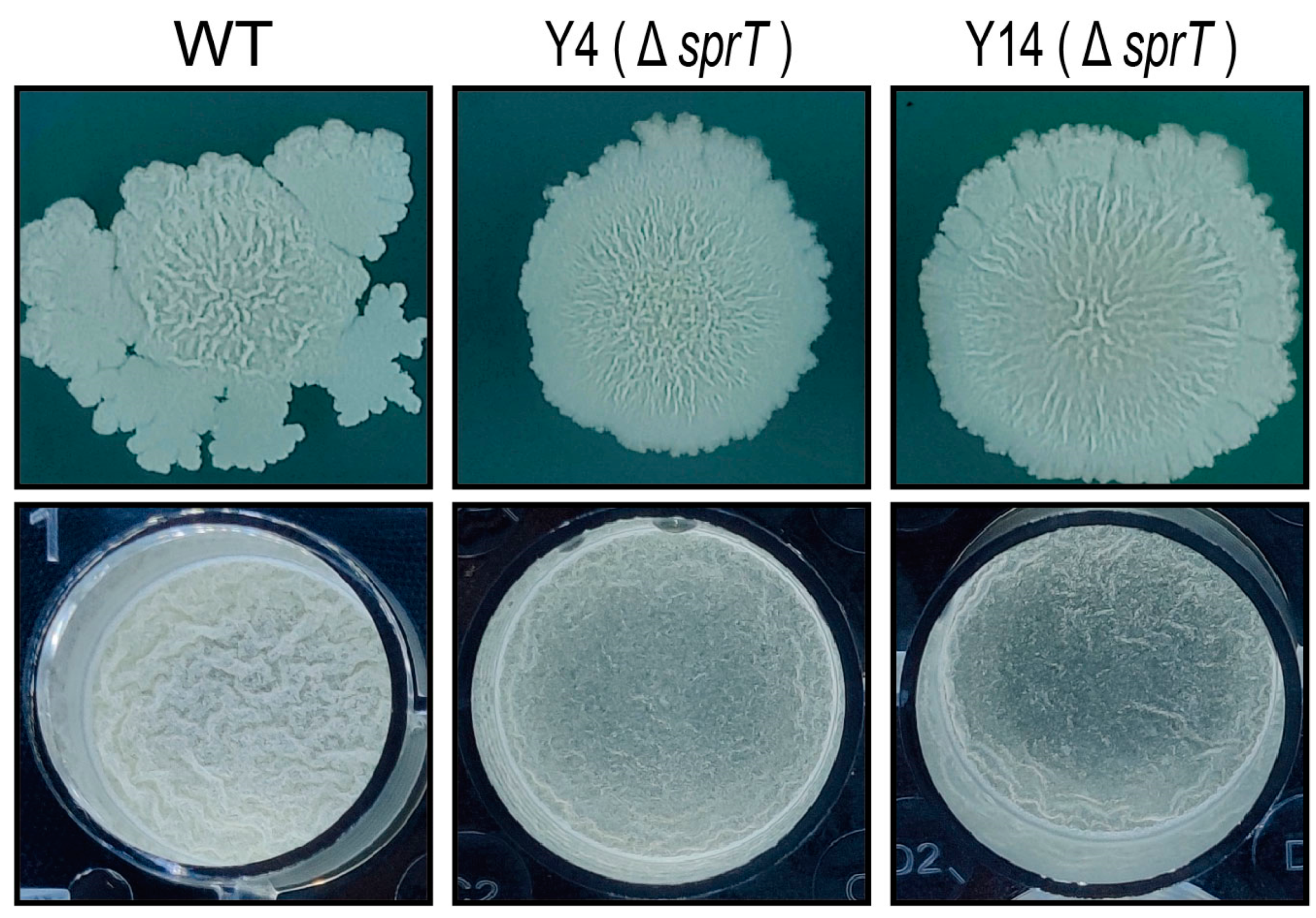
2.1. sprT Is Involved in the Biofilm Formation of B. velezensis FZB42

In a previous piece of work, we screened the TnYLB-1 transposon library of B. velezensis FZB42 [21] and noticed that the insertional mutant Y4 has a slightly different colony morphology (Figure 1), indicating that Y4 may be affected in biofilm formation. We examined the pellicle formation of Y4 in LB medium. The result showed that, after 34 h incubation, Y4 formed a less wrinkled pellicle than the wild-type FZB42. This confirmed that Y4 has a defected ability of forming biofilm. We analyzed the position of TnYLB-1 insertion and found it inserted in the sprT gene (Figure 2A). Since we were unable to transform a plasmid for sprT complementation in Y4, we independently constructed a new mutant by knocking out sprT using the homologous recombination method to avoid the possibility that other point mutations during transposition caused the phenotype change in Y14. The generated knockout strain was named Y14 and examined for its ability to form biofilm. We found that Y14 displayed a similar biofilm result to Y4 at either a solid–gas interface or a liquid–gas interface. These results confirmed that sprT should be responsible for the biofilm defects of Y4 and Y14.

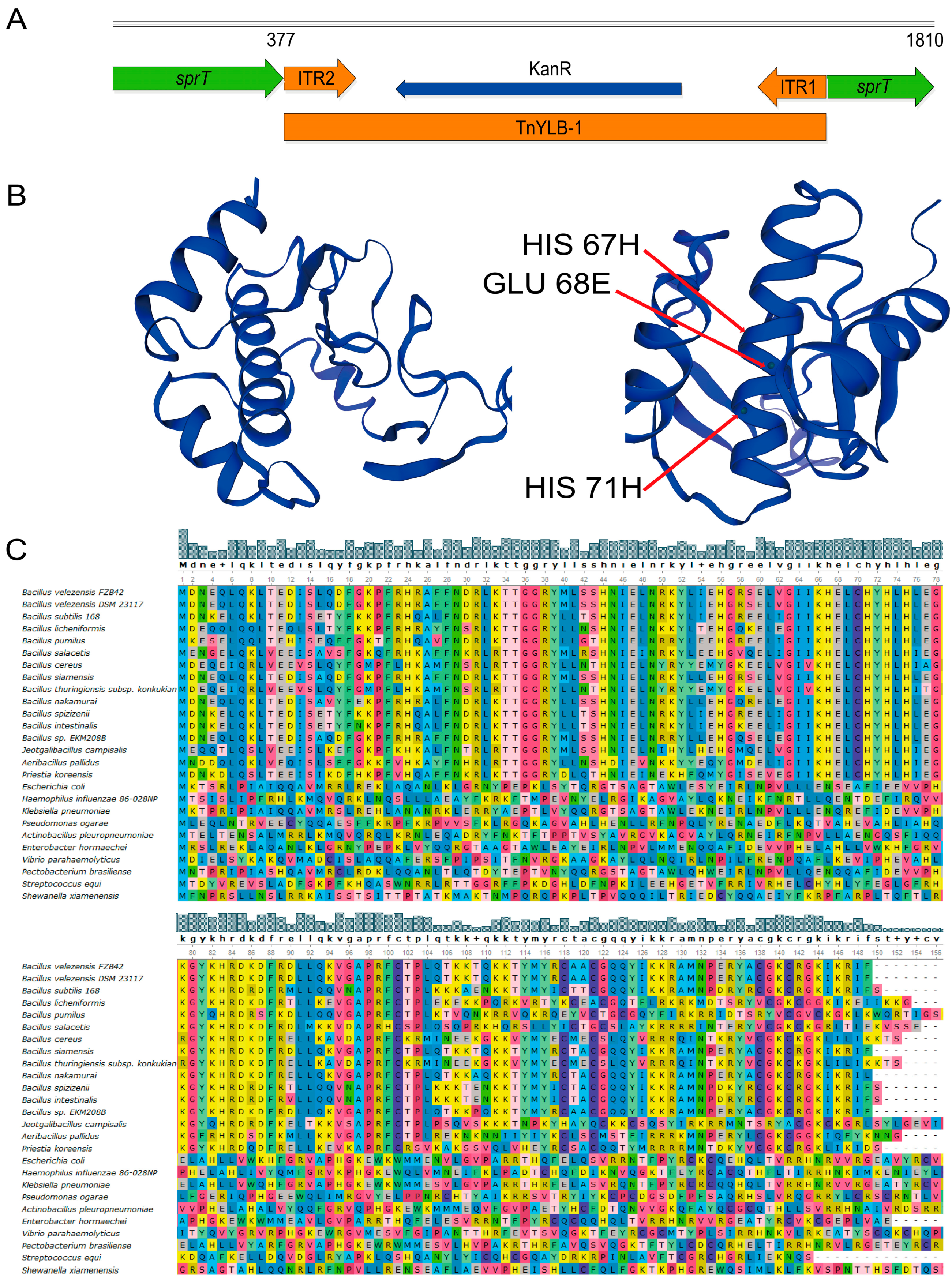
2.2. Bioinformatic Analysis Shows sprT Is Conserved among Bacillus Species

Before deep exploration of the function of sprT, we performed a bioinformatic analysis. The gene was predicted to encode a polypeptide (SprT) consisting of 149 amino acids, with a molecular weight of 17.709 kDa, belonging to the metalloprotease family. The SprT protein has an isoelectric point of 9.84, a half-life of 30 h, and an instability index of 56.01, which is greater than the threshold value of 40. Its fat coefficient is 65.50, and its average hydrophilicity is −0.909 (negative value). Therefore, it is predicted that SprT is an unstable hydrophilic protein. No threonine phosphorylation site was predicted in SprT, but there were two phosphorylation sites (43 S and 59 S) on serine residues. No signaling peptide was predicted, indicating that SprT is a non-secretory protein.
In the SprT protein, the α-helix has 52 amino acids, accounting for 34.90%; the β-sheet has 12 amino acids, accounting for 8.05%; the random coil has 59 amino acids, accounting for 39.60%; and the extended chain has 26 amino acids, accounting for 17.45% (Figure 2B). Therefore, α-helix and random coils are the most abundant secondary structures in SprT. The amino acids 67H, 68E, and 71H located on one α-helix were predicted to be the active sites, and among which, 67H and 71H may have a zinc ion binding function (Figure 2B).
The sequence alignment analysis showed that the SprT protein sequence was conserved in the Bacillus species, and especially highly conserved at 67H, 68E, and 71H but less conserved in other phylogenetically distant species (Table S2, Figure 2C). This indicates that the function of sprT may only be specific to the Bacillus species.

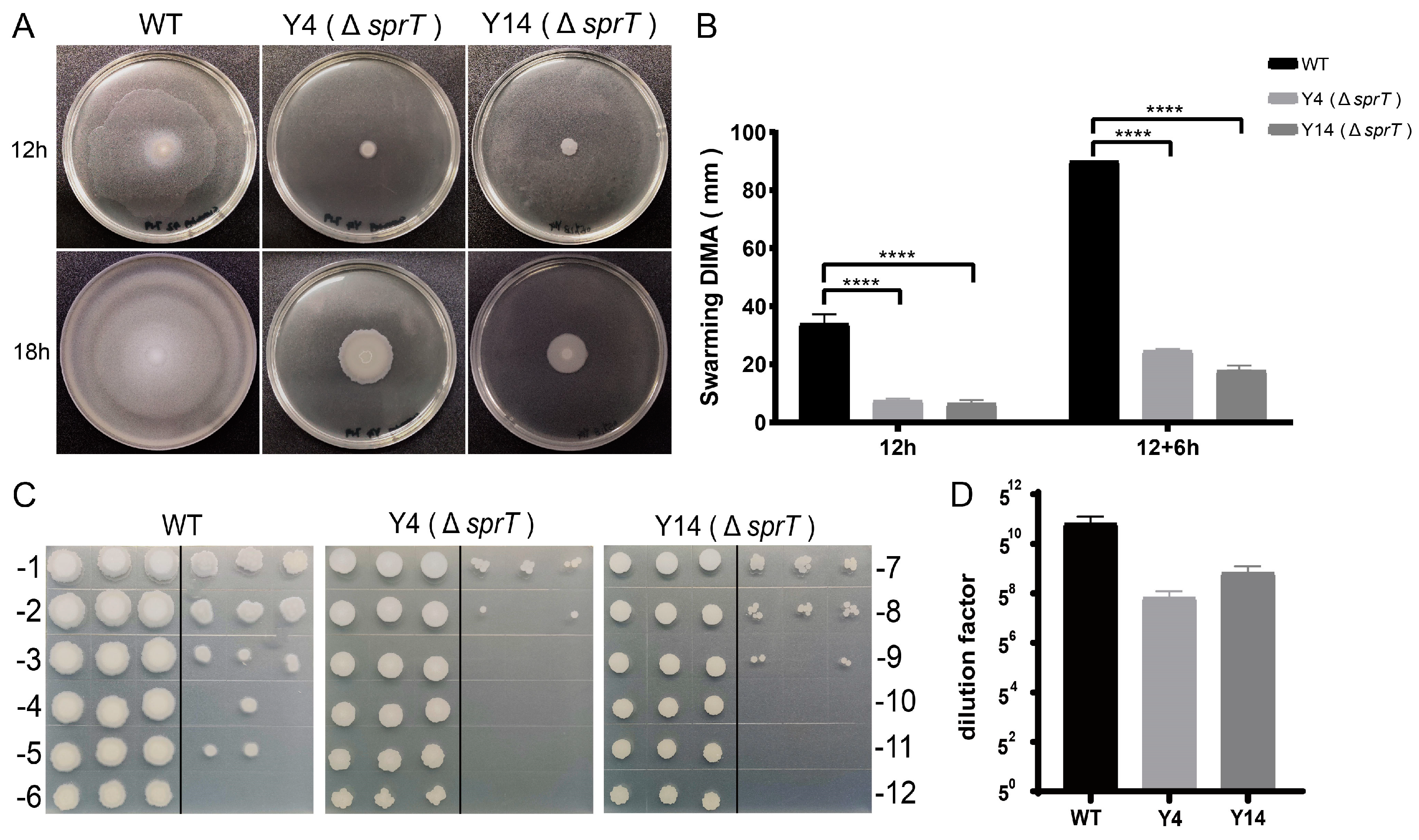
2.3. sprT Deletion Resulted in a Defect in Swarming Ability and Sporulation of FZB42

Motility and sporulation are closely linked to biofilm formation in Bacillus. To test whether sprT also affects the motility of FZB42, we examined their swarming ability on 0.5% LB solid medium. The mean diameter of colonies of the FZB42 wild type was 34.25 mm 12 h after incubation, while the mean diameters of Y4 and Y14 were 7.43 mm and 6.43 mm, respectively (Figure 3A,B), whereby both of which were significantly smaller than that of the wild type. When the FZB42 wild-type colonies reached 90 mm in diameter after 18 h, the mean diameters of Y4 and Y14 were 24.6 mm and 17.8 mm, respectively (Figure 3A,B). They were still smaller than those of the wild type. These results indicated that deletion of sprT reduced the swarming ability of FZB42.
To determine whether ΔsprT affects the spore formation of FZB42, we collected the cultures after 24 h of incubation of the assays. After heat treatment and five rounds of serial dilution, the endospores were spotted on plates for quantification. The results showed that the sporulation rate of the FZB42 wild type was maintained at the dilution of 5−9, which is approximately 25 times that of the sporulation rates of Y4 and Y14 (Figure 3C,D). The result indicated that sprT is also involved in the sporulation of FZB42.

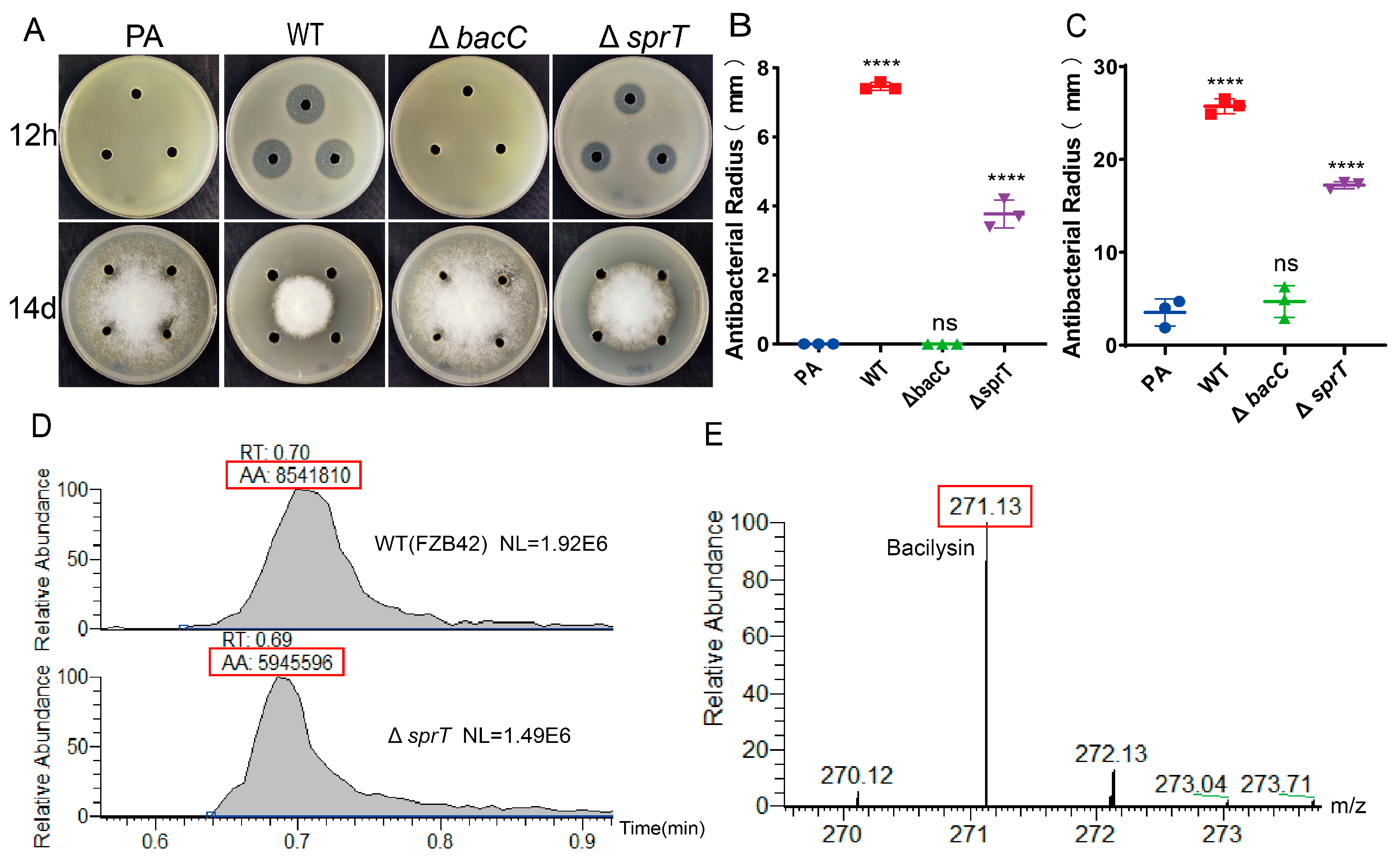
2.4. sprT Deletion Reduces Production of the Antibiotic Bacilysin

In FZB42, regulations of antibiotic production, biofilm formation, and sporulation share some similarities. To test whether sprT is also involved in the antibiotic production of FZB42, we used Staphylococcus aureus ATCC-9144 and Phytophthora sojae as indicator strains, whereby both of which are sensitive to the antibiotic bacilysin [2,22]. The results showed that the sterile supernatants of the wild-type FZB42 cultures displayed strong antagonistic ability toward S. aureus ATCC-9144 and P. sojae, while the supernatants of the ΔbacC mutant (the negative control) and PA medium (the blank) had no inhibitory effect. However, the ΔsprT mutant demonstrated a significantly smaller inhibitory zone than the wild type against ATCC-9144 (Figure 4A,B). Similarly, the ΔsprT mutant showed a significantly reduced inhibitory ability against P. sojae (Figure 4A,C). Furthermore, we compared the yield of bacilysin in the strains using the HPLCMS method. The result showed that the wild-type strain produced 1.43 times the amount of bacilysin that ΔsprT produced (Figure 4D,E). Taken together, it can be concluded that sprT plays a positive regulatory role in the production of bacilysin.

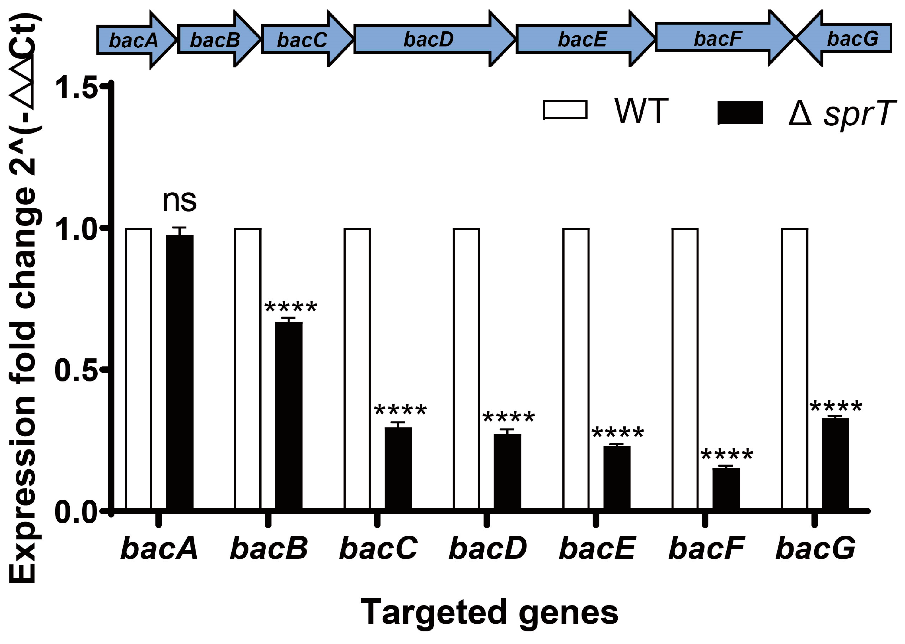
2.5. Transcription of Bac Operon Is Down-Regulated via ΔsprT Deletion

The production of bacilysin in the ΔsprT mutant may result from lowered biosynthesis and exportation. To pinpoint which process is affected in the ΔsprT mutant, we first examined the transcription of the bac operon which encodes the synthetases for bacilysin production. The bac operon is composed of bacA, bacB, bacC, bacD, bacE, bacF, and bacG genes. Using qPCR, we found that almost all of the genes were significantly down-regulated in ΔsprT, except for the bacA gene. In particular, the transcript levels of the downstream parts of the bac operon, that is, bacCbacG, were no more than half the level of what was expressed in the wild type (Figure 5). These results suggest that ΔsprT affects the production of bacilysin at the level of transcription.

3. Discussion

With a pyramid of previous studies, B. velezensis FZB42 has become one of the most well-studied PGPR strains [11,23]. In this work, we observed different biofilm morphologies of the mutant Y4 obtained from a transposon library. The insertion site of Y4 was determined to be the sprT gene. We found that the deletion of sprT had a negative effect on the biofilm formation, motility, sporulation, and antibiotic production of FZB42. In Bacillus spp., the SprT protein is predicted to be a metalloproteinase and is conserved. The association between metalloprotease and biofilm has been reported in pathogenic bacteria. For example, a serine alkaline metalloprotease from Halobacillus karajensis was effective for biofilm reduction in Pseudomonas aeruginosa and S. aureus [24]. In addition, a metalloprotease aureolysin from Staphylococcus warneri inhibited biofilms of S. warneri, and the aureolysin-inactivated mutant formed biofilm about twice as much as the wild type [25]. In contrast to the pathogenic bacteria, FZB42 is a plant-beneficial bacterium. Its effect of the sprT gene on biofilm and other related phenotypes provides new insights into the functions of metalloprotease.
Biofilm formation requires bacteria to first develop surface attachment, and studies have shown that motility is critical for initial attachment and biofilm formation [26]. Moreover, biofilms and motility share common regulation, for example, both biofilm formation and swarming motility are activated by low and very low levels of DegU~P, but inhibited by high levels of DegU~P [27]. We tested the swarming ability of ∆sprT and the results showed that the ∆sprT strain nearly lost its motility. This may be one reason for impaired biofilm formation of the ∆sprT strain. To date, there has been little research on the link between metalloproteases and bacterial motility. One extracellular metalloprotease, Zmp1, has been found in Clostridium difficile and is involved in the regulation of adhesion motility [28]. The specific mechanism for this link remains unclear. The results we discovered here may provide a new way to study the relationship between bacterial motility and metalloproteases in the future.
Sporulation is closely linked to biofilm formation and motility in Bacillus. The research by Shankweiler found that loss of a neutral metalloprotease had no detectable effect on growth, morphology, or sporulation in B. subtilis, indicating that the metalloproteinase is not essential for the development of endospores [29]. However, our results showed that sporulation of FZB42 decreased by about 25 times after the deletion of sprT. Sporulation initiation in B. subtilis is controlled by the phosphorylated form of the master regulator Spo0A, which controls the transcription of a multitude of sporulation genes [30]. The metalloprotease InhA from Bacillus thuringiensis has been shown to be related to the sporulation gene spo0A [31]. We cannot exclude the possibility that there may be also a correlation between spo0A and sporulation.
In FZB42, biofilm formation, motility, and sporulation are connected with antibiotic synthesis, since they are regulated by a network with some shared regulators. Bacilysin, produced by FZB42, is a rather simple dipeptide composed of only one L-alanine and one unusual amino acid called anticapsin [32]. Our results showed that ∆sprT reduced the yield of bacilysin. The bac operon, a gene cluster responsible for bacilysin synthesis and secretion, was detected via qPCR, showing that almost all bac genes were significantly down-regulated. The effect of metalloprotease genes on the yield of bacilysin was reported for the first time in FZB42.
Heterotrophic bacteria usually secrete proteases, mainly serine proteases and metalloprotease, outside of their cells to degrade environmental proteins for nutrition purposes [33]. Therefore, we also believe that metalloprotease plays a role in providing substrates for the synthesis of antibiotics. The deletion of the metalloprotease gene sprT leads to a reduction in the raw material for antibiotic synthesis, which in turn reduces the production of bacilysin. On the other hand, we suggest that there is a relationship between motility and antibiotic synthesis in FZB42. We speculate that the deletion of the sprT gene first affects the motility of FZB42, causing some unknown changes, which in turn affects the yield of bacilysin [34,35]. However, further research is needed to test this hypothesis.
In summary, our results have demonstrated the importance of the metalloprotease gene sprT for biofilm formation, swarming, sporulation, and antibiotic production in FZB42. Understanding the role of this metalloprotease in the synthesis of FZB42 antibiotics can help in the use of PGPR strains, such as FZB42, to play a better role in biocontrol. In the future, in-depth studies can be performed to uncover the molecular mechanisms underlying these effects.

4. Materials and Methods

4.1. Strains, Growth Conditions, and Primers

The bacterial cells, both S. aureus and FZB42, were routinely grown in LB medium (Tryptone 10 g, yeast extract 5 g, NaCl 10 g [36]). FZB42 was grown in PA medium (KH2PO4 1.1 g, MgSO4·7H2O 0.55 g, KCl 0.55 g, glutamic acid 3.828 g, sucrose 13.7 g, Na3C6H5O7·2H2O 0.1 g, FeCl3 0.1 g) at 37 °C for bacilysin production. P. sojae was cultured on solid V8 agar medium [37] (10% tomato juice, 0.01% CaCO3, and 1.5% agar [38]) at 25 °C in the dark. The bacterial supernatant was prepared from PA culture via careful removal of cell pellets after centrifugation followed by passage through a 0.22 µm sterile filter. S. aureus was obtained from the Guangdong Microbial Culture Collection Center (GDMCC). P. sojae was obtained from the Institute of Forest Protection of Nanjing Forestry University. All primers used in this study are shown in Table S1.

4.2. Strain Construction

The genomic DNA of the Y4 mutant strain was used as a template to amplify the DNA fragment containing the KmR cassette flanked by the upstream and downstream sequences of the sprT gene. Then, the PCR product was transformed into the FZB42 wild type [10]. The transformants were verified via colony PCR and DNA sequencing.

4.3. Transposon TnYLB-1 Insertion Site Mapping

The genomic DNA of the mutants was digested using TaqI. Then, the digested products were ligated using T4 DNA Ligase. The circularized products were then inverse-amplified and sent to Shanghai Biological Company for sequencing. The sequencing results were found by blasting the FZB42 genome to locate the insertion site of the transposon TnYLB-1. The primers used are shown in Table S1.

4.4. Biofilm Formation Test

The bacteria were grown in LB until their optical density at 600 nm (OD600) reached 1.0. Then, for the colony surface assay, 2 μL culture was added on the center of a solid LB plate. The plates were dried for 10 min under sterile air in a biosafety cabinet; then, the plates were incubated at 30 °C for 84 h. For the pellicle assay, the cultures were inoculated to 1 mL LB medium contained in a 24-well plate at a 1:100 dilution. After being mixed well, the plates were incubated 25 °C and observed every 12 h. Then, the state of biofilm formation was observed, based on the colony’s ability to produce wrinkles on its surface. Three biological replicates were performed, with each culture being technically replicated in three wells.

4.5. Bioinformatics Analysis

The sprT gene sequence was inputted in the Uniprot website to obtain basic information. The protein sequences of SprT from different species were downloaded from the NCBI protein database. Unipro UGENE software was used for protein sequence conservation analysis of SprT. SOPMA and SWISS-MODEL were used to analyze and model secondary and tertiary structures of the SprT of FZB42.

4.6. Swarming Test

The bacteria were grown in LB until reaching an OD600 of ~1.0. Then, 2 μL of the cultures was spotted onto the center of LB containing 0.5% agar (the medium should be used immediately after preparation). After being dried for 30 min in a clean bench, the plates were incubated at 25 °C for 12 h in an incubator with a moisturizing function, and the diameters of the colonies were measured. Three biological replicates were performed, with each culture being technically replicated in three plates.

4.7. Sporulation Test

The strains were grown in LB for 24 h, and 1 mL bacterial solution was taken and added to a 2 mL centrifuge tube. The centrifuge tube containing bacterial solution was treated at 85 °C for 10 min. The heat-treated cultures were 5-fold serially diluted. A total of 3 μL of each dilution was spotted on LB plates and then dried for 10 min before being incubated at 37 °C for 24 h. Three biological replicates were performed, with each culture being technically replicated in three spots.

4.8. Antagonistic Test of Antibiotic Bacilysin

The method is as follows: Pick fresh FZB42 and Y14 colonies and inoculate them into 10 mL of LB liquid culture. Shake at 37 °C and 200 rpm for 6–8 h. Adjust the OD600 of the pre-culture to 1 and transfer it to 30 mL of PA culture medium at a ratio of 1%. Shake at 37 °C and 200 rpm for 24 h. Transfer the bacterial solution to a 2 mL centrifuge tube and centrifuge at 12,000 rpm for 10 min. Use a syringe to collect the supernatant, filter it through a 0.22 μm filter membrane, and obtain the supernatant containing the antibiotic after 24 h of bacterial growth.
For S. aureus as the indicator strain, the culture of S. aureus ATCC-9144 at an OD600 of ~1.0 was diluted with LB medium at a 1:100 ratio and poured onto LB agar. Holes were made using a sterile punch and filled with 100 μL of bacterial culture supernatants. The plates were incubated at 37 °C for 12 h before the radii of the antagonistic zones were measured. For S. aureus, the antagonism radius was defined as the center of the hole to the farthest end of the transparent inhibition zone. For P. sojae as the indicator strain, fresh P. sojae cake was inoculated onto V8 solid medium. Holes were similarly made and filled with 100 μL supernatants. The plates were incubated at 25 °C for 14 days before the antagonistic effects were observed. The antagonism radius was defined as the nearest distance from the edge of P. sojae to the hole center. Three biological replicates were performed, with each culture being technically replicated in three holes.

4.9. HPLC-MS

A positive detection mode of HPLC detection of bacterial cultural supernatant was used with 0.1% formic acid added to buffer solution A (H2O) and buffer solution B (acetonitrile). Liquid chromatography was performed using a 50 × 2.1 mm 5 μm Hypercarb column with a flow rate of 0.4 mL/min. The sample injection volume was 3 μL, and the LC program was as follows: 0% buffer B for 1 min, linear gradient of buffer B from 0% to 95% within 10 min, linear gradient of buffer B from 95% to 0% within 2 min, re-equilibration with 0% buffer B for 10 min, and then the next sample was loaded. The samples were re-run after dilution with water to avoid saturation of the mass signal detector. The mass spectrometer was set to the following parameters: capillary voltage of 2500 V, drying gas at 350 °C with a flow rate of 10 L/min, nebulizer pressure at 30 psi, and segmented voltage at 125 V.

4.10. Real-Time Quantitative PCR

The bacterial cells cultured in PA medium for 24 h were collected for RNA preparation using the Trizol method. Reverse transcription for cDNAs was performed using TAKARA PrimeScript™ RT Master Mix (TaKaRa, Maebashi, Japan), according to the instructions. qPCR was carried out using TAKARA TB Green® Premix Ex Taq™ II (TaKaRa, Maebashi, Japan), according to the instructions. The fluorescence quantitative PCR results were calculated using the 2−∆∆Ct method. The housekeeping gene gyrA was used as the internal standard.

Supplementary Materials

The following supporting information can be downloaded at https://www.mdpi.com/article/10.3390/ijms242316815/s1.

Author Contributions

Conceptualization, B.F.; validation, Y.Y. and R.C.; formal analysis, Y.Y., R.C. and C.W.; writing—original draft preparation, Y.Y. and R.C.; writing—review and editing, M.U.R. and B.F. All authors have read and agreed to the published version of the manuscript.

Funding

This work was supported by the National Natural Science Foundation of China (no. 31970097).

Institutional Review Board Statement

Not applicable.

Informed Consent Statement

Not applicable.

Data Availability Statement

Data are contained within the article and Supplementary Materials.

Conflicts of Interest

The authors declare no conflict of interest.

References

  1. Singla, J.; Krattinger, S.G. Biotic Stress Resistance Genes in Wheat. In The Production and Genetics of Food Grains; Elsevier: Amsterdam, The Netherlands, 2016; pp. 388–392. [Google Scholar]
  2. Han, X.; Shen, D.; Xiong, Q.; Bao, B.; Zhang, W.; Dai, T.; Zhao, Y.; Borriss, R.; Fan, B. The Plant-Beneficial Rhizobacterium Bacillus velezensis FZB42 Controls the Soybean Pathogen Phytophthora sojae Due to Bacilysin Production. Appl. Environ. Microbiol. 2021, 87, e0160121. [Google Scholar] [CrossRef]
  3. Ricci, P.; Bonnet, P.; Huet, J.C.; Sallantin, M.; Beauvais-Cante, F.; Bruneteau, M.; Billard, V.; Michel, G.; Pernollet, J.C. Structure and activity of proteins from pathogenic fungi Phytophthora eliciting necrosis and acquired resistance in tobacco. Eur. J. Biochem. 1989, 183, 555–563. [Google Scholar] [CrossRef] [PubMed]
  4. Chowdhury, S.P.; Uhl, J.; Grosch, R.; Alquéres, S.; Pittroff, S.; Dietel, K.; Schmitt-Kopplin, P.; Borriss, R.; Hartmann, A. Cyclic Lipopeptides of Bacillus amyloliquefaciens subsp. plantarum Colonizing the Lettuce Rhizosphere Enhance Plant Defense Responses Toward the Bottom Rot Pathogen Rhizoctonia solani. Mol. Plant-Microbe Interact. 2015, 28, 984–995. [Google Scholar] [CrossRef] [PubMed]
  5. Fan, B.; Borriss, R.; Bleiss, W.; Wu, X. Gram-positive rhizobacterium Bacillus amyloliquefaciens FZB42 colonizes three types of plants in different patterns. J. Microbiol. 2012, 50, 38–44. [Google Scholar] [CrossRef]
  6. Liu, S.; Tian, Y.; Jia, M.; Lu, X.; Yue, L.; Zhao, X.; Jin, W.; Wang, Y.; Zhang, Y.; Xie, Z.; et al. Induction of Salt Tolerance in Arabidopsis thaliana by Volatiles from Bacillus amyloliquefaciens FZB42 via the Jasmonic Acid Signaling Pathway. Front Microbiol. 2020, 11, 562934. [Google Scholar] [CrossRef] [PubMed]
  7. Chen, X.H.; Koumoutsi, A.; Scholz, R.; Eisenreich, A.; Schneider, K.; Heinemeyer, I.; Morgenstern, B.; Voss, B.; Hess, W.R.; Reva, O.; et al. Comparative analysis of the complete genome sequence of the plant growth-promoting bacterium Bacillus amyloliquefaciens FZB42. Nat. Biotechnol. 2007, 25, 1007–1014. [Google Scholar] [CrossRef]
  8. Kim, S.Y.; Kim, J.Y.; Kim, S.H.; Bae, H.J.; Yi, H.; Yoon, S.H.; Koo, B.S.; Kwon, M.; Cho, J.Y.; Lee, C.E.; et al. Surfactin from Bacillus subtilis displays anti-proliferative effect via apoptosis induction, cell cycle arrest and survival signaling suppression. FEBS Lett. 2007, 581, 865–871. [Google Scholar] [CrossRef]
  9. Doornbos, R.F.; Van Loon, L.C.; Bakker, P.A.H.M. Impact of root exudates and plant defense signaling on bacterial communities in the rhizosphere. A review. Agron. Sustain. Dev. 2011, 32, 227–243. [Google Scholar] [CrossRef]
  10. Cao, X.; Li, Y.; Fan, J.; Zhao, Y.; Borriss, R.; Fan, B. Two Lysine Sites That Can Be Malonylated Are Important for LuxS Regulatory Roles in Bacillus velezensis. Microorganisms 2021, 9, 1338. [Google Scholar] [CrossRef]
  11. Chowdhury, S.P.; Hartmann, A.; Gao, X.; Borriss, R. Biocontrol mechanism by root-associated Bacillus amyloliquefaciens FZB42A review. Front. Microbiol. 2015, 6, 780. [Google Scholar] [CrossRef]
  12. Schneider, K.; Chen, X.H.; Vater, J.; Franke, P.; Nicholson, G.; Borriss, R.; Süssmuth, R.D. Macrolactin is the polyketide biosynthesis product of the pks2 cluster of Bacillus amyloliquefaciens FZB42. J. Nat. Prod. 2007, 70, 1417–1423. [Google Scholar] [CrossRef] [PubMed]
  13. Zhang, L.; Sun, C. Fengycins, Cyclic Lipopeptides from Marine Bacillus subtilis Strains, Kill the Plant-Pathogenic Fungus Magnaporthe grisea by Inducing Reactive Oxygen Species Production and Chromatin Condensation. Appl. Environ. Microbiol. 2018, 84, e00445-18. [Google Scholar] [CrossRef]
  14. Chen, X.H.; Vater, J.; Piel, J.; Franke, P.; Scholz, R.; Schneider, K.; Koumoutsi, A.; Hitzeroth, G.; Grammel, N.; Strittmatter, A.W.; et al. Structural and functional characterization of three polyketide synthase gene clusters in Bacillus amyloliquefaciens FZB 42. J. Bacteriol. 2006, 188, 4024–4036. [Google Scholar] [CrossRef] [PubMed]
  15. Chen, X.H.; Koumoutsi, A.; Scholz, R.; Schneider, K.; Vater, J.; Süssmuth, R.; Piel, J.; Borriss, R. Genome analysis of Bacillus amyloliquefaciens FZB42 reveals its potential for biocontrol of plant pathogens. J. Biotechnol. 2009, 140, 27–37. [Google Scholar] [CrossRef] [PubMed]
  16. Borriss, R.; Chen, X.H.; Rueckert, C.; Blom, J.; Becker, A.; Baumgarth, B.; Fan, B.; Pukall, R.; Schumann, P.; Spröer, C.; et al. Relationship of Bacillus amyloliquefaciens clades associated with strains DSM 7T and FZB42T: A proposal for Bacillus amyloliquefaciens subsp. amyloliquefaciens subsp. nov. and Bacillus amyloliquefaciens subsp. plantarum subsp. nov. based on complete genome sequence comparisons. Int. J. Syst. Evol. Microbiol. 2011, 61, 1786–1801. [Google Scholar]
  17. Wu, L.; Wu, H.; Chen, L.; Yu, X.; Borriss, R.; Gao, X. Difficidin and bacilysin from Bacillus amyloliquefaciens FZB42 have antibacterial activity against Xanthomonas oryzae rice pathogens. Sci. Rep. 2015, 5, 12975. [Google Scholar] [CrossRef]
  18. Wang, T.; Liu, X.H.; Wu, M.B.; Ge, S. Molecular insights into the antifungal mechanism of bacilysin. J. Mol. Model. 2018, 24, 118. [Google Scholar] [CrossRef]
  19. Vlamakis, H.; Chai, Y.; Beauregard, P.; Losick, R.; Kolter, R. Sticking together: Building a biofilm the Bacillus subtilis way. Nat. Rev. Microbiol. 2013, 11, 157–168. [Google Scholar] [CrossRef]
  20. Talukdar, P.K.; Olguín-Araneda, V.; Alnoman, M.; Paredes-Sabja, D.; Sarker, M.R. Updates on the sporulation process in species. Res. Microbiol. 2015, 166, 225–235. [Google Scholar] [CrossRef]
  21. Budiharjo, A.; Chowdhury, S.P.; Dietel, K.; Beator, B.; Dolgova, O.; Fan, B.; Bleiss, W.; Ziegler, J.; Schmid, M.; Hartmann, A.; et al. Transposon Mutagenesis of the Plant-Associated Bacillus amyloliquefaciens ssp. plantarum FZB42 Revealed That the nfrA and RBAM17410 Genes Are Involved in Plant-Microbe-Interactions. PLoS ONE 2014, 9, e98267. [Google Scholar] [CrossRef]
  22. Garrity, L.F.; Ordal, G.W. Chemotaxis in Bacillus subtilis: How bacteria monitor environmental signals. Pharmacol. Ther. 1995, 68, 87–104. [Google Scholar] [CrossRef] [PubMed]
  23. Fan, B.; Wang, C.; Song, X.; Ding, X.; Wu, L.; Wu, H.; Gao, X.; Borriss, R. Bacillus velezensis FZB42 in 2018: The Gram-Positive Model Strain for Plant Growth Promotion and Biocontrol. Front. Microbiol. 2018, 9, 2491. [Google Scholar] [CrossRef] [PubMed]
  24. Ghoreishi, F.S.; Roghanian, R.; Emtiazi, G. Inhibition of quorum sensing-controlled virulence factors with natural substances and novel protease, obtained from Halobacillus karajensis. Microb. Pathog. 2020, 149, 104555. [Google Scholar] [CrossRef]
  25. Yokoi, K.J.; Kuzuwa, S.; Iwasaki, S.; Yamakawa, A.; Taketo, A.; Kodaira, K. Aureolysin of Staphylococcus warneri M accelerates its proteolytic cascade, and participates in biofilm formation. Biosci. Biotechnol. Biochem. 2016, 80, 1238–1242. [Google Scholar] [CrossRef] [PubMed]
  26. Van Wolferen, M.; Orell, A.; Albers, S.V. Archaeal biofilm formation. Nat. Rev. Microbiol. 2018, 16, 699–713. [Google Scholar] [CrossRef]
  27. Verhamme, D.T.; Kiley, T.B.; Stanley-Wall, N.R. DegU co-ordinates multicellular behaviour exhibited by Bacillus subtilis. Mol. Microbiol. 2007, 65, 554–568. [Google Scholar] [CrossRef]
  28. Schacherl, M.; Pichlo, C.; Neundorf, I.; Baumann, U. Structural Basis of Proline-Proline Peptide Bond Specificity of the Metalloprotease Zmp1 Implicated in Motility of Clostridium difficile. Structure 2015, 23, 1632–1642. [Google Scholar] [CrossRef]
  29. Shankweiler, G.W.Z. Primary Subsite Mapping and Carbohydrate Analysis of the Serine Protease, Bacillopeptidase F, from Bacillus subttlis. Ph.D. Dissertation, New Mexico State University, Las Cruces, NM, USA, 1986. [Google Scholar]
  30. Vishnoi, M.; Narula, J.; Devi, S.N.; Dao, H.A.; Igoshin, O.A.; Fujita, M. Triggering sporulation in Bacillus subtilis with artificial two-component systems reveals the importance of proper Spo0A activation dynamics. Mol. Microbiol. 2013, 90, 181–194. [Google Scholar] [CrossRef]
  31. Grandvalet, C.; Gominet, M.; Lereclus, D. Identification of genes involved in the activation of the Bacillus thuringiensis inhA metalloprotease gene at the onset of sporulation. Microbiology 2001, 147, 1805–1813. [Google Scholar] [CrossRef]
  32. Kenig, M.; Abraham, E.P. Antimicrobial activities and antagonists of bacilysin and anticapsin. Microbiology 1976, 94, 37–45. [Google Scholar] [CrossRef]
  33. Wu, J.W.; Chen, X.L. Extracellular metalloproteases from bacteria. Appl. Microbiol. Biotechnol. 2011, 92, 253–262. [Google Scholar] [CrossRef]
  34. Diethmaier, C.; Chawla, R.; Canzoneri, A.; Kearns, D.B.; Lele, P.P.; Dubnau, D. Viscous drag on the flagellum activates Bacillus subtilis entry into the K-state. Mol. Microbiol. 2017, 106, 367–380. [Google Scholar] [CrossRef]
  35. Mariappan, A.; Makarewicz, O.; Chen, X.H.; Borriss, R. Two-component response regulator DegU controls the expression of bacilysin in plant-growth-promoting bacterium Bacillus amyloliquefaciens FZB42. J. Mol. Microbiol. Biotechnol. 2012, 22, 114–125. [Google Scholar] [CrossRef] [PubMed]
  36. Kanvinde, L.; Sastry, G.R. Agrobacterium tumefaciens Is a Diazotrophic Bacterium. Appl. Environ. Microbiol. 1990, 56, 2087–2092. [Google Scholar] [CrossRef] [PubMed]
  37. Perry, D.; Abraham, E.P. Transport and metabolism of bacilysin and other peptides by suspensions of Staphylococcus aureus. J. Gen. Microbiol. 1979, 115, 213–221. [Google Scholar] [CrossRef] [PubMed]
  38. Alves, S.H.; Linares, C.E.; Loreto, E.S.; Rodrigues, M.; Thomazi, D.I.; Souza, F.; Santurio, J.M. Utilization of tomato juice agar (V8 agar) in the presumptive identification of Candida dubliniensis. Rev. Da Soc. Bras. De Med. Trop. 2006, 39, 92–93. [Google Scholar] [CrossRef]
Figure 1. Effect of sprT deletion on biofilm formation of B. velezensis FZB42. Colonies or pellicles formed by the FZB42 wild type (WT), the sprT transposon insertional mutant Y4, and the sprT knockout mutant Y14 were evaluated on LB agar at 25 °C for 84 h and in LB liquid medium at 25 °C for 34 h. Each plate represents a representative sample from three replicates.
Ijms 24 16815 g001
Figure 2. Location of the TnYLB-1 transposon in the mutant Y4 (A) and bioinformatic analysis of SprT protein. (A) The transposon TnYLB-1 was determined to be inserted between the 377th and 378th nucleotides of the sprT gene in Y4. (B) The predicted tertiary structure of SprT of B. velezensis FZB42. (C) Conservation analysis of SprT proteins among different bacteria. Different colors represent different amino acids. The higher the conservation, the more consistent the colors are from top to bottom.
Ijms 24 16815 g002
Figure 3. Effects of sprT deletion on FZB42 swarming ability and sporulation. (A) Swarming of the FZB42 wild type and the sprT mutant strains Y4 and Y14 cultured at 25 °C for 12 h (top) and 18 h (bottom) on 0.5% LB agar. (B) Statistics of colony diameters of the WT, Y4, and Y14 after swarming, as described for (A). (C) Sporulation of the FZB42 wild type and the sprT mutant strains Y4 and Y14 cultured at 37 °C on LB solid medium for 24 h. (D) Statistics of sporulated cells of the WT, Y4, and Y14 after treatment. One representative plate out of six replicates is shown; T-test was performed, where **** p < 0.001.
Ijms 24 16815 g003
Figure 4. Effect of ΔsprT on the bacilysin production of B. velezensis FZB42. (A) Antagonism of the strains’ culture supernatant toward S. aureus ATCC-9144 and P. sojae. One representative plate out of three replicates is shown. S. aureus ATCC9144 is on the top; P. sojae is on the bottom. (B) Statistics of antagonism radii of the strains toward S. aureus ATCC-9144 incubated overnight (B) and P. sojae incubated at 25 °C for 14 d (C). In panel (C), the antagonism radius was defined as the nearest distance from the edge of P. sojae to the hole center (mm). (D,E) HPLC-MS analysis of bacilysin produced by the FZB42 wild type and ΔsprT. T-test was performed, where **** p < 0.001.
Ijms 24 16815 g004
Figure 5. Relative mRNA levels of bacilysin biosynthetic genes in the FZB42 WT and the ΔsprT mutant revealed via qPCR. RNA was extracted from bacterial cultures in PA medium 24 h after inoculation for qPCR. The expression fold change was normalized to the expression of the housekeeping gene gyrA in WT. Three biological replicates and three technical replicates were used for each gene. T-test was performed where **** p < 0.001.
Ijms 24 16815 g005
Disclaimer/Publisher’s Note: The statements, opinions and data contained in all publications are solely those of the individual author(s) and contributor(s) and not of MDPI and/or the editor(s). MDPI and/or the editor(s) disclaim responsibility for any injury to people or property resulting from any ideas, methods, instructions or products referred to in the content.

