12 pages, 1070 KiB  
Article
Action of Platelet-Rich Plasma on In Vitro Cellular Bioactivity: More than Platelets
by Maider Beitia 1,†, Diego Delgado 1,†, Jon Mercader 2,3, Pello Sánchez 1, Leonor López de Dicastillo 3 and Mikel Sánchez 1,3,*
1 Advanced Biological Therapy Unit, Hospital Vithas Vitoria, 01008 Vitoria-Gasteiz, Spain
2 Microfluidics Cluster UPV/EHU, BIOMICs Microfluidics Group, Lascaray Research Center, University of the Basque Country UPV/EHU, 01006 Vitoria-Gasteiz, Spain
3 Arthroscopic Surgery Unit, Hospital Vithas Vitoria, 01008 Vitoria-Gasteiz, Spain
These authors contributed equally to this work.
Int. J. Mol. Sci. 2023, 24(6), 5367; https://doi.org/10.3390/ijms24065367 - 10 Mar 2023
Cited by 11 | Viewed by 1742
Abstract
Platelet-rich plasma (PRP) is a biological therapy in which one of the mechanisms of action is the stimulation of biological processes such as cell proliferation. The size of PRP’s effect depends on multiple factors, one of the most important being the composition of [...] Read more.
Platelet-rich plasma (PRP) is a biological therapy in which one of the mechanisms of action is the stimulation of biological processes such as cell proliferation. The size of PRP’s effect depends on multiple factors, one of the most important being the composition of PRP. The aim of this study was to analyze the relationship between cell proliferation and the levels of certain growth factors (IGF-1, HGF, PDGF, TGF-β and VEG) in PRP. First, the composition and effect on cell proliferation of PRP versus platelet-poor plasma (PPP) were compared. Subsequently, the correlation between each growth factor of PRP and cell proliferation was evaluated. Cell proliferation was higher in cells incubated with lysates derived from PRP compared to those cultured with lysates derived from PPP. In terms of composition, the levels of PDGF, TGF-β, and VEGF were significantly higher in PRP. When analyzing the PRP growth factors, IGF-1 was the only factor that correlated significantly with cell proliferation. Of those analyzed, the level of IGF-1 was the only one that did not correlate with platelet levels. The magnitude of PRP’s effect depends not only on platelet count but also on other platelet-independent molecules. Full article
Show Figures

Figure 1

22 pages, 5169 KiB  
Article
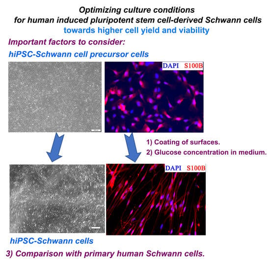
Culture Conditions for Human Induced Pluripotent Stem Cell-Derived Schwann Cells: A Two-Centre Study
by Zhong Huang 1,2,†, Rebecca Powell 3,4,†, Svenja Kankowski 1, James B. Phillips 3,4,*,‡ and Kirsten Haastert-Talini 1,2,*,‡
1 Institute of Neuroanatomy and Cell Biology, Hannover Medical School (MHH), 30623 Hannover, Germany
2 Center for Systems Neuroscience (ZSN) Hannover, 30559 Hannover, Germany
3 Department of Pharmacology, University College London (UCL) School of Pharmacy, 29-39 Brunswick Square, London WC1N 1AX, UK
4 UCL Centre for Nerve Engineering, UCL, London WC1H 0AL, UK
These authors contributed equally to this work.
Int. J. Mol. Sci. 2023, 24(6), 5366; https://doi.org/10.3390/ijms24065366 - 10 Mar 2023
Cited by 1 | Viewed by 2271
Abstract
Adult human Schwann cells represent a relevant tool for studying peripheral neuropathies and developing regenerative therapies to treat nerve damage. Primary adult human Schwann cells are, however, difficult to obtain and challenging to propagate in culture. One potential solution is to generate Schwann [...] Read more.
Adult human Schwann cells represent a relevant tool for studying peripheral neuropathies and developing regenerative therapies to treat nerve damage. Primary adult human Schwann cells are, however, difficult to obtain and challenging to propagate in culture. One potential solution is to generate Schwann cells from human induced pluripotent stem cells (hiPSCs). Previously published protocols, however, in our hands did not deliver sufficient viable cell numbers of hiPSC-derived Schwann cells (hiPSC-SCs). We present here, two modified protocols from two collaborating laboratories that overcome these challenges. With this, we also identified the relevant parameters to be specifically considered in any proposed differentiation protocol. Furthermore, we are, to our knowledge, the first to directly compare hiPSC-SCs to primary adult human Schwann cells using immunocytochemistry and RT-qPCR. We conclude the type of coating to be important during the differentiation process from Schwann cell precursor cells or immature Schwann cells to definitive Schwann cells, as well as the amounts of glucose in the specific differentiation medium to be crucial for increasing its efficiency and the final yield of viable hiPSC-SCs. Our hiPSC-SCs further displayed high similarity to primary adult human Schwann cells. Full article
(This article belongs to the Special Issue Tissue Engineering and Cell Therapy)
Show Figures

Graphical abstract

14 pages, 759 KiB  
Review
Models of Congenital Adrenal Hyperplasia for Gene Therapies Testing
by Olga Glazova 1,2,*, Asya Bastrich 1, Andrei Deviatkin 1,2,*, Nikita Onyanov 2, Samira Kaziakhmedova 2, Liudmila Shevkova 1,2, Nawar Sakr 1,2, Daria Petrova 1, Maria V. Vorontsova 1,2 and Pavel Volchkov 1,2
1 Gene Editing Laboratory, Endocrinology Research Centre, 117292 Moscow, Russia
2 Genome Engineering Laboratory, Moscow Institute of Physics and Technology, 141700 Dolgoprudniy, Russia
Int. J. Mol. Sci. 2023, 24(6), 5365; https://doi.org/10.3390/ijms24065365 - 10 Mar 2023
Cited by 1 | Viewed by 2259
Abstract
The adrenal glands are important endocrine organs that play a major role in the stress response. Some adrenal glands abnormalities are treated with hormone replacement therapy, which does not address physiological requirements. Modern technologies make it possible to develop gene therapy drugs that [...] Read more.
The adrenal glands are important endocrine organs that play a major role in the stress response. Some adrenal glands abnormalities are treated with hormone replacement therapy, which does not address physiological requirements. Modern technologies make it possible to develop gene therapy drugs that can completely cure diseases caused by mutations in specific genes. Congenital adrenal hyperplasia (CAH) is an example of such a potentially treatable monogenic disease. CAH is an autosomal recessive inherited disease with an overall incidence of 1:9500–1:20,000 newborns. To date, there are several promising drugs for CAH gene therapy. At the same time, it remains unclear how new approaches can be tested, as there are no models for this disease. The present review focuses on modern models for inherited adrenal gland insufficiency and their detailed characterization. In addition, the advantages and disadvantages of various pathological models are discussed, and ways of further development are suggested. Full article
Show Figures

Figure 1

22 pages, 1238 KiB  
Review
The Role of Regulated Programmed Cell Death in Osteoarthritis: From Pathogenesis to Therapy
by Suqing Liu 1,2,†, Yurong Pan 1,2,†, Ting Li 1,3, Mi Zou 1,3, Wenji Liu 1,3, Qingqing Li 1,3, Huan Wan 1,3, Jie Peng 3,* and Liang Hao 1,*
1 Department of Orthopedics, Second Affifiliated Hospital of Nanchang University, Nanchang 330006, China
2 Queen Marry College, Nanchang University, Nanchang 330006, China
3 The Second Clinical Medical College, Nanchang University, Nanchang 330006, China
These authors have contributed equally to this work.
Int. J. Mol. Sci. 2023, 24(6), 5364; https://doi.org/10.3390/ijms24065364 - 10 Mar 2023
Cited by 10 | Viewed by 3026
Abstract
Osteoarthritis (OA) is a worldwide chronic disease that can cause severe inflammation to damage the surrounding tissue and cartilage. There are many different factors that can lead to osteoarthritis, but abnormally progressed programmed cell death is one of the most important risk factors [...] Read more.
Osteoarthritis (OA) is a worldwide chronic disease that can cause severe inflammation to damage the surrounding tissue and cartilage. There are many different factors that can lead to osteoarthritis, but abnormally progressed programmed cell death is one of the most important risk factors that can induce osteoarthritis. Prior studies have demonstrated that programmed cell death, including apoptosis, pyroptosis, necroptosis, ferroptosis, autophagy, and cuproptosis, has a great connection with osteoarthritis. In this paper, we review the role of different types of programmed cell death in the generation and development of OA and how the different signal pathways modulate the different cell death to regulate the development of OA. Additionally, this review provides new insights into the radical treatment of osteoarthritis rather than conservative treatment, such as anti-inflammation drugs or surgical operation. Full article
(This article belongs to the Special Issue Focus on Cartilage Biology)
Show Figures

Figure 1

20 pages, 2804 KiB  
Article
The Regulatory Roles of Ezh2 in Response to Lipopolysaccharide (LPS) in Macrophages and Mice with Conditional Ezh2 Deletion with LysM-Cre System
by Areerat Kunanopparat 1,2, Asada Leelahavanichkul 2,3,4,†, Peerapat Visitchanakun 2, Patipark Kueanjinda 2, Pornpimol Phuengmaung 2, Kritsanawan Sae-khow 2, Atsadang Boonmee 1,5, Salisa Benjaskulluecha 1,5, Tanapat Palaga 1,5,† and Nattiya Hirankarn 1,2,*,†
1 Center of Excellence in Immunology and Immune-Mediated Diseases, Chulalongkorn University, Bangkok 10330, Thailand
2 Department of Microbiology, Faculty of Medicine, Chulalongkorn University, Bangkok 10330, Thailand
3 Center of Excellence in Translational Research in Inflammation and Immunology (CETRII), Faculty of Medicine, Chulalongkorn University, Bangkok 10330, Thailand
4 Division of Nephrology, Department of Medicine, Faculty of Medicine, Chulalongkorn University, Bangkok 10330, Thailand
5 Department of Microbiology, Faculty of Science, Chulalongkorn University, Bangkok 10330, Thailand
These authors contributed equally to this work.
Int. J. Mol. Sci. 2023, 24(6), 5363; https://doi.org/10.3390/ijms24065363 - 10 Mar 2023
Cited by 6 | Viewed by 1571
Abstract
The responses of macrophages to lipopolysaccharide (LPS) might determine the direction of clinical manifestations of sepsis, which is the immune response against severe infection. Meanwhile, the enhancer of zeste homologue 2 (Ezh2), a histone lysine methyltransferase of epigenetic regulation, might interfere [...] Read more.
The responses of macrophages to lipopolysaccharide (LPS) might determine the direction of clinical manifestations of sepsis, which is the immune response against severe infection. Meanwhile, the enhancer of zeste homologue 2 (Ezh2), a histone lysine methyltransferase of epigenetic regulation, might interfere with LPS response. Transcriptomic analysis on LPS-activated wild-type macrophages demonstrated an alteration of several epigenetic enzymes. Although the Ezh2-silencing macrophages (RAW264.7), using small interfering RNA (siRNA), indicated a non-different response to the control cells after a single LPS stimulation, the Ezh2-reducing cells demonstrated a less severe LPS tolerance, after two LPS stimulations, as determined by the higher supernatant TNF-α. With a single LPS stimulation, Ezh2 null (Ezh2flox/flox; LysM-Crecre/−) macrophages demonstrated lower supernatant TNF-α than Ezh2 control (Ezh2fl/fl; LysM-Cre−/−), perhaps due to an upregulation of Socs3, which is a suppressor of cytokine signaling 3, due to the loss of the Ezh2 gene. In LPS tolerance, Ezh2 null macrophages indicated higher supernatant TNF-α and IL-6 than the control, supporting an impact of the loss of the Ezh2 inhibitory gene. In parallel, Ezh2 null mice demonstrated lower serum TNF-α and IL-6 than the control mice after an LPS injection, indicating a less severe LPS-induced hyper-inflammation in Ezh2 null mice. On the other hand, there were similar serum cytokines after LPS tolerance and the non-reduction of serum cytokines after the second dose of LPS, indicating less severe LPS tolerance in Ezh2 null mice compared with control mice. In conclusion, an absence of Ezh2 in macrophages resulted in less severe LPS-induced inflammation, as indicated by low serum cytokines, with less severe LPS tolerance, as demonstrated by higher cytokine production, partly through the upregulated Socs3. Full article
(This article belongs to the Special Issue Macrophages, Immunity and Inflammation in Health and Disease)
Show Figures

Figure 1

18 pages, 3792 KiB  
Article
Schiedeella bajaverapacensis (Orchidaceae, Spiranthinae), a New Orchid Species from Guatemala
by Fredy L. Archila Morales 1,2, Monika M. Lipińska 3,4,*, Magdalena Dudek 3 and Dariusz L. Szlachetko 3
1 Estación Experimental de Orquídeas de la Familia Archila, Cobán 16001, Guatemala
2 Herbario BIGU, Escuela de Biología, Facultad de Ciencias Químicas y Farmacia, Universidad de San Carlos de Guatemala, Guatemala City 01012, Guatemala
3 Department of Plant Taxonomy and Nature Conservation, Faculty of Biology, University of Gdańsk, 80-308 Gdansk, Poland
4 Foundation Polish Orchid Association, 81-825 Sopot, Poland
Int. J. Mol. Sci. 2023, 24(6), 5362; https://doi.org/10.3390/ijms24065362 - 10 Mar 2023
Viewed by 1212
Abstract
Guatemala is recognized for its diverse and rich flora and fauna. It is estimated that over 1200 orchid species, classified in 223 genera, are known to occur in this rather small, yet megadiverse country. While studying the diversity of this plant group in [...] Read more.
Guatemala is recognized for its diverse and rich flora and fauna. It is estimated that over 1200 orchid species, classified in 223 genera, are known to occur in this rather small, yet megadiverse country. While studying the diversity of this plant group in the department of Baja Verapaz, we found individuals that clearly belonged to the genus Schiedeella, but whose features did not fit any previously known species. At that time, nine terrestrial taxon representatives were known to occur in Guatemala. We conducted the morphological analysis in accordance with the standard procedures of classical taxonomy. For phylogenetic reconstruction, 59 sequences of the ITS region and 48 of the trnL-trnF marker were applied. The topology of trees was obtained based on the Bayesian inference. Schiedeella bajaverapacensis was described and illustrated based on morphological evidence, and its taxonomic position was confirmed by phylogenetic analyses. The new entity is the 10th Schiedeella representative known from Guatemala. Full article
(This article belongs to the Section Molecular Plant Sciences)
Show Figures

Figure 1

14 pages, 3355 KiB  
Article
FapydG in the Shadow of OXOdG—A Theoretical Study of Clustered DNA Lesions
by Bolesław T. Karwowski
DNA Damage Laboratory of Food Science Department, Faculty of Pharmacy, Medical University of Lodz, ul. Muszynskiego 1, 90-151 Lodz, Poland
Int. J. Mol. Sci. 2023, 24(6), 5361; https://doi.org/10.3390/ijms24065361 - 10 Mar 2023
Cited by 3 | Viewed by 1123
Abstract
Genetic information, irrespective of cell type (normal or cancerous), is exposed to a range of harmful factors, which can lead to more than 80 different types of DNA damage. Of these, oxoG and FapyG have been identified as the most abundant [...] Read more.
Genetic information, irrespective of cell type (normal or cancerous), is exposed to a range of harmful factors, which can lead to more than 80 different types of DNA damage. Of these, oxoG and FapyG have been identified as the most abundant in normoxic and hypoxic conditions, respectively. This article considers d[AFapyGAOXOGA]*[TCTCT] (oligo-FapyG) with clustered DNA lesions (CDLs) containing both the above types of damage at the M06-2x/6-31++G** level of theory in the condensed phase. Furthermore, the electronic properties of oligo-FapyG were analysed in both equilibrated and non-equilibrated solvation–solute interaction modes. The vertical/adiabatic ionization potential (VIP, AIP) and electron affinity (VEA, AEA) of the investigated ds-oligo were found as follows in [eV]: 5.87/5.39 and −1.41/−2.09, respectively. The optimization of the four ds-DNA spatial geometries revealed that the transFapydG was energetically privileged. Additionally, CDLs were found to have little influence on the ds-oligo structure. Furthermore, for the FapyGC base-pair isolated from the discussed ds-oligo, the ionization potential and electron affinity values were higher than those assigned to OXOGC. Finally, a comparison of the influence of FapyGC and OXOGC on charge transfer revealed that, in contrast to the OXOGC base-pair, which, as expected, acted as a radical cation/anion sink in the oligo-FapyG structure, FapyGC did not significantly affect charge transfer (electron–hole and excess–electron). The results presented below indicate that 7,8-dihydro-8-oxo-2′-deoxyguanosine plays a significant role in charge transfer through ds-DNA containing CDL and indirectly has an influence on the DNA lesion recognition and repair process. In contrast, the electronic properties obtained for 2,6-diamino-4-hydroxy-5-foramido-2′deoxypyrimidine were found to be too weak to compete with OXOG to influence charge transfer through the discussed ds-DNA containing CDL. Because increases in multi-damage site formation are observed during radio- or chemotherapy, understanding their role in the above processes can be crucial for the efficiency and safety of medical cancer treatment. Full article
(This article belongs to the Special Issue Future Challenges and Trends of Nucleic Acids)
Show Figures

Figure 1

14 pages, 592 KiB  
Review
Organophosphate-Pesticide-Mediated Immune Response Modulation in Invertebrates and Vertebrates
by Karime Guadalupe Bernal-González 1, Carlos Eduardo Covantes-Rosales 2, Milton Rafael Camacho-Pérez 1, Ulises Mercado-Salgado 2,3, Victor Wagner Barajas-Carrillo 2, Daniel Alberto Girón-Pérez 2, Ashley Carolina Montoya-Hidalgo 4, Karina Janice Guadalupe Díaz-Resendiz 2, Rocío Guadalupe Barcelos-García 2, Gladys Alejandra Toledo-Ibarra 2,* and Manuel Iván Girón-Pérez 2,*
1 Maestría en Ciencias Biológico Agropecuarias, Universidad Autónoma de Nayarit, Xalisco 63780, Nayarit, Mexico
2 Laboratorio Nacional de Investigación para la Inocuidad Alimentaria (LANIIA)-Unidad Nayarit, Universidad Autónoma de Nayarit, Tepic 63173, Nayarit, Mexico
3 Doctorado en Ciencias Biológico Agropecuarias, Universidad Autónoma de Nayarit, Xalisco 63780, Nayarit, Mexico
4 Licenciatura en Biología, Universidad Autónoma de Nayarit, Xalisco 63780, Nayarit, Mexico
Int. J. Mol. Sci. 2023, 24(6), 5360; https://doi.org/10.3390/ijms24065360 - 10 Mar 2023
Cited by 5 | Viewed by 1781
Abstract
Organophosphate pesticides (OPs) have greatly facilitated food production worldwide, and their use is not limited to agriculture and the control of pests and disease vectors. However, these substances can directly affect the immune response of non-target organisms. In this sense, exposure to OPs [...] Read more.
Organophosphate pesticides (OPs) have greatly facilitated food production worldwide, and their use is not limited to agriculture and the control of pests and disease vectors. However, these substances can directly affect the immune response of non-target organisms. In this sense, exposure to OPs can have negative effects on innate and adaptive immunity, promoting deregulation in humoral and cellular processes such as phagocytosis, cytokine expression, antibody production, cell proliferation, and differentiation, which are crucial mechanisms for host defense against external agents. This review focuses on the scientific evidence of exposure to OPs and their toxic effects on the immune system of non-target organisms (invertebrates and vertebrates) from a descriptive perspective of the immuno-toxic mechanisms associated with susceptibility to the development of bacterial, viral, and fungal infectious diseases. During the exhaustive review, we found that there is an important gap in the study of non-target organisms, examples of which are echinoderms and chondrichthyans. It is therefore important to increase the number of studies on other species directly or indirectly affected by Ops, to assess the degree of impact at the individual level and how this affects higher levels, such as populations and ecosystems. Full article
Show Figures

Figure 1

14 pages, 3902 KiB  
Article
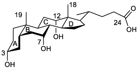
Analysis of the Electron Density of a Water Molecule Encapsulated by Two Cholic Acid Residues
by María Pilar Vázquez-Tato 1, Julio A. Seijas 1,*, Francisco Meijide 2, Santiago de Frutos 2 and José Vázquez Tato 2
1 Departamento de Química Orgánica, Facultade de Ciencias, Universidade de Santiago de Compostela, Campus Terra, 27080 Lugo, Spain
2 Departamento de Química Física, Facultade de Ciencias, Universidade de Santiago de Compostela, Campus Terra, 27080 Lugo, Spain
Int. J. Mol. Sci. 2023, 24(6), 5359; https://doi.org/10.3390/ijms24065359 - 10 Mar 2023
Cited by 1 | Viewed by 948
Abstract
Cholic acid is a trihydroxy bile acid with a nice peculiarity: the average distance between the oxygen atoms (O7 and O12) of the hydroxy groups located at C7 and C12 carbon atoms is 4.5 Å, a value which perfectly matches with the O/O [...] Read more.
Cholic acid is a trihydroxy bile acid with a nice peculiarity: the average distance between the oxygen atoms (O7 and O12) of the hydroxy groups located at C7 and C12 carbon atoms is 4.5 Å, a value which perfectly matches with the O/O tetrahedral edge distance in Ih ice. In the solid phase, they are involved in the formation of hydrogen bonds with other cholic acid units and solvents. This fact was satisfactorily used for designing a cholic dimer which encapsulates one single water molecule between two cholic residues, its oxygen atom (Ow) being exactly located at the centroid of a distorted tetrahedron formed by the four steroid hydroxy groups. The water molecule participates in four hydrogen bonds, with the water simultaneously being an acceptor from the 2 O12 (hydrogen lengths are 2.177 Å and 2.114 Å) and a donor towards the 2 O7 (hydrogen bond lengths are 1.866 Å and 1.920 Å). These facts suggest that this system can be a nice model for the theoretical study of the formation of ice-like structures. These are frequently proposed to describe the water structure found in a plethora of systems (water interfaces, metal complexes, solubilized hydrophobic species, proteins, and confined carbon nanotubes). The above tetrahedral structure is proposed as a reference model for those systems, and the results obtained from the application of the atoms in molecules theory are presented here. Furthermore, the structure of the whole system allows a division into two interesting subsystems in which water is the acceptor of one hydrogen bond and the donor of another. The analysis of the calculated electron density is performed through its gradient vector and the Laplacian. The calculation of the complexation energy used correction of the basis set superposition error (BSSE) with the counterpoise method. As expected, four critical points located in the H…O bond paths were identified. All calculated parameters obey the proposed criteria for hydrogen bonds. The total energy for the interaction in the tetrahedral structure is 54.29 kJ/mol, while the summation obtained of the two independent subsystems and the one between the alkyl rings without water is only 2.5 kJ/mol higher. This concordance, together with the calculated values for the electron density, the Laplacian of the electron density, and the lengths of the oxygen atom and the hydrogen atom (involved in the formation of each hydrogen bond) to the hydrogen bond critical point, suggests that each pair of hydrogen bonds can be considered independent of each other. Full article
(This article belongs to the Special Issue Nano-Materials and Methods 4.0)
Show Figures

Figure 1

15 pages, 991 KiB  
Review
Xerostomia and Its Cellular Targets
by Yoon-Jung Kim
Department of Physiology and Neuroscience, Dental Research Institute, Seoul National University School of Dentistry, Seoul 03080, Republic of Korea
Int. J. Mol. Sci. 2023, 24(6), 5358; https://doi.org/10.3390/ijms24065358 - 10 Mar 2023
Cited by 3 | Viewed by 3756
Abstract
Xerostomia, the subjective feeling of a dry mouth associated with dysfunction of the salivary glands, is mainly caused by radiation and chemotherapy, various systemic and autoimmune diseases, and drugs. As saliva plays numerous essential roles in oral and systemic health, xerostomia significantly reduces [...] Read more.
Xerostomia, the subjective feeling of a dry mouth associated with dysfunction of the salivary glands, is mainly caused by radiation and chemotherapy, various systemic and autoimmune diseases, and drugs. As saliva plays numerous essential roles in oral and systemic health, xerostomia significantly reduces quality of life, but its prevalence is increasing. Salivation mainly depends on parasympathetic and sympathetic nerves, and the salivary glands responsible for this secretion move fluid unidirectionally through structural features such as the polarity of acinar cells. Saliva secretion is initiated by the binding of released neurotransmitters from nerves to specific G-protein-coupled receptors (GPCRs) on acinar cells. This signal induces two intracellular calcium (Ca2+) pathways (Ca2+ release from the endoplasmic reticulum and Ca2+ influx across the plasma membrane), and this increased intracellular Ca2+ concentration ([Ca2+]i) causes the translocation of the water channel aquaporin 5 (AQP5) to the apical membrane. Consequently, the GPCR-mediated increased [Ca2+]i in acinar cells promotes saliva secretion, and this saliva moves into the oral cavity through the ducts. In this review, we seek to elucidate the potential of GPCRs, the inositol 1,4,5-trisphosphate receptor (IP3R), store-operated Ca2+ entry (SOCE), and AQP5, which are essential for salivation, as cellular targets in the etiology of xerostomia. Full article
(This article belongs to the Special Issue Recent Advances in Salivary Gland and Their Function 2.0)
Show Figures

Figure 1

21 pages, 1507 KiB  
Article
Water from Nitrodi’s Spring Induces Dermal Fibroblast and Keratinocyte Activation, Thus Promoting Wound Repair in the Skin: An In Vitro Study
by Filomena Napolitano 1, Loredana Postiglione 1,2, Ilaria Mormile 1, Valentina Barrella 3, Amato de Paulis 1,2, Nunzia Montuori 1,2 and Francesca Wanda Rossi 1,2,*
1 Department of Translational Medical Sciences, University Federico II, 80131 Naples, Italy
2 Center for Basic and Clinical Immunology Research (CISI), WAO Center of Excellence, University Federico II, 80131 Naples, Italy
3 CEINGE, Advanced Biotechnologies, 80145 Naples, Italy
Int. J. Mol. Sci. 2023, 24(6), 5357; https://doi.org/10.3390/ijms24065357 - 10 Mar 2023
Cited by 3 | Viewed by 1511
Abstract
The Romans knew of Nitrodi’s spring on the island of Ischia more than 2000 years ago. Although the health benefits attributed to Nitrodi’s water are numerous, the underlying mechanisms are still not understood. In this study, we aim to analyze the physicochemical properties [...] Read more.
The Romans knew of Nitrodi’s spring on the island of Ischia more than 2000 years ago. Although the health benefits attributed to Nitrodi’s water are numerous, the underlying mechanisms are still not understood. In this study, we aim to analyze the physicochemical properties and biological effects of Nitrodi’s water on human dermal fibroblasts to determine whether the water exerts in vitro effects that could be relevant to skin wound healing. The results obtained from the study indicate that Nitrodi’s water exerts strong promotional effects on dermal fibroblast viability and a significant stimulatory activity on cell migration. Nitrodi’s water induces alpha-SMA expression in dermal fibroblasts, thus promoting their transition to myofibroblast-protein ECM deposition. Furthermore, Nitrodi’s water reduces intracellular reactive oxygen species (ROS), which play an important role in human skin aging and dermal damage. Unsurprisingly, Nitrodi’s water has significant stimulatory effects on the cell proliferation of epidermal keratinocytes and inhibits the basal ROS production but enhances their response to the oxidative stress caused by external stimuli. Our results will contribute to the development of human clinical trials and further in vitro studies to identify inorganic and/or organic compounds responsible for pharmacological effects. Full article
Show Figures

Figure 1

19 pages, 3564 KiB  
Article
Combinatorial Network of Transcriptional and miRNA Regulation in Colorectal Cancer
by Rupesh Kumar 1, Maged Mostafa Mahmoud 2,3,4, Hanaa M. Tashkandi 5, Shafiul Haque 6,7,8, Steve Harakeh 9, Kalaiarasan Ponnusamy 10,* and Shazia Haider 1,*
1 Department of Biotechnology, Jaypee Institute of Information Technology, A-10, Sector 62, Noida 201309, India
2 King Fahd Medical Research Center, King Abdulaziz University, Jeddah 21589, Saudi Arabia
3 Department of Medical Laboratory Sciences, Faculty of Applied Medical Sciences, King Abdulaziz University, Jeddah 21589, Saudi Arabia
4 Molecular Genetics and Enzymology Department, Human Genetics and Genome Research Institute, National Research Centre, Cairo 12622, Egypt
5 Department of General Surgery, Faculty of Medicine, King Abdulaziz University, Jeddah 21589, Saudi Arabia
6 Research and Scientific Studies Unit, College of Nursing and Allied Health Sciences, Jazan University, Jazan 45142, Saudi Arabia
7 Gilbert and Rose-Marie Chagoury School of Medicine, Lebanese American University, Beirut 13-5053, Lebanon
8 Centre of Medical and Bio-Allied Health Sciences Research, Ajman University, Ajman P.O. Box 346, United Arab Emirates
9 King Fahd Medical Research Center, and Yousef Abdullatif Jameel Chair of Prophetic Medicine Application, Faculty of Medicine, King Abdulaziz University, Jeddah 21589, Saudi Arabia
10 Biotechnology Division, National Centre for Disease Control, New Delhi 110054, India
Int. J. Mol. Sci. 2023, 24(6), 5356; https://doi.org/10.3390/ijms24065356 - 10 Mar 2023
Viewed by 1770
Abstract
Colorectal cancer is one of the leading causes of cancer-associated mortality across the worldwide. One of the major challenges in colorectal cancer is the understanding of the regulatory mechanisms of biological molecules. In this study, we aimed to identify novel key molecules in [...] Read more.
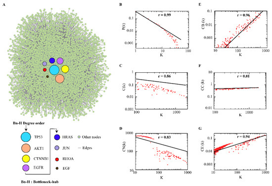
Colorectal cancer is one of the leading causes of cancer-associated mortality across the worldwide. One of the major challenges in colorectal cancer is the understanding of the regulatory mechanisms of biological molecules. In this study, we aimed to identify novel key molecules in colorectal cancer by using a computational systems biology approach. We constructed the colorectal protein–protein interaction network which followed hierarchical scale-free nature. We identified TP53, CTNBB1, AKT1, EGFR, HRAS, JUN, RHOA, and EGF as bottleneck-hubs. The HRAS showed the largest interacting strength with functional subnetworks, having strong correlation with protein phosphorylation, kinase activity, signal transduction, and apoptotic processes. Furthermore, we constructed the bottleneck-hubs’ regulatory networks with their transcriptional (transcription factor) and post-transcriptional (miRNAs) regulators, which exhibited the important key regulators. We observed miR-429, miR-622, and miR-133b and transcription factors (EZH2, HDAC1, HDAC4, AR, NFKB1, and KLF4) regulates four bottleneck-hubs (TP53, JUN, AKT1 and EGFR) at the motif level. In future, biochemical investigation of the observed key regulators could provide further understanding about their role in the pathophysiology of colorectal cancer. Full article
(This article belongs to the Special Issue Molecular Advances in Colorectal Cancer)
Show Figures

Figure 1

15 pages, 6656 KiB  
Article
Metformin Attenuates the Inflammatory Response via the Regulation of Synovial M1 Macrophage in Osteoarthritis
by Meng Zheng 1,†, Yuanli Zhu 2,†, Kang Wei 1, Hongxu Pu 1, Renpeng Peng 1, Jun Xiao 1, Changyu Liu 1 and Xuying Sun 1,*
1 Department of Orthopaedic Surgery, Tongji Hospital, Tongji Medical College, Huazhong University of Science and Technology, Wuhan 430030, China
2 Department of Pathology, Tongji Hospital, Tongji Medical College, Huazhong University of Science and Technology, Wuhan 430030, China
These authors contributed equally to this work.
Int. J. Mol. Sci. 2023, 24(6), 5355; https://doi.org/10.3390/ijms24065355 - 10 Mar 2023
Cited by 5 | Viewed by 1929
Abstract
Osteoarthritis (OA), the most common chronic inflammatory joint disease, is characterized by progressive cartilage degeneration, subchondral bone sclerosis, synovitis, and osteophyte formation. Metformin, a hypoglycemic agent used in the treatment of type 2 diabetes, has been evidenced to have anti-inflammatory properties to treat [...] Read more.
Osteoarthritis (OA), the most common chronic inflammatory joint disease, is characterized by progressive cartilage degeneration, subchondral bone sclerosis, synovitis, and osteophyte formation. Metformin, a hypoglycemic agent used in the treatment of type 2 diabetes, has been evidenced to have anti-inflammatory properties to treat OA. It hampers the M1 polarization of synovial sublining macrophages, which promotes synovitis and exacerbates OA, thus lessening cartilage loss. In this study, metformin prevented the pro-inflammatory cytokines secreted by M1 macrophages, suppressed the inflammatory response of chondrocytes cultured with conditional medium (CM) from M1 macrophages, and mitigated the migration of M1 macrophages induced by interleukin-1ß (IL-1ß)-treated chondrocytes in vitro. In the meantime, metformin reduced the invasion of M1 macrophages in synovial regions brought about by the destabilization of medial meniscus (DMM) surgery in mice, and alleviated cartilage degeneration. Mechanistically, metformin regulated PI3K/AKT and downstream pathways in M1 macrophages. Overall, we demonstrated the therapeutic potential of metformin targeting synovial M1 macrophages in OA. Full article
Show Figures

Figure 1

17 pages, 3133 KiB  
Article
Ternary Mixture of Azoxystrobin, Boscalid and Pyraclostrobin Disrupts the Gut Microbiota and Metabolic Balance of Honeybees (Apis cerana cerana)
by Jie Dong 1, Minjie Huang 1,*, Haikun Guo 2, Jiawen Zhang 1, Xiaodong Tan 1 and Deqian Wang 1,*
1 Institute of Animal Husbandry and Veterinary Science, Zhejiang Academy of Agricultural Sciences, Hangzhou 310021, China
2 Institute of Agro-Product Safety and Nutrition, Zhejiang Academy of Agricultural Sciences, Hangzhou 310021, China
Int. J. Mol. Sci. 2023, 24(6), 5354; https://doi.org/10.3390/ijms24065354 - 10 Mar 2023
Cited by 3 | Viewed by 1539
Abstract
There is a growing risk of pollinators being exposed to multiple fungicides due to the widespread use of fungicides for plant protection. A safety assessment of honeybees exposed to multiple commonly used fungicides is urgently required. Therefore, the acute oral toxicity of the [...] Read more.
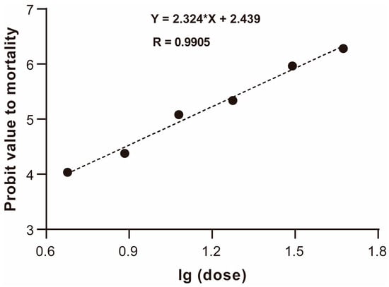
There is a growing risk of pollinators being exposed to multiple fungicides due to the widespread use of fungicides for plant protection. A safety assessment of honeybees exposed to multiple commonly used fungicides is urgently required. Therefore, the acute oral toxicity of the ternary mixed fungicide of ABP (azoxystrobin: boscalid: pyraclostrobin = 1:1:1, m/m/m) was tested on honeybees (Apis cerana cerana), and its sublethal effect on foragers’ guts was evaluated. The results showed that the acute oral median lethal concentration (LD50) of ABP for foragers was 12.6 μg a.i./bee. ABP caused disorder of the morphological structure of midgut tissue and affected the intestinal metabolism; the composition and structure of the intestinal microbial community was perturbed, which altered its function. Moreover, the transcripts of genes involved in detoxification and immunity were strongly upregulated with ABP treatment. The study implies that exposure to a fungicide mixture of ABP can cause a series of negative effects on the health of foragers. This work provides a comprehensive understanding of the comprehensive effects of common fungicides on non-target pollinators in the context of ecological risk assessment and the future use of fungicides in agriculture. Full article
(This article belongs to the Special Issue Pesticides Exposure and Toxicity)
Show Figures

Figure 1

15 pages, 1845 KiB  
Article
Transcriptomic Signatures of Single-Suture Craniosynostosis Phenotypes
by Samantha Lapehn 1, Jonas A. Gustafson 1, Andrew E. Timms 1, Michael L. Cunningham 1,2 and Alison G. Paquette 1,2,*
1 Center for Developmental Biology & Regenerative Medicine, Seattle Children’s Research Institute, Seattle, WA 98101, USA
2 Department of Pediatrics, University of Washington, Seattle, WA 98195, USA
Int. J. Mol. Sci. 2023, 24(6), 5353; https://doi.org/10.3390/ijms24065353 - 10 Mar 2023
Cited by 2 | Viewed by 1421
Abstract
Craniosynostosis is a birth defect where calvarial sutures close prematurely, as part of a genetic syndrome or independently, with unknown cause. This study aimed to identify differences in gene expression in primary calvarial cell lines derived from patients with four phenotypes of single-suture [...] Read more.
Craniosynostosis is a birth defect where calvarial sutures close prematurely, as part of a genetic syndrome or independently, with unknown cause. This study aimed to identify differences in gene expression in primary calvarial cell lines derived from patients with four phenotypes of single-suture craniosynostosis, compared to controls. Calvarial bone samples (N = 388 cases/85 controls) were collected from clinical sites during reconstructive skull surgery. Primary cell lines were then derived from the tissue and used for RNA sequencing. Linear models were fit to estimate covariate adjusted associations between gene expression and four phenotypes of single-suture craniosynostosis (lambdoid, metopic, sagittal, and coronal), compared to controls. Sex-stratified analysis was also performed for each phenotype. Differentially expressed genes (DEGs) included 72 genes associated with coronal, 90 genes associated with sagittal, 103 genes associated with metopic, and 33 genes associated with lambdoid craniosynostosis. The sex-stratified analysis revealed more DEGs in males (98) than females (4). There were 16 DEGs that were homeobox (HOX) genes. Three TFs (SUZ12, EZH2, AR) significantly regulated expression of DEGs in one or more phenotypes. Pathway analysis identified four KEGG pathways associated with at least one phenotype of craniosynostosis. Together, this work suggests unique molecular mechanisms related to craniosynostosis phenotype and fetal sex. Full article
(This article belongs to the Section Molecular Informatics)
Show Figures

Figure 1