Article

Polyphasic Characterization and Taxonomic Evaluation of a Bloom-Forming Strain Morphologically Resembling Radiocystis fernandoi (Chroococcales, Cyanobacteria) from Lake Erhai, China

1
Research Center of Erhai Lake, Dali 671000, China
2
Zhejiang Provincial Key Laboratory for Subtropical Water Environment and Marine Biological Resources Protection, Wenzhou University, Wenzhou 325035, China
3
National and Local Joint Engineering Research Center of Ecological Treatment Technology for Urban Water Pollution, Wenzhou University, Wenzhou 325035, China
4
Wuhan Britain-China School, Gutian Sideroad 10, Wuhan 430034, China
*
Authors to whom correspondence should be addressed.
Diversity 2022, 14(10), 816; https://doi.org/10.3390/d14100816
Submission received: 9 August 2022 / Revised: 27 September 2022 / Accepted: 27 September 2022 / Published: 29 September 2022
(This article belongs to the Special Issue Diversity and Ecology of Algae in China)

Abstract

:
Microcystis-dominated blooms cause environmental and ecological impacts worldwide. However, it is sometimes challenging to correctly identify Microcystis and its related genera. Radiocystis fernandoi Komárek et Komáková-Legnerová 1993, characterized by large cells and correspondingly large colonies with gas vesicles, has been frequently found in cyanobacterial blooms in Brazil. However, its molecular and phylogenetic identity is unknown because no valuable DNA sequences are available in GenBank. In this study, a strain with R. fernandoi morphology was successfully isolated from a cyanobacterial bloom sample in Lake Erhai, a subtropical plateau lake in China. Molecular characterization and phylogenetic analyses based on 16S rRNA and cpcBA genes revealed that the strain is different from the type species of the genus Radiocystis but formed a well-supported clade with the Microcystis species. The comparative results of the ITS region between 16S–23S rRNA genes also showed that the strain had very similar secondary structures to the species of Microcystis, supporting it as a new member of the genus Microcystis. Therefore, we propose the transfer of Radiocystis fernandoi into Microcystis fernandoi comb. nov. The correct identification and further taxonomic procedure for the bloom-forming cyanobacterial genera, such as Microcystis, Radiocystis, and Sphaerocavum, are also discussed in this study.

1. Introduction

Cyanobacterial blooms, occurring worldwide along with global warming and intense eutrophication [1,2,3], have become a challenging problem due to their severe ecological impacts and associated health threats [4,5]. Among the bloom-forming cyanobacterial groups, such as Microcystis, Dolichospermum, Planktothrix, Aphanizomenon, Raphidiopsis and Cuspidiothrix, Microcystis has been always in the spotlight because it is the most frequently and intensively occurring genus. Further, Microcystis spp. are of major concern because many strains produce cyclic heptapeptide toxins called microcystins [6]. Therefore recognizing and identifying Microcystis species in the blooms of field waters, preferably at species level, have become an important task and an initial step for further monitoring and research on the Microcystis-dominated blooms.
Taxonomically, more than 50 Microcystis species have been morphologically described based on some essential characteristics, such as variations in colony form, mucilage structure, cell size, cell arrangement within a colony, and the presence or absence of phycoerythrin [7]. DNA sequences, such as the 16S rRNA gene, ITS between 16S–23S rRNA, cpcBA, and even whole genomic DNA showed high similarities among Microcystis species/strains, indicating that the Microcystis genus is monophyletic and difficult to distinguish at the species level. However, some coccoid cyanobacterial genera with morphological and ecological similarities to Microcystis species have been less characterized for their DNA sequences and molecular phylogeny; thus, the genetic distinction from the genus Microcystis is not sufficient.
Radiocystis is a coccoid cyanobacterial genus that is morphologically similar to Microcystis, and both genera belong to the family Microcystaceae [8]. This genus was established by Skuja in 1948 with the type species R. geminata [9] and is characterized by free-floating, microscopic, more or less spherical colonies, with radial arrangement of the cells, which are slightly distant from one another. To date, five species have been described in the genus, three of which have small cell sizes (2(2.5)–4(5) μm): R. geminata, R. aphanothecoidea Hindák 1996, and R. elongata Hindák 1996 [10]. The type species is facultative with gas vesicles, but the latter two species are complete without gas vesicles. Another species of the genus, R. fernandoi Komárek et Komáková-Legnerová 1993, is characterized by large cells and correspondingly large colonies with gas vesicles [11]. Molecular characterization of this genus has been rarely performed since only three 16S rRNA gene sequences with >1300 bp were found in GenBank, including one from the type species and two from unclassified species in the genus. The monophyly of this genus remains unknown. However, later studies on the cultured strains revealed that R. fernandoi share many similarities, such as morphology, formation of blooms, and microcystin production, with Microcystis aeruginosa [12], but surprisingly no 16S rRNA gene sequences are available in these strains of R. fernandoi.
In this study, R. fernandoi colonies were found in cyanobacterial blooms from Lake Erhai, a plateau lake defined as an early preliminary eutrophic lake. Through the isolation of the strain possessing the morphologies of R. fernandoi, DNA sequences, including the 16S rRNA gene and ITS between 16S–23S, were successfully measured, and molecular phylogeny based on 16S rRNA gene sequences was constructed to evaluate the systematic position of the R. fernandoi strain. The obtained results indicated that R. fernandoi was highly similar to Microcystis spp. in molecular DNA sequences and should taxonomically belong to the genus Microcystis, and therefore renamed Microcystis fernandoi.

2. Materials and Methods

2.1. Sample Collection and Strain Isolation

Lake Erhai, located at 25.6–25.9° N and 100.1–100.3° E with a water surface area of 249 km2, is the second-largest lake in southwestern China and an important drinking water source for Dali City, Yunnan Province. A water sample was collected in September 2020 from the south of Lake Erhai, and a cyanobacterial strain was isolated from the bloom sample using the Pasteur pipette method under a dissecting microscope (Carl Zeiss STEMI 508, Oberkochen, Germany). The obtained unialgal strain WZU 1501 was kept in screw-capped tubes containing 10 mL of BG-11 medium [13] at 25 °C under a 12 h:12 h light/dark cycle with a photon flux density of 35 μmol.m−2·s−1.

2.2. Morphological Observation

Morphological characteristics of the field bloom sample and strain were observed according to the description by Komárek et al. [11]. The images were captured using a high-resolution camera (1000KPA 4/3” Sony sensor 12MP). The photographs were analyzed and adjusted using the LightTools software x64, 4.8.15957 (Hangzhou ToupTek Photonics Co., Hangzhou, China).

2.3. DNA Extraction and PCR Amplification

Fresh cells of the strain WZU 1501 were collected and washed three times using sterile phosphorus-free sterilized modified liquid BG-11 medium to avoid contamination with other bacteria. Total genomic DNA from the strain was extracted using the modified cetyltrimethylammonium bromide (CTAB) method adopted from Neilan et al. [14]. The primers used for amplification of 16S rRNA were PA (5′-AGAGTTTGATCCTGGCTCAG-3′) [15] and B23S (5′-CTTCGCCTCTGTGTGCCT AGG T-3′) [16]. The primers used for the amplification of the phycocyanin operon (cpcBA gene) were PCβF (5′-GGCTGCTTGTTTACGCGACA-3′) and PCαR (5′-CCAGTACCACCAGCAACTAA-3′) [14]. The 16S rRNA PCR with a volume of 50 μL comprised 1 μL of template DNA, 1 μL of each primer (10 μmoL/L), 22 μL of sterile water, and 25 μL of 2× Taq Plus Master Mix (Dye Plus) (Vazyme Biotech Co., Ltd., Nanjing, China). PCR amplification was performed using a SimpliAmpTM Thermal Cycler (Carlsbad, CA, USA) with a PCR profile of an initial denaturation at 95 °C for 5 min, 30 cycles of 30 s at 95 °C, 30 s at 55 °C, 2 min at 72 °C, and a final 10 min elongation step at 72 °C. The cpcBA gene reaction was as follows: initial denaturation at 95 °C for 5 min, 30 cycles of 30 s at 95 °C, 30 s at 55 °C, and 1 min at 72 °C, and a final 5 min elongation step at 72 °C. PCR products were purified using a TIANgel Midi Purification kit (Tiangen Biotech Co., Ltd., Beijing, China) and then cloned using the pClone007 Versatile Simple Vector Kit (Beijng Tsingke Biotech Co., Ltd., Beijing, China). The plasmids containing inserts duplicated in chemically competent Escherichia coli TrelidfTM 5α cells (Beijng Tsingke Biotech Co., Ltd., Beijing, China) were cultured for 12–14 h at 37 °C. Clones, including the target fragment, were sequenced bidirectionally using the standard plasmid primers M13F (5′-TGTAAAACGACGGCCAGT-3′) and M13R (5′-CAGGAAACAGCTATGACC-3′) by Wuhan Tianyi Huayu Gene Technology Co., Ltd. (Wuhan, China).

2.4. Detection for Microystins Synthesis Genes

Genomic DNA from the strain WZU 1501 was used to detect the genes associated with microcystin synthesis (mcyA, mcyB, mcyD and mcyE). The primers and PCR procedures were according to the methods of previous studies by Zuo et al. (2018), Briand et al. (2009), Rinta-Kanto et al. (2005), and Zuo et al. (2018), respectively [17,18,19].

2.5. Phylogenetic Analyses

The 16S rRNA gene and cpcBA gene sequences obtained in this study were compared using the NCBI website, and the highly similar sequences downloaded from GenBank were used for phylogenetic analyses. All downloaded and obtained sequences were aligned using MAFFT v7.463 [20], and both ends were cut using BioEdit v7.0.9 [21]. The sequences of Gloeobaacter violaceus PCC7421 and Gloeobaacter kilaueensis JS1 were used as outgroups. After multiple sequence alignment, a data matrix of 1436 bp sequences of 16S rRNA gene with 54 nucleotide sites was generated. Another data matrix of 499 bp sequences with 15 nucleotide sites was based on the cpcBA gene. The ModelFinder program in PhyloSuite v1.2.2 molecular phylogenetic platform [22] was used to select the best substitution model for phylogenetic analyses. Based on the Akaike Information Criterion (AIC), the standard selection nucleic acid substitution model, general time reversible (GTR+I+G), was selected as the best fitting model for Bayesian inference (BI) and maximum likelihood (ML) analyses. The specific operational parameters were individually estimated using IQ-TREE v2.1.3 [23] and MrBayes v3.2.7 [24]. In the ML analysis, a thorough bootstrap with 1000 pseudoreplications was performed under standard option to obtain relative support. The BI analysis comprised two parallel runs for 10,000,000 generations, sampling every 100th generation, in which the initial 25% of the sampled data were discarded as burn-in. Neighbor-joining (NJ) analysis was performed using the Kimura 2-parameter model with 1000 bootstrap replications in MEGA 11 [25]. Genetic distances of the similarity matrices of the 16S rRNA gene sequences were estimated using the Kimura 2-parameter model in MEGA 11 to calculate the p-distance with pairwise deletion of gaps. Phylogenetic trees were visualized in FigTree v1.4.3, edited using Tree View 1.6.6 software [26] and adjusted using the Adobe Illustrator 2021 software.

2.6. Analyses of 16S–23S Internal Transcribed Spacer (ITS)

The presence of tRNA gene sequences was tested using the tRNAscan-SE2.0 web Server [27]. The 16S–23S rRNA ITS secondary structures (D1–D1′, Box–B, and V3 helices) of WZU 1501 and other closed Microcystis species were determined using the S-fold website (https://sfold.wadsworth.org/ (accessed on 8 August 2022)). All the sequences obtained in this study have been deposited in the GenBank database under accession numbers—OP279644 and OP288980.

3. Results

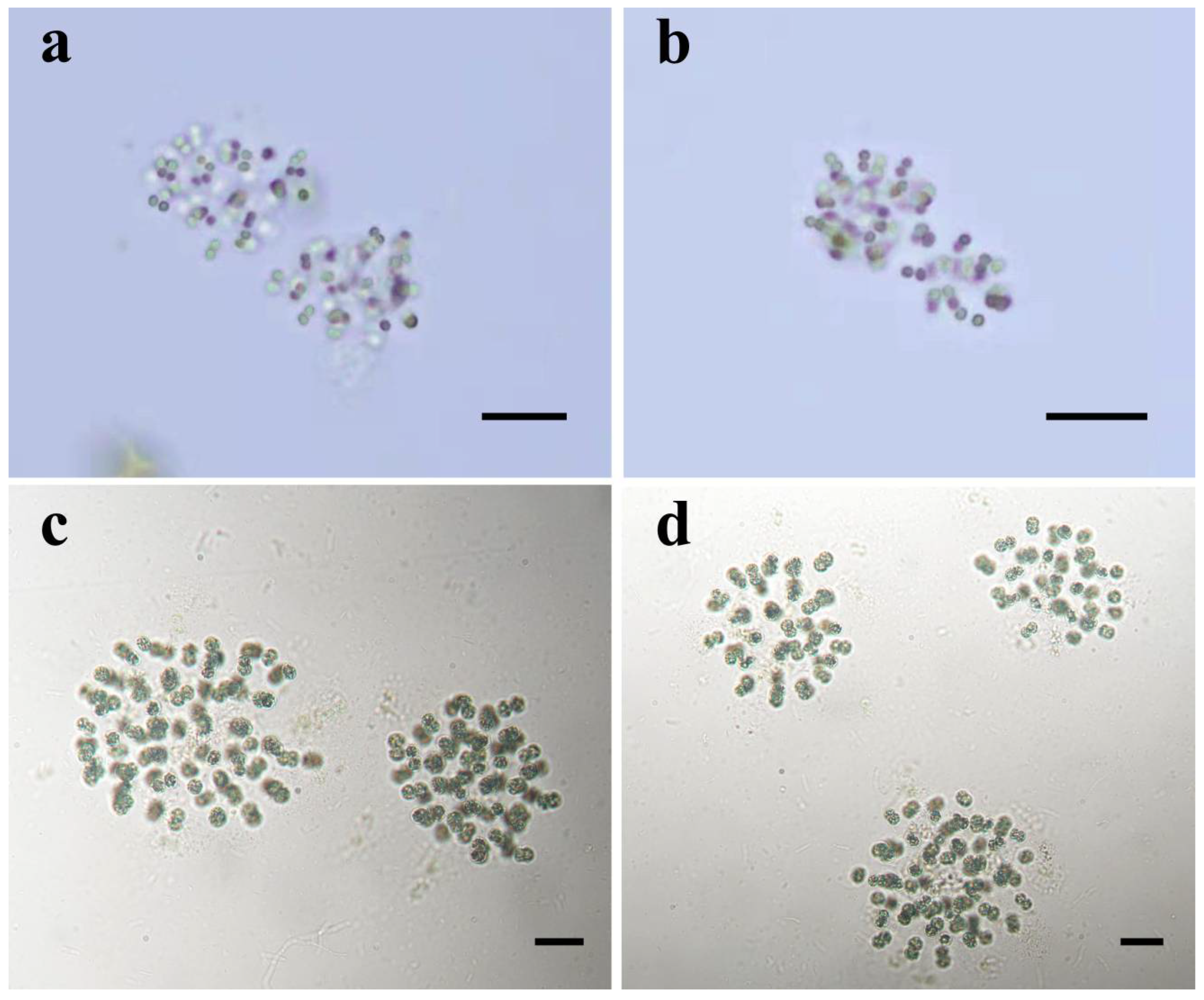
3.1. Morphological Description

Microcystis fernandoi (Komárek and Komárková-Legnerová) X. Lv, R. Geng and R. Li comb. nov. (Figure 1).
Basionym: Radiocystis fernandoi Komárek and Komárková-Legnerová 1993.
Diagnosis: This species is morphologically similar to the genus Radiocystis. Colonies are formed by unicells surrounded by fine, homogeneous, colourless and diffluent mucliage; cells radially arranged, spherical, widely oval (before division), and slightly distant from each other. Phylogenetic analyses revealed that the strain was clustered into an evolutionary branch with the genus Microcystis and different from the Radiocystis clade.
Description: In nature, colonies are microscopic, blue-green; cells usually spherical to elliptical, slightly distant from one another, with loose arrangement between cells, sometimes appearing in pairs, with a diameter of 4.71–(6.67)–8.05 μm, free-floating in water; aerotopes (groups of gas vesicles) facultatively occurring in cells (Figure 1a,b). In liquid culture, colonies are formed by unicells, small, more or less spherical or slightly irregularly oval; large colonies are usually composed of subcolonies, free-living in water. Colonies are enveloped by fine, homogeneous, colorless, and diffluent mucilage, with radially oriented cells; variable rows are usually slightly disordered, sometimes not clearly distinct in the center of colonies or in old colonies. Cells spherical or widely oval before division, slightly distant from each other, sometimes appear in pairs after division, with a diameter of 5.53–(6.98)–9.43 μm; content granular, blue-green, bright blue-green or olive green, and yellow in decline phase, usually with gas vesicles. In subsequent generations, cells divide horizontally in one plane to form radial rows, and at later stages of growth, divide vertically in rows perpendicular to form new rows. The daughter cells usually remain close to one another for a very long time after division (Figure 1c,d).
Reference strain: WZU 1501.
Holotype: designated as iconotype Figure 1a–d in Komárek et al. (1993).
Isotype: designated as dry material of Microcystis fernandoi WZU 1501 preserved in the Herbarium of Wenzhou University (WZUH), Wenzhou, Zhejiang Province, China, as specimen No. WZUH-ZLYN202001.
Isotype locality: In Lake Erhai, Yunnan Province, China.
Etymology: Radiocystis fernandoi Komárek and Komárková-Legnerová 1993.
Habitat: free-living in water.

3.2. Molecular and Phylogenetic Analyses

Molecular analyses used 54 sequences of 16S rRNA gene (53 sequences downloaded from the NCBI database) after multiple sequence alignment to construct phylogenetic trees based on NJ, ML, and Bayesian methods. The 16S rRNA gene phylogenetic tree (Figure 2) showed that the strain WZU 1501 and Microcystis strains, including the type species Microcystis aeruginosa, were clustered into a large evolutionary branch (Figure 2, clade A). It was different from the type species of the genus Radiocystis (Figure 2, clade B), with high bootstrap values of 100%/100%/1.00 according to NJ/ML/BI calculations, respectively. The sequence similarities between the strain WZU 1501 and the strains of Microcystis were >99% (Table 1), which is higher than the threshold of bacterial species classification (97%) [28,29,30], indicating that this strain is likely to be a new member of the genus Microcystis.
Furthermore, the cpcBA gene sequences of the two clones of WZU 1501 obtained in this study shared 100% similarity with each other. Thirteen reference sequences downloaded from the NCBI database and two WZU 1501 clones were used to construct NJ/ML/BI phylogenetic trees (Figure 3). As shown in the cpcBA phylogeny tree, strain WZU 1501 formed an independent clade with M. aeruginosa (Figure 3, red clade) and further clustered with other Microcystis strains into a large well-supported clade (Figure 3, gray clade), far away from the type species of Radiocystis genus (Figure 3, green clade), supported by high bootstrap values (100%/100%/1.00). This result was consistent with the conclusion obtained above using the 16S rRNA gene sequences, indicating the need that Radiocystis fernandoi should be transferred from Radiocystis to Microcystis.
In addition, the genes associated with synthesis of microcystins (mcyA, mcyB, mcyD, and mcyE) were not detected in the strain WZU 1501.

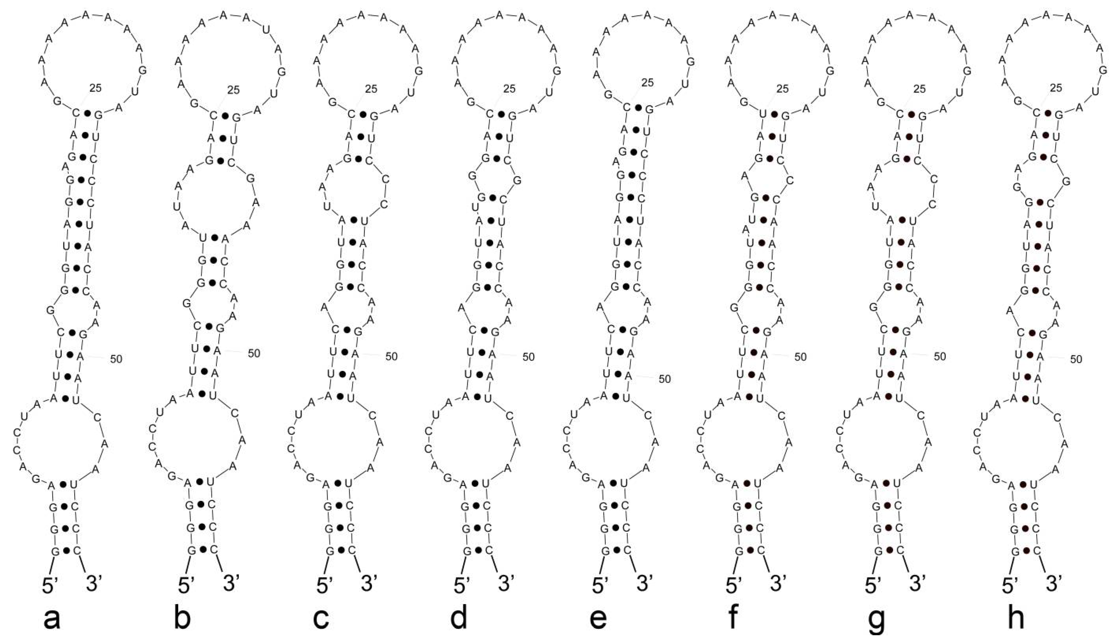
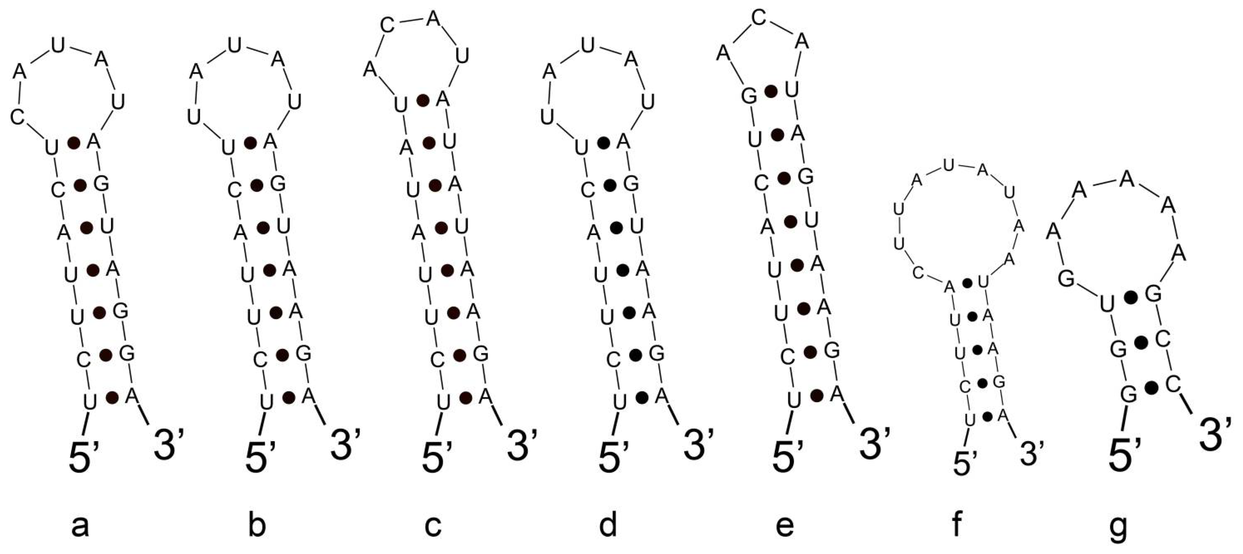
3.3. Analyses of ITS between 16S and 23S rRNA Gene and Secondary Structures

The full-length of ITS sequence of strain WZU 1501 was 357 bp (Table 2), containing only one tRNAIle. Seven different Microcystis species (eight strains) together with the strain WZU 1501 were selected to compare and analyze ITS secondary structures in this study because there is no available ITS sequence of Radiocystis in the current public database; these included Microcystis wesenbergii NIES111, Microcystis viridis NIES 103, Microcystis novacekii VN517, Microcystis ichthyoblabe VN512, Microcystis panniformis VN516, Microcystis pseudofilamentosa VN511, Microcystis aeruginosa PCC 7941, and Microcystis aeruginosa IPPAS B-1527. We calculated the similarity values for the ITS sequences (Table 3) and the results showed that the p-distance of WZU 1501 against strains of Microcystis ranged from 2.56–5.13%, which is in the ambiguous range. As the most conserved structure, the D1–D1′ (Figure 4) helices showed similar stem-loop structures between WZU 1501 and other Microcystis species, forming eight shapes based on tiny differences in few bases and stem-loop structures. Additionally, WZU 1501 and Microcystis ichthyoblabe VN512 had exactly the same Box–B helix secondary structure in the base sequence, sequence length, and stem-loop structure (Figure 5a), and all the nine strains used in this study shared the exact same V3 secondary configuration (Figure 5g). These results provided strong evidence for the phylogenetic conclusion obtained above, further indicating that this strain belongs to the genus Microcystis.

4. Discussion

Cyanobacterial blooms dominated by Microcystis species have frequently been documented worldwide [7,31]. Thus, the correct identification of Microcystis species has become an important basis for further monitoring of the blooms. Currently, more than 50 species of the genus Microcystis are taxonomically accepted [6]. Six morphospecies, such as M. aeruginosa, M. viridis, M. wesenbergii, M. flos-aquae, M. ichthyoblabe, and M. novacekii, were regarded as the main species that occur in water blooms according to investigations in Japanese waters [32]. However, twelve Microcystis morphospecies were reported as the main species in China, including the above six and M. smithii, M. botrys, M. firma, M. pseudofilamentosa, and M. panniformis [33,34]. Based on a current revision, Komárek and Johansen (2015) summarized approximately 25 species of Microcystis as planktonic species worldwide, and the others live benthic in moors, epiphytic or epilithic in different types of stagnant and running waters, and on wet rocks [35]. These non-planktonic species, lacking the ability to form gas vesicles, were also considered to belong to the genus Aphanocapsa [36]. Such heterogeneity may challenge the monophyly of the genus Microcystis because there are some Microcystis-related genera with a niche similar to that of Microcystis species, and Radiocystis is one such case. The colonial genus Radiocystis Skuja currently contains five species, three of which are restricted to temperate zones, and the other two occur in tropical areas [35]. The new revision of the taxonomic system of cyanobacteria by Komárek et al. (2014) has already transferred the genus Radiocystis into the family Microcystaceae from Synechococcaceae, indicating the similarities between Radiocystis and Microcystis [8]. In particular, Radiocystis fernandoi is the most common species of the genus in tropical regions, and some strains of this species in Brazil have been shown to form heavy blooms and produce microcystins in freshwaters in different tropical and subtropical regions of the continent [37,38,39,40,41,42]. R. fernandoi has been reported to contribute more than 70% of the total cyanobacterial biomass in some blooms [38,42,43,44]; therefore, this species is regarded as having great ecological importance in tropical regions. All these characteristics indicate high similarities between R. fernandoi and Microcystis aeruginosa. The morphological uniqueness of Radiocystis fernandoi, such as colonial form, radial cellular arrangement, and distance between cells within the colony, can be used to distinguish it from M. aeruginisa; however, molecular and phylogenetic differences between these two genera/species remain unknown. In addition, M. botrys is another bloom-forming cyanobacteria which is morphologically similar to R. fernandoi. This species usually doesn’t form blooms alone, but often occurs with other bloom-forming species. It shared a similar radial arrangements of peripheral cells of R. fernandoi, but the sheaths formed by M. botrys are not close to cells, and the tight arrangement of the cells and the smaller cell diameter are also important features that distinguish it from R. fernandoi.
In the present study, we used a strain of R. fernandoi isolated from Lake Erhai, a subtropical plateau lake in Yunnan Province. This is the first time to record R. fernandoi and obtain the strain of this species in China. Morphological examination of both field samples and cultivated strains revealed the presence of R. fernandoi in Lake Erhai (Figure 1). Further molecular detection using 16S rRNA gene, ITS between 16S–23S, and cpcBA revealed that the strain had high molecular homology to the strains of Microcystis. Phylogenetic analyses also showed that the strain formed a tight cluster with all Microcystis strains used in the study, indicating a high likelihood combining and moving R. fernandoi to the Microcystis genus.
The results and conclusions obtained seemed direct and simple. At the genus level, Radiocystis is heterogeneous because it comprises five species with different climate zones, presence of gas vesicles reflecting the ability to form cyanobacterial blooms, and differences in cell sizes. Currently, very few DNA sequences are available in GenBank for Radiocystis species. Only three 16S rRNA sequences obtained from the type species and two unidentified strains were homogenous. However, the molecular results in this study, from both sequence similarity and phylogenetic grouping, indicate that R. fernandoi is molecularly different from the R. geminnata. This study proved that the genus Radiocystis is not monophyletic, and the treatment for such a genus without monophyly is to move R. fernandoi out of the genus Radiocystis based on the advantage of the type species.
Within the family Microcystaceae, Sphaerocavum is a planktonic coccoid cyanobacterium that is commonly found in blooms, and the genus is characterized by several distinct morphological features from Microcystis, including hollow colony formation throughout its life cycle, loose cell disposal on the mucilage surface, and two-plane cell division along successive generations [45]. However, Sphaerocavum has repeatedly been reported to co-occur with Microcystis in several South American countries and New Zealand [46]. Rigonato et al. (2018) revealed, based on molecular and phylogenetic analyses, that Sphaerocavum and Microcystis formed a close and well-established cluster, and they proposed that Sphaerocavum must be included within the genus Microcystis and the type species Sphaerocavum brasiliensis is renamed Microcystis brasiliensis [47]. Based on these two studies, that is M. fernandoi in this study and Microcystis brasiliensis by Rigonato et al. (2018), future taxonomic evaluation and revision of the cyanobacterial taxa related to the Microcystis genus should use the polyphasic approach [47]. Without molecular characters, such as sequences of marked genes, the taxonomic positions of these cyanobacteria should not be determined even though some distinct morphological features from Microcystis are apparent.
In conclusion, the strain WZU 1501, isolated from a cyanobacterial bloom sample in Lake Erhai, a subtropical plateau lake in southwestern China, was morphologically identified as R. fernandoi with a typical radial arrangement of cells within the colony and two division planes. However, molecular characterization and phylogenetic analyses based on 16S rRNA, ITS, and cpcBA revealed the strain to be far away from the type species of the genus Radiocystis but formed a tight cluster with Microcystis species; therefore, we propose a new combination that Radiocystis fernandoi be renamed as Microcystis fernandoi. We are expecting to obtain more strains from different waters in future to verify this taxonomic transfer in this study. It also provides an example for further discussions in future taxonomic revisions in Microcystis-like cyanobacteria.

Author Contributions

Author Contributions: Conceptualization, X.L. and R.L.; methodology, Z.H.; software, X.L. and S.Z.; formal analysis, Y.C. and R.G.; investigation, S.Z. and P.X.; resources, H.Z.; data curation, Y.C. and R.G.; writing—original draft preparation, R.L., X.L., R.G. and Y.C.; writing—review and editing, R.L. and R.G.; visualization, Y.C. All authors have read and agreed to the published version of the manuscript.

Funding

This work was supported by “National Observation and Research Station of Erhai Lake Ecosystem in Yunnan” from Shanghai Jiao Tong University.

Institutional Review Board Statement

Not applicable.

Informed Consent Statement

Not applicable.

Data Availability Statement

The 16S rRNA, 16S–23S ITS, and cpcBA sequences are available in the GenBank database.

Conflicts of Interest

The authors declare no conflict of interest.

References

  1. Heisler, J.; Glibert, P.M.; Burkholder, J.M.; Anderson, D.M.; Cochlan, W.; Dennison, W.C.; Dortch, Q.; Gobler, C.J.; Heil, C.A.; Humphries, E.; et al. Eutrophication and harmful algal blooms: A scientific consensus. Harmful Algae 2008, 8, 3–13. [Google Scholar] [CrossRef] [PubMed]
  2. Paerl, H.W.; Huisman, J. Climate—Blooms like it hot. Science 2008, 320, 57–58. [Google Scholar] [CrossRef] [PubMed]
  3. Paerl, H.W.; Paul, V.J. Climate change: Links to global expansion of harmful cyanobacteria. Water Res. 2012, 46, 1349–1363. [Google Scholar] [CrossRef] [PubMed]
  4. O’Neil, J.M.; Davis, T.W.; Burford, M.A.; Gobler, C.J. The rise of harmful cyanobacteria blooms: The potential roles of eutrophication and climate change. Harmful Algae 2012, 14, 313–334. [Google Scholar] [CrossRef]
  5. Rigosi, A.; Carey, C.C.; Ibelings, B.W.; Brookes, J.D. The interaction between climate warming and eutrophication to promote cyanobacteria is dependent on trophic state and varies among taxa. Limnol. Oceanogr. 2014, 59, 99–114. [Google Scholar] [CrossRef]
  6. Huo, D.; Gan, N.; Geng, R.; Cao, Q.; Song, L.; Yu, G.; Li, R. Cyanobacterial blooms in China: Diversity, distribution, and cyanotoxins. Harmful Algae 2021, 109, 102106. [Google Scholar] [CrossRef] [PubMed]
  7. Komárek, J.; Komáková, J. Review of the European Microcystis-morphospecies (Cyanoprokaryotes) from nature. Czech Phycol. Olomouc 2002, 2, 1–24. [Google Scholar]
  8. Komárek, J.; Katovsk, J.; Mare, J.; Johansen, J.R. Taxonomic classification of cyanoprokaryotes (cyanobacterial genera) 2014, using a polyphasic approach. Preslia 2014, 86, 295–335. [Google Scholar]
  9. Skuja, H. Taxonomie des Phytoplanktons einiger Seen in Uppland, Schweden. Symb. Bot. Ups. 1948, 9, 1–399. [Google Scholar]
  10. Hinoák, F. Two new species of the genus Radiocystis (Chroococcales, Cyanophyta/Cyanobacteria). Eur. J. Pediatr. Surg. 1996, 19, 2, 113–116. [Google Scholar] [CrossRef]
  11. Komárek, J.; Komárková-Legnerová, J. Radiocystis fernandoii, a new planktic cyanoprokariotic from tropical freshwater reservoirs. Preslia 1993, 65, 355–357. [Google Scholar]
  12. Rios, J.F.; Retz, D.C.L.; Leal, C.R.; Sant’Anna, C.L. The influence of temperature on Radiocystis fernandoi strain (cyanobacteria) growth and microcystin production. Braz. J. Bot. 2018, 41, 675–680. [Google Scholar]
  13. Stanier, R.Y.; Kunisawa, R.; Mandel, M.; Cohen–Bazire, G. Purification and properties of unicellular blue-green algae (order Chroococcales). Bacteriol. Rev. 1971, 35, 171–205. [Google Scholar] [CrossRef] [PubMed]
  14. Neilan, B.A.; Jacobs, D.; Goodman, A.E. Genetic diversity and phylogeny of toxic cyanobacteria determined by DNA polymorphisms within the phycocyanin locus. Appl. Environ. Microbiol. 1995, 61, 3875–3883. [Google Scholar] [CrossRef]
  15. Edwards, U.; Rogall, T.; Blocker, H.; Emde, M.; Bottger, E.C. Isolation and direct complete nucleotide determination of entire genes. Characterization of a gene coding for 16S ribosomal RNA. Nucleic Acids Res. 1989, 17, 7843–7853. [Google Scholar] [CrossRef] [Green Version]
  16. Gkelis, S.; Rajaniemi, P.; Vardaka, E.; Moustaka-gouni, M.; Lanaras, T.; Sivonen, K. Limnothrix redekei (Van Goor) Meffert (Cyanobacteria) strains from Lake Kastoria, Greece form a separate phylogenetic group. Microb. Ecol. 2005, 49, 176–182. [Google Scholar] [CrossRef]
  17. Briand, E.; Escoffier, N.; Straub, C.; Sabart, M.; Quiblier, C.; Humbert, J.F. Spatiotemporal changes in the genetic diversity of a bloom-forming Microcystis aeruginosa (cyanobacteria) population. ISME J. 2009, 3, 4, 419–429. [Google Scholar] [CrossRef]
  18. Rinta-Kanto, J.M.; Ouellette, A.J.A.; Boyer, G.L.; Twiss, M.R.; Bridgeman, T.B.; Wilhelm, S.W. Quantification of toxic Microcystis spp. during the 2003 and 2004 blooms in western Lake Erie using quantitative real-time PCR. Environ. Sci. Technol. 2005, 39, 4198–4205. [Google Scholar] [CrossRef]
  19. Zuo, J.; Chen, L.; Shan, K.; Hu, L.; Song, L.; Gan, N. Assessment of different mcy genes for detecting the toxic to non-toxic Microcystis ratio in the field by multiplex qPCR. J. Oceanol. Limnol. 2018, 36, 1132–1144. [Google Scholar] [CrossRef]
  20. Katoh, K.; Standley, D.M. MAFFT multiple sequence alignment software version 7: Improvements in performance and usability. Mol. Biol. Evol. 2013, 30, 772–780. [Google Scholar] [CrossRef]
  21. Hall, T.A. BioEdit: A User-Friendly Biological Sequence Alignment Editor and Analysis Program for Windows 95/98/NT. Nuclc-Acids Symp. Ser. 1999, 41, 95–98. [Google Scholar]
  22. Zhang, D.; Gao, F.; Jakovli, I.; Zou, H.; Wang, G.T. PhyloSuite: An integrated and scalable desktop platform for streamlined molecular sequence data management and evolutionary phylogenetics studies. Mol. Ecol. Resour. 2020, 20, 348–355. [Google Scholar] [CrossRef] [PubMed]
  23. Nguyen, L.T.; Schmidt, H.A.; Von Haeseler, A.; Minh, B.Q. IQ-TREE: A fast and effective stochastic algorithm for estimating maximum-likelihood phylogenies. Mol. Biol. Evol. 2015, 32, 268–274. [Google Scholar] [CrossRef] [PubMed]
  24. Ronquist, F.; Teslenko, M.; Van der Mark, P.; Ayres, D.L.; Darling, A.; Höhna, S.; Larget, B.; Liu, L.; Suchard, M.A.; Huelsenbeck, J.P. MrBayes 3.2: Efficient Bayesian phylogenetic inference and model choice across a large model space. Syst. Biol. 2012, 61, 539–542. [Google Scholar] [CrossRef]
  25. Tamura, K.; Stecher, G.; Kumar, S. MEGA11: Molecular Evolutionary Genetics Analysis Version 11. Mol. Biol. Evol. 2021, 38, 7, 3022–3027. [Google Scholar] [CrossRef]
  26. Page, R.D.M. TreeView: An application to display phylogenetic trees on personal computers. Comput. Appl. Biosci. 1996, 12, 357–358. [Google Scholar]
  27. Lowe, T.M.; Chan, P.P. tRNAscan-SE On-line: Integrating search and context for analysis of transfer RNA genes. Nucleic Acids-Res. 2016, 44, W54–W57. [Google Scholar] [CrossRef]
  28. Wayne, L.G.; Brenner, D.J.; Colwell, R.R.; Grimont, P.A.D.; Kandler, O.; Krichevsky, M.I.; Starr, M.P. Report of the ad hoc committee on reconciliation of approaches to bacterial systematics. Int. J. Syst. Evol. Microbiol. 1987, 37, 463–464. [Google Scholar] [CrossRef]
  29. Stackebrandt, E.; Goebel, B.M. Taxonomic note: A placefor DNA–DNA reassociation and 16S rRNA sequence analysisin the present species definition in bacteriology. Int. J. Syst. Bacteriol. 1994, 44, 846–849. [Google Scholar] [CrossRef]
  30. Stackebrandt, E.; Ebers, J. Taxonomic parameters revisited: Tarnished gold standards. Microbiol. Today 2006, 33, 152–155. [Google Scholar]
  31. Xu, Y.; Wu, Z.; Yu, B.; Peng, X.; Yu, G.; Wei, Z.; Wang, G.; Li, R. Non-microcystin producing Microcystis wesenbergii (Komárek) Komárek (Cyanobacteria) representing a main water bloom-forming species in Chinese waters. Environ. Pollut. 2008, 156, 162–167. [Google Scholar] [CrossRef] [PubMed]
  32. Komárek, J. A review of water-bloom forming Microcystis species, with regard to population from Japan. Arch. Hydrobiol. (Suppl.)/Algol. Stud. 1991, 73, 115–127. [Google Scholar]
  33. Yu, G.L.; Song, L.R.; Li, R.H. Taxonomic notes on water bloom forming Microcystis species (Cyanophyta) from China—An example from samples of the Dianchi Lake. Acta Phytotaxon. Sin. 2007, 45, 727–741. [Google Scholar] [CrossRef]
  34. Zhang, J.; Zhu, B.; Wu, Z.; Xu, T.; Lu, Z. Microcystis panniformis—A newly recorded species of Microcystis in China. Sci. Limnol. Sin. 2012, 24, 647–650. [Google Scholar]
  35. Komárek, J.; Johansen, J. Freshwater Algae of North America; Wehr, J.D., Sheath, R., Kociolek, J.P., Eds.; Academic Press: Cambridge, MA, USA, 2015; p. 1050. [Google Scholar]
  36. Nägeli, C. Gattungen einzelliger Algen, physiologisch und systematisch bearbeitet. Neue Denkschr. Der Allg. Schweiz. Ges. Für Die Gesammten Nat. 1849, 10, 1–139. [Google Scholar]
  37. Vieira, J.; Azevedo, M.; Azevedo, S.; Honda, R.Y.; Corrêa, B. Microcystin production by Radiocystis fernandoi (Chroococcales, cyanobacteria) isolated from a drinking water reservoir in the city of belém, pa, brazilian amazonia region. Toxicon 2003, 42, 709–713. [Google Scholar] [CrossRef]
  38. Vieira, J.M.D.S.; Azevedo, M.T.D.P.; Azevedo, S.M.F.D.O.; Honda, R.Y.; Corrêa, B. Toxic cyanobacteria and microcystin concentrations in a public water supply reservoir in the Brazilian Amazonia region. Toxicon 2005, 45, 7, 901–909. [Google Scholar] [CrossRef]
  39. Sant’Anna, C.L.A.; Werner, V.R.T.P.; Dogo, C.R.; Rios, F.R.D.C.; Azevedo, M.T.D.P.; De Carvalho, L.R. Review of toxic species of Cyanobacteria in Brazil. Algol. Stud. 2008, 126, 251–265. [Google Scholar] [CrossRef]
  40. Borges, P.A.F.; Train, S.; Dias, J.D.; Bonecker, C.C. Effects of fish farming on plankton structure in a Brazilian tropical reservoir. Hydrobiologia 2010, 649, 279–291. [Google Scholar] [CrossRef]
  41. Ferrari, G.; Pérez, M.D.C.; Dabezies, M.; Míguez, D.; Saizar, C. Planktic Cyanobacteria in the Lower Uruguay River, South America. J. Czech Phycol. Soc. 2011, 11, 225–234. [Google Scholar] [CrossRef]
  42. Fonseca, I.A.; Maniglia, T.C.; Rodrigues, L.; Prioli, A.J.; Prioli, S. Identification of the mcyA gene in natural blooms of Radiocystis fernandoi from a tributary of the Rosana reservoir, Brazil. Acta Sci. Biol. Sci. 2011, 33, 319–324. [Google Scholar] [CrossRef] [Green Version]
  43. Lombardo, M.; Pinto, F.C.R.; Vieira, J.M.S.; Honda, R.Y.; Pimenta, A.M.; Bemquerer, M.P.; Carvalho, L.; Kiyota, S. Isolation and structural characterization of microcystin-LR and three minor oligopeptides simultaneously produced by Radiocystis fernandoi (Chroococcales, Cyanobacteriae): A Brazilian toxic cyanobacterium. Toxicon Off. J. Int. Soc. Toxinol. 2006, 47, 560–566. [Google Scholar] [CrossRef] [PubMed]
  44. Borges, P.A.F.; Train, S.; Rodrigues, L.C. Estrutura do fitoplâncton, em curto período de tempo, em um braço do reservatório de Rosana (ribeirão do Corvo, Paraná, Brasil). Acta Sci. Anim. Sci. 2008, 30, 57–65. [Google Scholar]
  45. Azevedo, M.T.D.P.; Sant’Anna, C.L. Sphaerocavum, a new genus of planktic cyanobacteria from continental water bodies in Brazil. Algol. Stud. 2003, 109, 79–92. [Google Scholar]
  46. Wood, S.; Crowe, A.L.M.; Ruck, J.G.; Wear, R.G. New records of planktonic cyanobacteria in New Zealand freshwaters. N. Zeal. J. Bot. 2005, 43, 479–492. [Google Scholar] [CrossRef]
  47. Rigonato, J.; Sant’Anna, C.L.; Giani, A.; Azevedo, M.T.P.; Gama, W.A.; Viana, V.F.L.; Fiore, M.F.; Werner, V.R. Sphaerocavum: A coccoid morphogenus identical to Microcystis in terms of 16S-rDNA and ITS sequence phylogenies. Hydrobiologia 2018, 811, 35–48. [Google Scholar] [CrossRef]
Figure 1. Images of Microcystis fernandoi under the light microscopy. (a,b): Microcystis fernandoi colonies from filed samples in Lake Erhai; (c,d): Microcystis fernandoi colonies from the strain WZU 1501 in liquid BG-11 medium. All scale bars are 20 μm.
Diversity 14 00816 g001
Figure 2. Bayesian inference (BI) phylogenetic tree of WZU 1501 based on 16S rRNA gene sequences (1436 bp). Bootstrap values greater than 50% are showed on the BI tree for NJ/ML methods and Bayesian posterior probabilities. The studied strain was indicated in bold. Bar, 0.02.
Diversity 14 00816 g002
Figure 3. Phylogenetic tree based on cpc-BA gene sequences (499 bp) with maximum-likelihood (ML) analysis. Bootstrap values of NJ/ML/BI methods greater than 50% are showed on the ML tree. The studied species was indicated in bold.
Diversity 14 00816 g003
Figure 4. D1–D1′ helices of Microcystis. (a). WZU1501. (b). Microcystis wesenbergii NIES111. (c). Microcystis viridis NIES 103. (d). Microcystis novacekii VN517. (e). Microcystis ichthyoblabe VN512. (f). Microcystis panniformis VN516. (g). Microcystis pseudofilamentosa VN511. (h). Microcystis aeruginosa PCC 7941 and Microcystis aeruginosa IPPAS B-1527.
Diversity 14 00816 g004
Figure 5. Box–B helices and V3 helices of Microcystis. (a). Box–B helix of WZU1501 and Microcystis ichthyoblabe VN512. (b). Box–B helix of Microcystis wesenbergii NIES111. (c). Box–B helix of Microcystis viridis NIES 103 and Microcystis pseudofilamentosa VN511. (d). Box–B helix of Microcystis novacekii VN517 and Microcystis panniformis VN516. (e). Box–B helix of Microcystis aeruginosa PCC 7941. (f). Box–B helix of Microcystis aeruginosa IPPAS B-1527. (g). V3 helix of all strains.
Diversity 14 00816 g005
Table 1. Sequence similarity comparison of the 16S rRNA gene between Microcystis fernandoi WZU 1501 and other related taxa. Similarity = [1 − (p-distance)] × 100.
Strains123456789101112131415
1. Microcystis fernandoi WZU1501
2. Microcystis aeruginosa BKP SB6999.85
3. Microcystis aeruginosa FACHB-90599.5499.70
4. Microcystis wesenbergii TAC5299.7099.8599.85
5. Microcystis wesenbergii NRERC-21299.6299.7799.7799.92
6. Microcystis viridis NIES-10299.7099.8599.7099.7099.62
7. Microcystis viridis TAC1799.7099.8599.7099.7099.62100.00
8. Microcystis ichthyoblabe VN32599.7099.8598.7099.8599.7799.7099.70
9. Microcystis ichthyoblabe NRERC-21799.7799.9299.7799.9299.8599.7799.7799.92
10. Microcystis flos-aquae NRERC 21899.6299.7799.6299.7799.7099.6299.6299.9299.85
11. Microcystis flos-aquae NRERC-21199.6299.7799.6299.7799.7099.6299.6299.9299.8599.85
12. Microcystis novacekii CHAB144499.5499.7099.7099.8599.7799.5499.5499.7099.7799.6299.62
13. Microcystis novacekii TAC6599.4799.6299.4799.6299.5499.6299.6299.6299.7099.5499.5499.62
14. Microcystis bengalensis VN48699.6299.7799.6299.6299.5499.7799.7799.6299.7099.5499.5499.6299.70
15. Radiocystis sp. JJ30-391.0591.2291.3191.3191.2291.2291.2291.3191.2291.2291.2291.1490.8890.96
16. Radiocystis geminata TAU-MAC 121490.7190.8890.9790.9790.8890.8890.8890.9790.8890.8890.8890.8090.5390.6499.45
Table 2. Analyses on ITS of 16S–23S region for Microcystis fernandoi WZU 1501 and other related strains.
StrainGenbankComplete ITS (nt)D1-D1′
Helix
(nt)
tRNAIletRNAAlaBox-B
Helix (nt)
Box-A
Helix (nt)
D4V3
Helix (nt)
D5
1. Microcystis fernandoi WZU1501OP27964435759+-191471217
2. Microcystis wesenbergii NIES111AB015388.136159+-191471217
3. Microcystis viridis NIES 103AB254444.135859+-191471217
4. Microcystis novacekii VN517AB665999.135958+-191471217
5. Microcystis ichthyoblabe VN512AB666031.136259+-191471217
6. Microcystis panniformis VN516AB666041.135959+-191471217
7. Microcystis pseudofilamentosa VN511AB666052.135859+-191471217
8. Microcystis aeruginosa PCC 7941MH620004.135959+-191471217
9. Microcystis aeruginosa IPPAS B-1527MW396712.136459+-191471217
Table 3. Sequence similarity comparison of the ITS between Microcystis fernandoi WZU 1501 and other related taxa. Similarity = [1 − (p-distance)] × 100.
Strain12345678
1. Microcystis fernandoi WZU1501
2. Microcystis wesenbergii NIES11195.44
3. Microcystis viridis NIES 10395.7398.86
4. Microcystis novacekii VN51796.3096.3096.58
5. Microcystis ichthyoblabe VN51294.8796.8796.8798.29
6. Microcystis panniformis VN51695.4496.3096.3097.7297.72
7. Microcystis pseudofilamentosa VN51197.1594.8794.8794.5994.3094.87
8. Microcystis aeruginosa PCC 794196.8795.1695.1694.8794.5994.5999.72
9. Microcystis aeruginosa IPPAS B-152797.4495.1695.1694.8795.1695.7397.4497.15
Publisher’s Note: MDPI stays neutral with regard to jurisdictional claims in published maps and institutional affiliations.

