Article

Hydrolithon farinosum and Lithophyllum epiphyticum sp. nov. (Corallinaceae, Corallinales, Rhodophyta), Two Epiphytic Crustose Coralline Algae from the Abrolhos Archipelago, Brazil, Southwestern Atlantic

by
Manoela B. Lyra
1,
Ricardo G. Bahia
1,*,
Michel B. Jesionek
1,
Rodrigo T. Carvalho
1,
Fernando C. Moraes
1,
Adele S. Harvey
2,
Renato C. Pereira
3,
Fabiano Salgueiro
4 and
Leonardo T. Salgado
1,*
1
Research Institute of the Botanical Garden of Rio de Janeiro, Rua Pacheco Leão, 915, Jardim Botânico, Rio de Janeiro 22460-030, Brazil
2
Department of Ecology, Environment and Evolution, La Trobe University, Bundoora, VIC 3083, Australia
3
Department of Marine Biology, Institute of Biology, Federal Fluminense University, Niterói, Rio de Janeiro 24220-900, Brazil
4
Department of Botany, Center for Biological and Health Sciences, Federal University of the State of Rio de Janeiro, Av. Pasteur 458, Rio de Janeiro 22290-240, Brazil
*
Authors to whom correspondence should be addressed.
Diversity 2023, 15(9), 1013; https://doi.org/10.3390/d15091013
Submission received: 21 June 2023 / Revised: 23 August 2023 / Accepted: 23 August 2023 / Published: 12 September 2023
(This article belongs to the Special Issue Diversity and Ecology of Marine Benthic Communities)

Abstract

:
The aim of this study was to elucidate the taxonomy of the common but overlooked epiphytic coralline algae species from shallow reefs and seagrass meadows of the Abrolhos Archipelago, Brazil. Two thin (two vegetative cells thick) epiphytic coralline species were recorded: Lithophyllum epiphyticum sp. nov. and Hydrolithon farinosum. Molecular analysis from psbA genetic marker confirmed the position of L. epiphyticum into Lithophylloideae and revealed a phylogenetic relationship with an undescribed Lithophyllum from Italy. Thin thallus (2–3 cells thick) and cells lining the pore of tetrasporangial conceptacles protruding laterally occluding the canal, either partially or totally, are its main diagnostic characteristics. Hydrolithon farinosum is herein described in detail for Brazil, and its worldwide distribution is then discussed.

1. Introduction

Research efforts to provide a more accurate understanding of coralline algae taxonomy have steadily increased over the past decade [1,2,3,4]. This ongoing scientific interest on coralline algae can be mainly attributed to their ecological importance (reviewed by [5]), sensitivity to climate changes [6,7,8,9,10], as well as their increasing application as paleoecological proxies and paleoenvironmental recorders [11,12].
Coralline algae are distributed worldwide in a variety of marine habitats (only one freshwater species is known, [13]), occurring in areas as diverse as polar regions, tropical latitudes, intertidal shores, and the upper dysphotic zone, and can be encountered up to approximately 300 m deep [5]. They can be found attached to numerous substrates, including rocks, with epilithic forms encompassing the majority of coralline algae. They are also found attached to other algae or seagrasses (epiphytic forms); animals, such as mollusks, sponges, or echinoderms (epizoic forms); and glass, plastics, metal, and other artificial anthropogenic substrates (epigenous forms). Some species may also grow as unattached (free-living) rhodoliths, while others can grow wholly or partially inside another alga (parasitic or semi-endophytic forms) [14].
The Abrolhos Bank comprises much of the coralline algal diversity of the Brazilian marine area [15,16,17]. This region encompasses a mosaic of 8.844 km2 biogenic reefs comprehending the largest and richest coral reefs of the South Atlantic and one of the world’s largest rhodolith beds (20.904 km2) along the Southern Australian bed (>20.000 km2) [18,19]. Non-geniculate coralline algae constitute one of the main reef builders on the Abrolhos Bank [18,20]. Fifteen rhodolith-forming non-geniculate coralline algae species are documented from the Abrolhos Bank [15,17,21], representing the world’s richest rhodolith bed in terms of coralline algae species. Attached non-geniculate coralline algae are represented by 9–11 documented species (depending on taxonomic interpretation) recognized from the shallow coral reefs of Abrolhos [17,22], three of which are common to the Abrolhos’ rhodolith beds.
Diving expeditions to Abrolhos Archipelago led us to note many thin, small crusts of coralline algae growing as epiphytes on seaweeds and seagrasses, a substrate that has been overlooked in previous studies of this region [17,22,23,24]. Thus, the aim of this study was to increase the knowledge about the diversity of common epiphytic coralline algae species, especially those found growing on algal host from shallow reefs and local seagrass meadows of the Abrolhos Archipelago.

2. Materials and Methods

Algal hosts with epiphytic coralline algae were photographed in situ and sampled through free diving and SCUBA at 3–5 m deep at the Abrolhos Archipelago, which is located within the Abrolhos Marine National Park, a No-Take Marine Protected Area in the Eastern Brazilian continental shelf, about 65 km from mainland. The hosts macroalgae were sampled from shallow reefs, adjacent soft-bottom communities and seagrass meadows (Halodule wrightii), and included chlorophyceans (Caulerpa, Halimeda, Udotea, Valonia) as well as the phaeophycean Sargassum. Samples were air dried, preserved in silica gel, and deposited in the Herbarium RB of the Rio de Janeiro Botanical Garden Research Institute.
For light microscopy, samples were prepared according to [25], with few modifications. The dry coralline algae crusts were decalcified in 10% nitric acid, rinsed in water, dehydrated in a sequence of 70%, 90%, and 100% ethanol solutions (60 min each), and immersed in Leica Historesin infiltration medium (12 h). After that, a hardening solution (Leica Historesin kit component) was added to the infiltration medium, and samples were placed in an oven at 60 °C for approximately 30 min for resin hardening.
Specimens were then sectioned (7 µm thickness) using a Reichert Jung 1130 rotary microtome. Sequential sections were removed from the microtome knife using a fine sable brush and transferred to a Petri dish containing toluidine blue (0.25 g borax 100 mL−1 and 0.06 g toluidine blue 100 mL−1) for staining (2–5 min). The stained sections were transferred to another Petri dish containing distilled water, and they were subsequently opened in slides covered with distilled water drops. The slides were then kept in an electronic laboratory desiccator for at least 24 h, allowing the sections to adhere homogeneously to the slide. Finally, the slides were covered with coverslips using Entellan (Entellan; Merck, Darmstadt, Germany).
In cell measurements, length denotes the distance between primary pit connections and diameter, with the maximum width of the cell lumen at right angles to this. Conceptacle measurements follow [26], thallus anatomical terminology follows [27], and morphological (growth forms) terminology follows [28].
For SEM imaging, samples were prepared according to [29], and a JEOL JSM-6490LV scanning electron microscope was used (operated at 15 kV) from the Centro Brasileiro de Pesquisas Físicas (CBPF).
Molecular analysis were carried out at the Instituto de Pesquisas Jardim Botânico do Rio de Janeiro, Brazil. For DNA extraction, the thallus of the epiphytic algae was carefully scraped with a blade, removing only the surface layers in order to avoid host contamination. The DNA was extracted using Qiagen DNeasy Blood and Tissue Kit® (Qiagen, Crawley, UK), following the protocol of [30]. For psbA plastid-encoded markers, the stages of amplification and purification were performed following [30], including the pair of primers used. Purified products were sequenced by Macrogen Inc. (Seoul, South Korea) in both directions, using the same PCR primers, through the traditional capillary sequencing method.
Consensus sequences were built using both BioEdith [31] and Sequencher® (v5.4.6, Gene Codes Corporation), chromatograms were visually checked for ambiguities, and the sequence generated was 888 base pairs. Alignment was performed first using MEGA6, and was then run through MAFFT Version 7 [32] and GBLOCKS to guarantee their quality. The extra sequences used were downloaded from GenBank, and only those with more than 750 bp and published were considered for the main tree (accession numbers in the phylogenetic tree—Figure 4). Sporolithon was picked out as an outgroup. The phylogenetic tree was inferred through the Maximum Likelihood method based on the General Time Reversible model (gamma distribution with invariant sites) at MEGA6 with 1000 bootstrap replicates [33], which were set as complete deletion. The analysis was counted with 71 sequences 751 bp in the final dataset. The intra and interspecific distance calculations were also conducted in MEGA6. The phylogenetic relationships were also examined using Bayesian inference (BI) with MrBayes 3.2.7 [34]. To sample across nucleotide substitution models, the command “lset nst = mixed” was used before running the analysis. Markov Chain Monte Carlo procedure consisted of four independent trials with four chains each. Each chain was run for 2000,000 generations. Posterior probability (PP) values for the resulting 50% majority rule consensus tree were estimated after discarding the first 20% of trees as burn-in.

3. Results

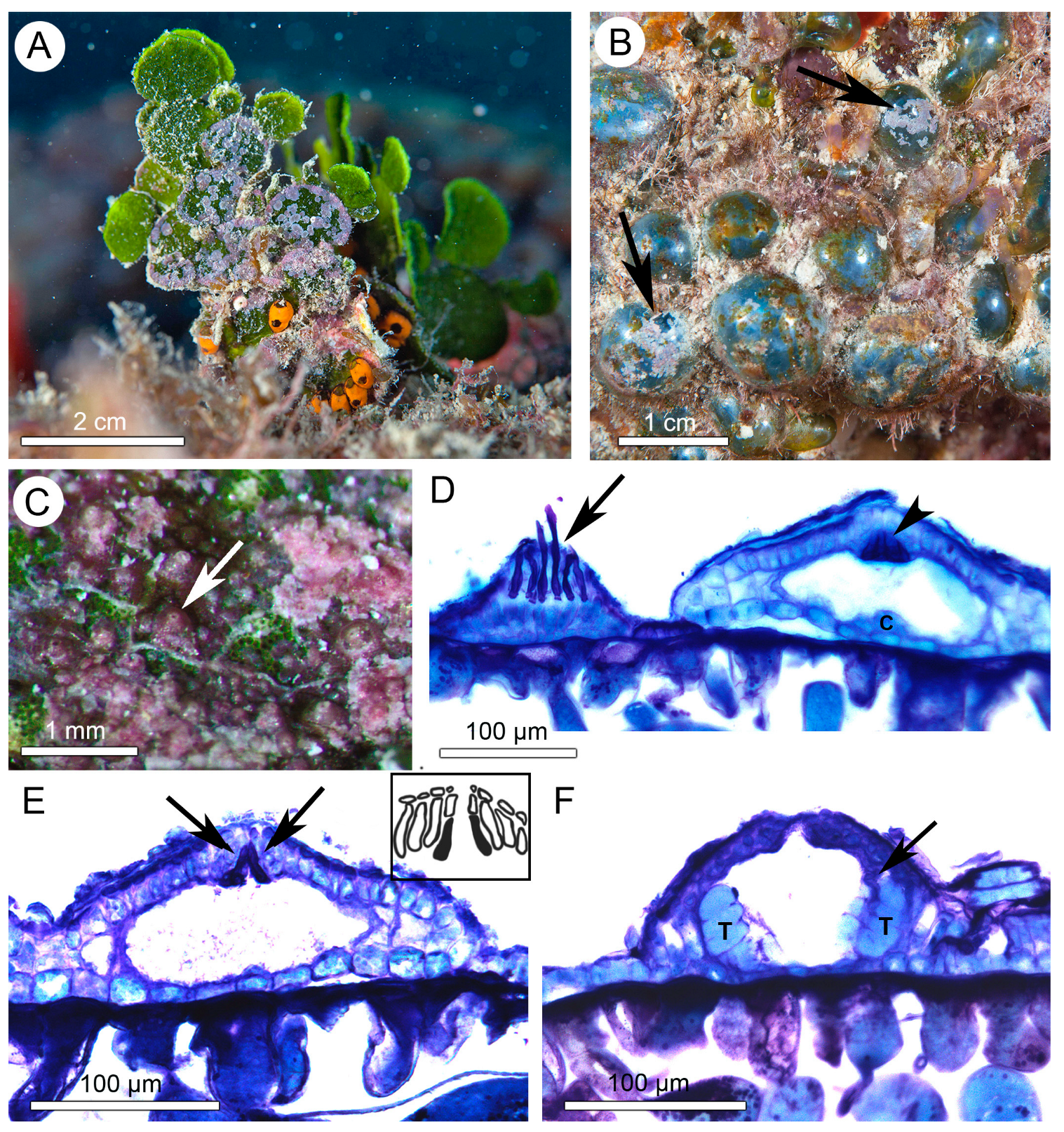
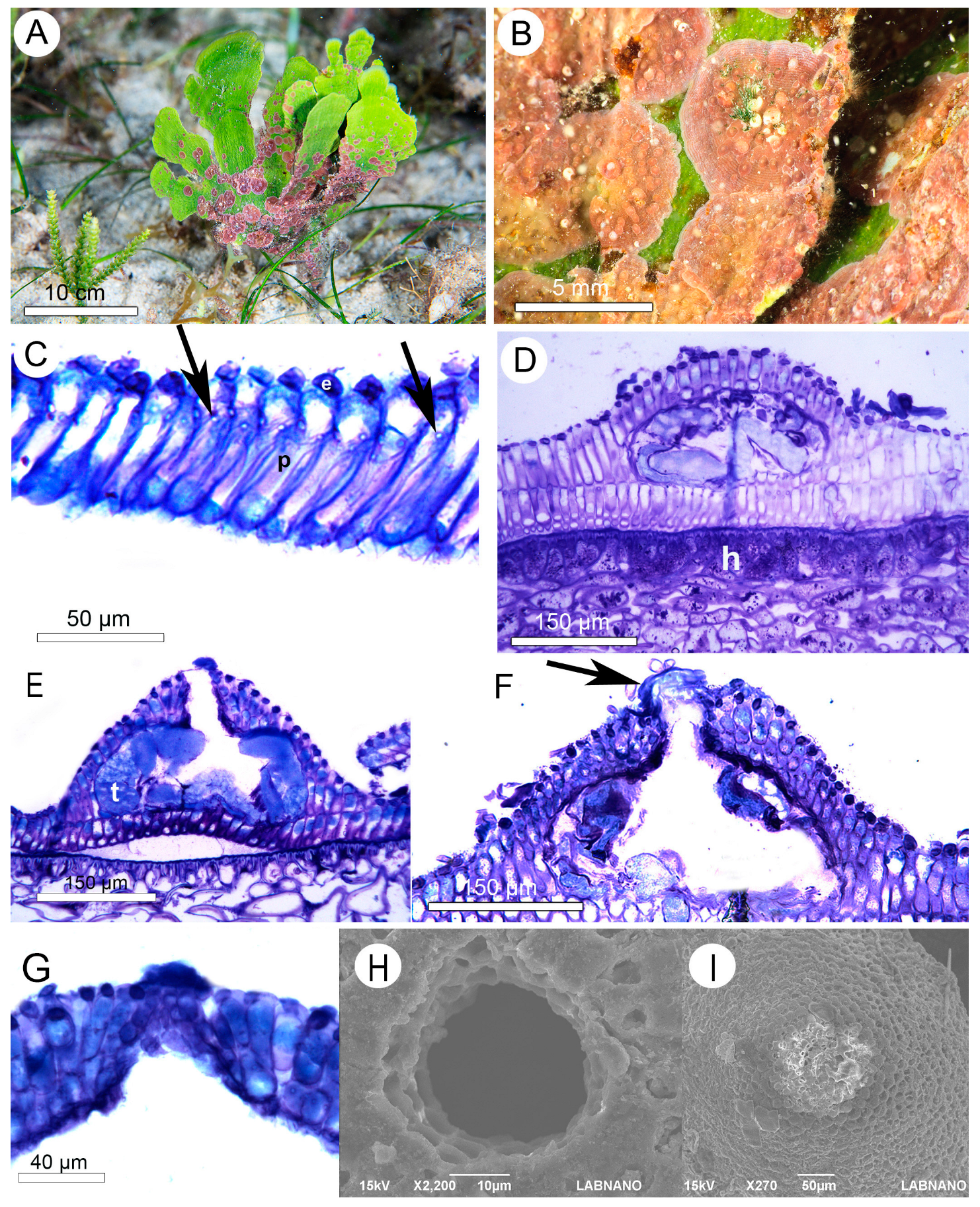
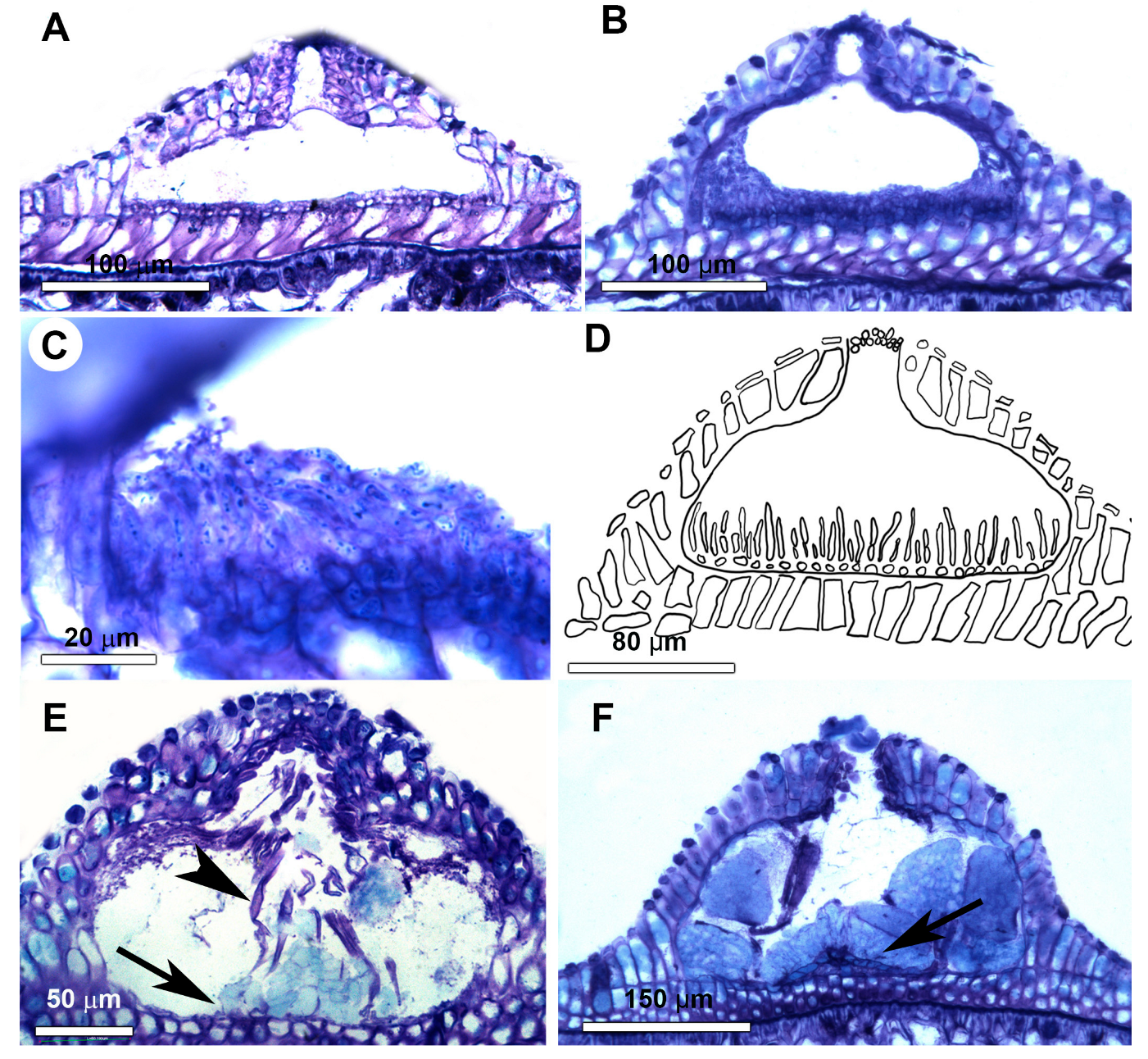
3.1. Morfoanatomical Results

Two species were identified and are described below. Additional illustrations are available in the Supplementary Material.
Hydrolithon farinosum (J.V. Lamouroux) Penrose & Y.M. Chamberlain 1993: 295.
Basionym:Melobesia farinosa Lamouroux 1816: 315.
Lectotype: CN unnumbered (Mediterranean, unspecified locality, Lamouroux, epiphytic on Sargassum linifolium).
Specimens examined:
Brazil, Bahia State, Abrolhos Archipelago, Porto Norte (17°57′42′′ S, 38°41′47′′ W), at 3–5 m depth, with multiple specimens on Halimeda (RG Bahia, 03.iii.2016, RB 784872).
Observations:
Thalli attached ventrally by cell adhesion on a variety of algal hosts, including Caulerpa, Halimeda, Sargassum, and Valonia (Figure 1A,B). Colour of living thalli whitish or pink to dark red (Figure 1A–C) Growth-form encrusting with a flour-like, powdery covering appearance (Figure 1A–C). Thallus organization dorsiventral, vegetative thallus composed of a single layer of ventral filaments with cells measuring 5–15 µm in diameter and 10–20 µm high, and a single layer of flattened or rounded epithallial cells. Ventral filaments connected laterally by cell fusions. Secondary pit connections and trichocytes not found.
Tetrasporangial conceptacles protruding above surrounding thallus surface (Figure 1D–F). Conceptacle chambers measuring 130–180 µm in diameter and 50–90 µm high, and they usually contain two peripheral tetrasporangia (18–30 µm in diameter and 45–55 µm high) with zonately-divided tetraspores (Figure 1F). Central columella either present (Figure 1D) or absent (Figure 1E,F) within conceptacle chambers. Conceptacle floors were observed right above the surface of the surrounding vegetative thallus (Figure 1D–F). Roofs 2–3 cells thick (including epithallial cells) formed by filaments surrounding and interspersed within the fertile area (Figure 1F). Pore canals lined by a ring of conspicuous and enlarged cells, not protruding laterally into the canal, and oriented nearly perpendicularly to the roof surface (Figure 1D,E). Gametangial and carposporangial thalli not found.
Lithophyllum epiphyticum Lyra, Bahia & A.S.Harv. sp. nov.
Holotype: RB 784870 collected 03 March 2016, leg RG Bahia & FC Moraes, psbA—GenBank OP186068.
Isotype: RB 784871, collected 03 March 2016, leg RG Bahia & FC Moraes.
Type Locality: Porto Norte, Abrolhos Archipelago, Bahia State, Brazil (17°57′42″ S, 38°41′37″ W), subtidal (3–5 m deep), epiphytic on Udotea flabellum (J. Ellis & Solander) M. Howe 1904: 94.
Etymology: ‘epiphyticum’ (from Latin, meaning epiphyte), referring to the epiphytic habit of the species.
Additional specimens examined: Brazil, Bahia State, Abrolhos Archipelago, Porto Norte (17°57′42″ S, 38°41′37″ W), at 5 m depth, specimens on Udotea flabellum (J. Ellis & Solander) M. Howe 1904: 94 (FC Moraes, 08.v.2016, RB 784871).
Distribution: Northeast Brazil, Bahia State (present study).
Observations:
Thalli attached ventrally by cell adhesion forming disk-like crusts, mostly on Udotea flabellum (Figure 2A), with growth-form encrusting, colour whitish to pink (Figure 2A,B). Thallus organization dorsiventral, construction dimerous (Figure 2C). Vegetative thallus composed of a single basal layer of sinuate palisade cells measuring 10–15 µm in diameter and 55–90 µm high and a single layer of flattened or rounded epithallial cells (Figure 2C). Basal filament connected laterally by secondary pit connections (Figure 2C). Cell fusions and trichocytes not found.
Tetrasporangial conceptacles viewed protruding above the surrounding surface of the thallus (Figure 2D–F). Conceptacle chambers measuring 240–300 µm in diameter and 100–140 µm high. Central columella either present or absent within the conceptacle chambers. Conceptacle chamber floor located one or two cells below the adjacent surface of the vegetative thallus (Figure 2D,E). Conceptacle roofs 2–4 cells (including epithallials) thick (Figure 2D,E). Cells lining the pore canals protrude laterally (Figure 2E,F) found either completely occluding the canal pore (Figure 2F,G,I) and unobstructed (Figure 2E,H). Pore measures are 4–7 µm in diameter and 6–10 µm high. Tetrasporangia (35–60 µm in diameter and 80–100 µm high) with four zonately divided tetraspores (Figure 2E).
Gametangial thalli are apparently monoecious, as male and carpogonial conceptacles seen forming the same crust. Whether these corresponded to only one individual or two fused ones, however, is uncertain. Male conceptacles protruding above the surrounding surface of the thallus; chambers measuring 135–285 µm in diameter and 45–75 µm high; floor located at the same level as the surrounding thallus surface; roofs composed of 2–3 layers of cells (Figure 3A,B). Spermatangial filaments unbranched and confined to the chamber floor (Figure 3B–D). Carpogonial conceptacles protruding above the surrounding surface of the thallus; floor located at same level as the surrounding thallus surface; chambers with 200–230 µm in diameter and 85–105 µm high; roofs composed of 2–3 layers of cells (Figure 3E). Carpogonial filaments arising from chamber floor, usually 2 cells long and bearing carpogonia with elongate trichogyne (Figure 3E). Carposporangial conceptacles larger than carpogonial ones, chambers with 265–340 µm in diameter and 100–165 µm high (Figure 3F). Carposporophytes comprises a central fusion cell (Figure 3E,F), and several-celled gonimoblast filaments arise peripherally with terminal orbicular carposporangia, 40–90 µm in diameter (Figure 3F).

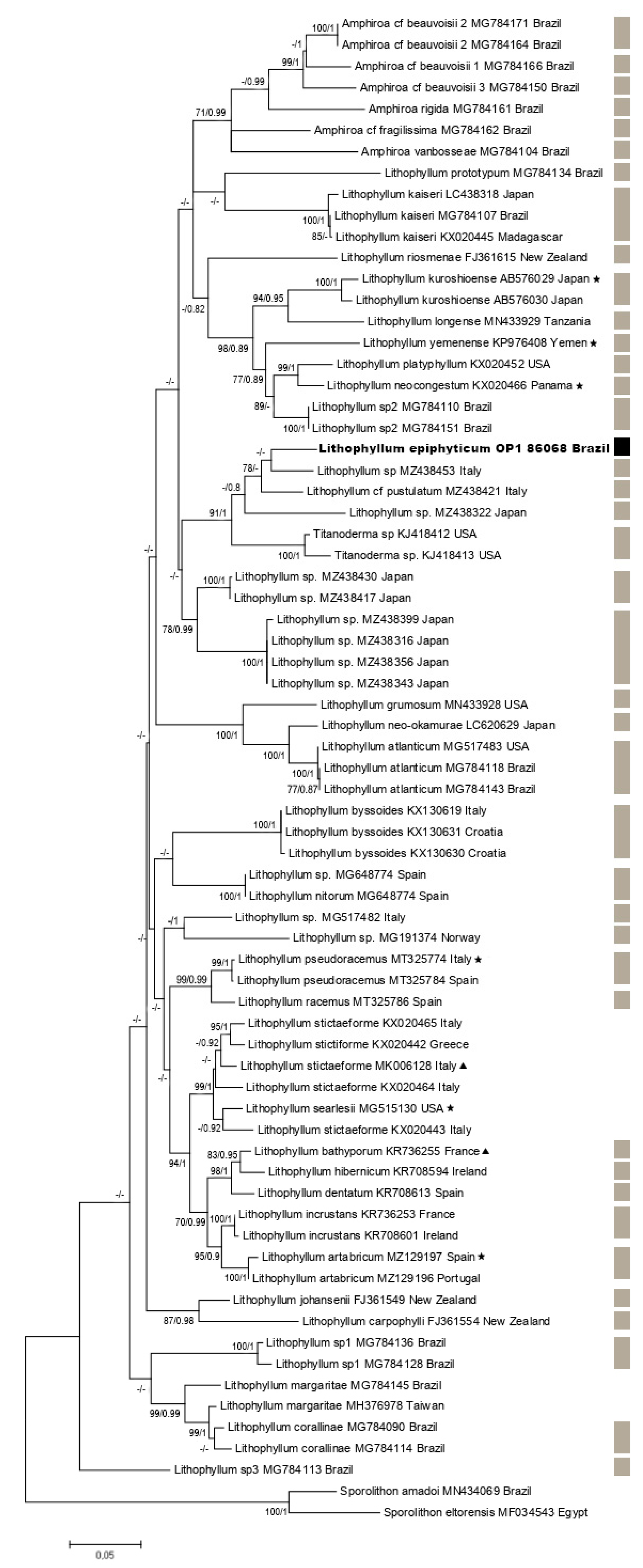
3.2. Molecular Results

DNA sequences were successfully obtained only for Lithophyllum epiphyticum. Exhaustive attempts at DNA extractions were made for H. farinosum, but all were unfruitful. The phylogenetic tree (Figure 4) shows that L. epiphyticum was resolved in a clade sister to undetermined deep water (54–80 m) Titanoderma (=Lithophyllum) specimens from the USA (Gulf of Mexico). L. epiphyticum formed a clade with three different species, two of which are not yet described in the literature, including the closest one, Lithophyllum sp. (MZ438453) from Italy [10], which diverges 5.3%. The second sequence in the clade is L. cf. pustutatum with 5.7%, and the most distant one is Lithophyllum. sp. (MZ438322) from Japan [10] with 8.7% of divergence.

4. Discussion

4.1. Ecological and Taxonomical Aspects

The epiphytic coralline algae identified in the present study were collected from large meadows of seagrass Halodule wrightii Ascherson intermixed with macroalgal communities at the Abrolhos Archipelago (see the description of these communities in [24]). Calcareous epibionts, such as coralline algae, have been demonstrated to be of paramount importance in the carbon cycle of seagrass ecosystems, as they are major contributors to CO2 fluxes through their high CaCO3 production and dissolution [35]. In addition, they make a significant contribution to calcareous sediment accumulation over geological timescales [36,37]. In respect to their ecological importance, calcareous epibionts may play an indirect role on host growth and proliferation once they reduce host photosynthetic capacity, increase the chance of host frond loss (breakage), increase the boundary layer at the host surface, and produce allelopathic substances that suppress the development of algal sporelings [38]. These corallines, however, are threatened by the global environmental changes that are expected in this century. Results obtained by [39] suggest that CaCO3 production by epiphytic coralline algae may decrease by more than 50% by the year 2100 due to anthropogenic ocean acidification. Specifically in Brazil, these algae are in extreme danger due to acidification, which has a higher % Mg substitution in the calcite crystals lattice that makes them more prone to dissolution in the near future [40].
Prior to this study, the published knowledge of Hydrolithon farinosum in Brazil was limited to isolated records without anatomical evidence (e.g., [41], as Fosliella farinosa). The subfamilial and generic position of H. farinosum remain unclear as long as no DNA sequences are available for this taxon. DNA sequencing of thin laminar thalli similar to our epiphytic species is challenging, as it is difficult to avoid DNA contamination from the algal host underneath. Therefore, the ascription of this species into Hydrolithoideae, with its single genus Hydrolithon, was based on morpho-anatomical features. According to [1], Hydrolithoideae is circumscribed by the following combination of features: (1) an outline of cell filaments entirely lost in large portions of the thallus due to pervasive and extensive cell fusions, giving it a distinctive and unique appearance in cross-section; (2) its primarily dimerous thallus construction; (3) its tetra/bisporangial conceptacle roof, formed by filaments surrounding and interspersed among tetrasporangial initials; and (4) its pore canals of uniporate tetra/bisporangial conceptacles, which are lined by a ring of elongate cells that do not protrude into the canal and are oriented more or less perpendicularly to the roof surface. All of these features were observed in H. farinosum from Abrolhos Archipelago, with exception of the first, which can only be observed in species with a thicker thallus [1].
Hydrolithon farinosum from the Abrolhos Archipelago closely matched the lectotype description provided by [42], with two notable exceptions: in the majority of cases, only two sporangia per chamber, and the absence of trichocytes. For the type material [42], a range of 3–8 sporangia is typically observed per chamber, however, the number of sporangia per chamber seen in the Australian specimens are similar to observed here. This feature is still somewhat variable, depending on the moment of life of the individual. As pointed out by [42,43], temperature and light may represent key environmental factors influencing the production or lack of trichocytes in coralline algae individuals, making both characteristics unreliable for taxa diagnosis. For this reason, we have decided to maintain the present determination as H. farinosum until molecular data are available to ensure a definitive identification.
In addition to H. farinosum, Hydrolithon boergesenii (Foslie) Foslie (1909: 56) is the only other species from the subfamily Hydrolithoideae that has been recorded in Brazil [17]. Hydrolithon farinosum can be distinguished from this species mainly by the presence of two features: (1) thalli that is exclusively epiphytic, and (2) vegetative thalli that is composed of only two cells. Taking into account these aspects, our specimen also resembles the alga Porolithon improcerum whose generic position is still unresolved [1]. P. improcerum also possess two layered thalli that can be epiphytic [44,45], but it is composed of several applanate branches that overgrowth one another and display trichocytes in large tightly packed horizontal fields [44,45], which have not been observed in the present samples identified as H. farinosum.
Lithophyllum epiphyticum Lyra, Bahia & A. S. Harv. sp. nov. is herein described as a new species for the Brazilian coast based on molecular data and phylogenetic analyses. Regarding the existent literature for this genus, the intraspecific divergence for Lithophyllum is considered lower than 2% for the psbA marker [46,47]. Here, the interspecific divergence with the most similar sequence, Lithophyllum sp. 9 [10], is 5.3%, and is significantly distinct.
L. epiphyticum resembles Lithophyllum chamberlainianum [48,49,50] in almost every morphoanatomical aspect but lacks an important diagnostic characteristic: completely occluded conceptacle pores by a cluster of overlapping tubular cells that usually project above the surrounding roof filaments. Due to the absence of molecular information regarding L. chamberlainianum, owing to the difficulty to obtain DNA sequences of type material being stored in formaldehyde, we can only confirm if they are different species when DNA data from topotypes become available.
Lithophyllum epiphyticum can be distinguished from the other five Lithophyllum species confirmed to occur in Brazil based on morpho-anatomical and molecular data [3], but mainly by the following combination of features: (1) an epiphytic habitat, and (2) a thin mature thallus (<10 layers of cells) (Table 1). [3] reported the occurrence of other four cryptic, as yet unnamed, Lithophyllum species from Brazil based on molecular data, but none correspond to epiphytic algae.
Based on morpho-anatomical data presented in Table 1, L. epiphyticum most closely resembles L. prototypum (Foslie) Foslie (1905:129) in Brazil. Both species exhibit a vegetative thallus composed only of palisade basal cells covered by a layer of epithallial cells in addition to other shared features related to tetrasporangial conceptacles. However, L. prototypum is commonly found in imbricate and numerous applanate branches with swirled margins in surface view [17,48], which have not been observed in L. epiphyticum from Abrolhos Archipelago, Brazil. In addition, the position of L. epiphyticum in psbA phylogram was in a distinct clade from L. prototypum.

4.2. Biogeographic Aspects

Hydrolithon farinosum is a cosmopolitan species (pending molecular confirmation) found on various seaweeds and seagrasses in tropical and temperate regions of the Pacific, Atlantic, and Indian Oceans [54]. Some of the common hosts of H. farinosum, such as Sargassum and Zostera [55], could function as dispersal vectors during algal rafting. The broad geographical distribution of H. farinosum, if confirmed through DNA analyses, could be related to rafting opportunities provided by its hosts, as observed for some invertebrates [56].
Ref. [57] demonstrated that debris containing benthic marine algae from the Great Tohoku Earthquake and Tsunami of March 2011 in Japan arrived on the coasts of Oregon and Washington, USA in June 2012, proving that dispersion over long distances can occur in a short period of time. In particular, smaller species may disperse more easily, and they might be more widespread globally. This feature was investigated by [58] in their search for traits that might predict introductions of species, concluding that size was influential for some taxonomic groups. Considering that coralline algae have been reported as having negative buoyancy and non-motile spores, which indicates short dispersal distances per generation [59], transoceanic dispersion through spores is unlikely.

4.3. Perspectives Biogeography x Taxonomy

Recent DNA sequence data is forcing taxonomists to question the practice of placing into synonymy specimens of coralline red algae that are widely geographically separated [60]. For example, [61] demonstrated that specimens labelled Mesophyllum erubescens (Foslie) Me.Lemoine (1928:252) based on morpho-anatomical characters in the Pacific Ocean are not the same molecular species as those reported from the Western Atlantic Ocean (including type locality), and it has been hypothesized that numerous cryptic species are apparently posing under that one name. On the other hand, there are increasing instances where morpho-anatomically related specimens from different ocean basins do belong to the same species. For example, DNA sequences of specimens of Hydrolithon boergesenii from the Caribbean and the Indo-Pacific (as H. reinboldii) could not be delimited by any single marker or combination of markers in the encompassing phylogenetic molecular analysis of the Corallinaceae provided by [1]. Similarly, [62] found identical rbcL and psbA sequences of Lithophyllum kaiseri (Heydrich) Heydrich (1897:412) from the Red Sea, the Caribbean, and Indo-West Pacific, while [17] also confirmed its occurrence through psbA sequence data in the Abrolhos Bank, Brazil.

Supplementary Materials

The following supporting information can be downloaded at: https://www.mdpi.com/article/10.3390/d15091013/s1. Figure S1: Illustrative diagrams depicting features of H. farinosum and L. epiphyticum, providing a more comprehensive visual representation of both species. (A,B) H. farinosum. (C,D) L. epiphyticum. (A) Tetrasporangial conceptacle with vestigial filaments (black) resulting in enlarged, domed cells lining the base of the pore canal. Note that the pore-canal filaments are oriented more-or-less vertically, and do not project into the pore. (B) Tetrasporangial conceptacle showing roof structure and two peripherally tetrasporangia with zonately divided tetraspores. (C) Vegetative thallus showing sinuate palisade cells with secondary pit connections. (D) Tetrasporangial conceptacle showing roof structure, with pore entirely open, two peripherally tetrasporangia with zonately divided tetraspores and a central columella (black cell).

Author Contributions

Conceptualization, M.B.L., R.G.B., M.B.J. and A.S.H.; methodology, M.B.L., R.G.B., M.B.J., R.T.C., F.C.M. and F.S.; software, M.B.L., M.B.J., R.T.C. and F.S.; validation, R.G.B., A.S.H., R.C.P. and L.T.S.; formal analysis, M.B.L., R.G.B., M.B.J., F.S., R.T.C. and A.S.H.; investigation, M.B.L., R.G.B., F.C.M., A.S.H. and M.B.J.; data curation, M.B.L., R.G.B. and M.B.J.; writing—original draft preparation, M.B.L., R.G.B. and A.S.H.; writing—review and editing, M.B.L., R.G.B. and R.C.P.; visualization, M.B.L., R.G.B. and F.C.M.; supervision, R.C.P. and L.T.S.; project administration, L.T.S.; funding acquisition, L.T.S. All authors have read and agreed to the published version of the manuscript.

Funding

This research received no external funding.

Institutional Review Board Statement

Not applicable.

Data Availability Statement

The authors can provide the data if needed.

Acknowledgments

M.B.L. acknowledges Coordenação de Aperfeiçoamento de Pessoal de Nível Superior (CAPES) for Master’s scholarship. Instituto Chico Mendes de Conservação da Biodiversidade (ICMBio) is acknowledged for research permits. All authors are much thankful to Áthila Bertoncini for providing the underwater photos used in this paper and to the Multiuser Laboratory of Nanoscience and Nanotechnology (LABNANO/CBPF) for enabling the use of SEM. RCP thanks Conselho Nacional de Desenvolvimento Científico e Tecnológico (CNPq) for Research Productivity fellowship. RCP and LTS thank FAPERJ for research productivity fellowships CNE and JCNE, respectively.

Conflicts of Interest

The authors declare no conflict of interest.

References

  1. Rosler, A.; Perfectti, F.; Peña, V.; Braga, J.C. Phylogenetic relationships of Corallinaceae (Corallinales, Rhodophyta): Taxonomic implications for reef-building corallines. J. Phycol. 2016, 52, 412–431. [Google Scholar] [CrossRef] [PubMed]
  2. Richards, J.L.; Sauvage, T.; Schmidt, W.E.; Frederiq, S.; Hughey, J.R.; Gabrielson, P.W. The coralline genera Sporolithon and Heydrichia clarified by sequencing type material of their generitypes and other species. J. Phycol. 2017, 53, 1044–1059. [Google Scholar] [CrossRef]
  3. Torrano-Silva, B.N.; Vieira, B.R.; Riosmena-Rodríguez, R.; Oliveira, M.C. Guidelines for DNA barcoding of coralline algae, focusing on Lithophylloideae (Corallinales) from Brazil. Bot. Mar. 2018, 61, 127–140. [Google Scholar] [CrossRef]
  4. Peña, V.; Torres, T.R.D.G. Lithophyllum artabricum V. Peña, sp. Nov.(Corallinales, Rhodophyta): A cryptic species in the Atlantic Iberian Peninsula hitherto assigned to Lithophyllum stictiforme (Areschoug) Hauck. Cryptogam. Algol. 2021, 42, 153–172. [Google Scholar] [CrossRef]
  5. Nelson, W.A. Calcified macroalgae—Critical to coastal ecosystems and vulnerable to change: A review. Mar. Freshw. Res. 2009, 60, 787–801. [Google Scholar] [CrossRef]
  6. Diaz-Pulido, G.; Anthony, K.R.N.; Kline, D.I.; Dove, S.; Hoegh-Guldberg, O. Interactions between ocean acidification and warming on the mortality and dissolution of coralline algae. J. Phycol. 2012, 48, 32–39. [Google Scholar] [CrossRef]
  7. Kamenos, N.A.; Burdett, H.L.; Aloisio, E.; Findlay, H.S.; Martin, S.; Longbone, C.; Dunn, J.; Widdicombe, S.; Calosi, P. Coralline algal structure is more sensitive to rate, rather than the magnitude, of ocean acidification. Glob. Chang. Biol. 2013, 19, 3621–3628. [Google Scholar] [CrossRef] [PubMed]
  8. Noisette, F.; Duong, G.; Six, C.; Davoult, D.; Martin, S. Effects of elevated pCO2 on the metabolism of a temperate rhodolith Lithothamnion corallioides grown under different temperatures. J. Phycol. 2013, 49, 746–757. [Google Scholar] [CrossRef] [PubMed]
  9. Martin, S.; Hall-Spencer, J.M. Effects of Ocean Warming and Acidification on Rhodolith/Maërl Beds. In Rhodolith/Maerl Beds: A Global Perspective; Riosmena Rodríguez, R., Nelson, W., Aguirre, J., Eds.; Springer International Publishing: Gewerbestrasse, Austria, 2017; Volume 15, pp. 55–85. [Google Scholar] [CrossRef]
  10. Peña, V.; Harvey, B.P.; Agostini, S.; Porzio, L.; Milazzo, M.; Horta, P.; Le Gall, L.; Hall-Spencer, J.M. Major loss of coralline algal diversity in response to ocean acidification. Glob. Chang. Biol. 2021, 27, 4785–4798. [Google Scholar] [CrossRef]
  11. McCoy, S.J.; Kamenos, N.A. Coralline algae (Rhodophyta) in a changing world: Integrating ecological, physiological, and geochemical responses to global change. J. Phycol. 2015, 51, 6–24. [Google Scholar] [CrossRef]
  12. Coletti, G.; Basso, D.; Corselli, C. Coralline algae as depth indicators in the Sommières Basin (early Miocene, Southern France). Geobios 2018, 51, 15–30. [Google Scholar] [CrossRef]
  13. Žuljević1, A.; Kaleb, S.; Peña, V.; Despalatović, M.; Cvitković, I.; De Clerck, O.; Le Gall, L.; Falace, A.; Vita, F.; Braga, J.C.; et al. First freshwater coralline alga and the role of local features in a major biome transition. Sci. Rep. 2016, 6, 19642. [Google Scholar] [CrossRef]
  14. Harvey, A.S.; Woelkerling, W.J.; Farr, T.J.; Neill, K.F.; Nelson, W.A. Coralline algae of central New Zealand: An identification guide to common ‘crustose’ species. NIWA Inf. Ser. 2005, 57, 1–145. [Google Scholar]
  15. Brasileiro, P.S.; Pereira-Filho, G.H.; Bahia, R.G.; Abrantes, D.P.; Guimarães, S.M.P.B.; Moura, R.L.; Francini-Filho, R.B.; Bastos, A.C.; Amado-Filho, G.M. Macroalgal composition and community structure of the largest rhodolith beds in the world. Mar. Biodivers. 2015, 46, 407–420. [Google Scholar] [CrossRef]
  16. Amado Filho, G.M.; Bahia, R.G.; Pereira Filho, G.H.; Longo, L.L. South Atlantic rhodolith beds: Latitudinal distribution, species composition, structure and ecosystem functions, threats and conservation status. In Rhodolith/Maerl Beds: A Global Perspective; Riosmena Rodríguez, R., Nelson, W., Aguirre, J., Eds.; Springer International Publishing: Gewerbestrasse, Austria, 2017; Volume 15, pp. 299–317. [Google Scholar] [CrossRef]
  17. Jesionek, M.B.; Bahia, R.G.; Hernández-Kantún, J.J.; Adey, W.H.; Yoneshigue-Valentin, Y.; Longo, L.L.; Amado-Filho, G.M. A taxonomic account of non-geniculate coralline algae (Corallinophycidae, Rhodophyta) from shallow reefs of the Abrolhos Bank, Brazil. Algae 2016, 31, 317–340. [Google Scholar] [CrossRef]
  18. Moura, R.L.; Secchin, N.A.; Amado-Filho, G.M.; Francini-Filho, R.B.; Freitas, M.O.; Minte-Vera, C.V.; Teixeira, J.B.; Thompson, F.L.; Dutra, G.F.; Sumida, P.Y.G.; et al. Spatial patterns of benthic megahabits and conservation planning in the Abrolhos Bank. Cont. Shelf Res. 2013, 70, 109–117. [Google Scholar] [CrossRef]
  19. Harvey, A.S.; Harvey, R.M.; Merton, E. The distribution, significance and vulnerability of Australian rhodolith beds: A review. Mar. Freshw. Res. 2016, 68, 411–428. [Google Scholar] [CrossRef]
  20. Amado-Filho, G.M.; Bahia, R.G.; Mariath, R.; Jesionek, M.B.; Moura, R.L.; Bastos, A.C.; Pereira-Filho, G.H.; Francini-Filho, R.B. Spatial and temporal dynamics of the abundance of crustose calcareous algae on the southernmost coral reefs of the western Atlantic (Abrolhos Bank, Brazil). Algae 2018, 33, 85–99. [Google Scholar] [CrossRef]
  21. Leão, L.A.S.; Bahia, R.G.; Jesionek, M.B.; Adey, W.H.; Johnson, G.; Salgado, L.T.; Pereira, R.C. Sporolithon franciscanum sp. nov. (Sporolithales, Rhodophyta), a New Rhodolith-Forming Species from Northeast Brazil. Diversity 2020, 12, 199. [Google Scholar] [CrossRef]
  22. Tâmega, F.T.; Riosmena-Rodriguez, R.; Mariath, R.; Figueiredo, M. Nongeniculate coralline red algae (Rhodophyta: Corallinales) in coral reefs from Northeastern Brazil and a description of Neogoniolithon atlanticum sp. nov. Phytotaxa 2014, 190, 277–298. [Google Scholar] [CrossRef]
  23. Figueiredo, M.D.O.; Steneck, R.S. Floristic and ecological studies of crustose coralline algae on Brazil’s Abrolhos reefs. In Proceedings of the 9th International Coral Reef Symposium 2002, Bali, Indonesia, 23–27 October 2000; Volume 1, pp. 493–498. [Google Scholar]
  24. Paula, A.F.; Figueiredo, M.A.O.; Creed, J.C. Structure of the macroalgal community associated with the seagrass Halodule wrightii Ascherson in the Abrolhos Marine National Park, Brazil. Bot. Mar. 2003, 46, 413–424. [Google Scholar] [CrossRef]
  25. Maneveldt, G.W.; Van der Merwe, E. Heydrichia cerasina sp. nov. (Sporolithales, Corallinophycidae, Rhodophyta) from the southernmost tip of Africa. Phycologia 2012, 51, 11–21. [Google Scholar] [CrossRef]
  26. Adey, W.H.; Adey, P.J. Studies on the biosystematics and ecology of the epilithic crustose Corallinaceae of the British Isles. Br. Phycol. J. 1973, 8, 343–407. [Google Scholar] [CrossRef]
  27. Chamberlain, Y.M. The genus Leptophytum (Rhodophyta, Corallinales) in the British Isles with descriptions of Leptophytum bornetii, L. elatum sp. nov., and L. laevae. Br. Phycol. J. 1990, 25, 179–199. [Google Scholar] [CrossRef]
  28. Woelkerling, W.J.; Irvine, L.M.; Harvey, A.S. Growthforms in non-geniculate coralline red algae (Corallinales, Rhodophyta). Aust. Syst. Bot. 1993, 6, 277–293. [Google Scholar] [CrossRef]
  29. Bahia, R.G.; Abrantes, D.P.; Brasileiro, P.S.; Pereira-Filho, G.H.; Amado-Filho, G.M. Rhodolith bed structure along a depth gradient on the northern coast of Bahia State, Brazil. Braz. J. Oceanogr. 2010, 58, 323–337. [Google Scholar] [CrossRef]
  30. Jesionek, M.B.; Bahia, R.G.; Lyra, M.B.; Leão, L.A.; Oliveira, M.C.; Amado-Filho, G.M. Newly discovered coralline algae in Southeast Brazil: Tectolithon fluminense gen. et sp. nov. and Crustaphytum atlanticum sp. nov.(Hapalidiales, Rhodophyta). Phycologia 2020, 59, 101–115. [Google Scholar] [CrossRef]
  31. Hall, T.A. BioEdit: A user-friendly biological sequence alignment editor and analysis program for Windows 95/98/NT. In Nucleic Acids Symposium Series; Oxford University Press: Oxford, UK, 1999; Volume 41, pp. 95–98. [Google Scholar]
  32. Katoh, K.; Rozewicki, J.; Yamada, K.D. MAFFT online service: Multiple sequence alignment, interactive sequence choice and visualization. Brief. Bioinform. 2019, 20, 1160–1166. [Google Scholar] [CrossRef]
  33. Felsenstein, J. Phylogenies and the Comparative Method. Am. Nat. 1985, 125, 1–15. [Google Scholar] [CrossRef]
  34. Ronquist, F.; Teslenko, M.; Van Der Mark, P.; Ayres, D.L.; Darling, A.; Hohna, S.; Larget, B.; Liu, L.; Suchard, M.A.; Huelsenbeck, J.P. Mrbayes 3.2: Efficient bayesian phylogenetic inference and model choice across a large model space. Syst. Biol. 2012, 61, 539–542. [Google Scholar] [CrossRef]
  35. Barrón, C.; Duarte, C.M.; Frankignoulle, M.; Borges, A.V. Organic carbon metabolism and carbonate dynamics in a Mediterranean seagrass (Posidonia oceanica), meadow. Estuaries Coasts 2006, 29, 417–426. [Google Scholar] [CrossRef]
  36. Walker, D.I.; Woelkerling, W.J. Quantative study of sediment contribution by epiphytic coralline red algae in seagrass meadows in Shark Bay, Western Australia. Mar. Ecol. Prog. Ser. 1988, 43, 71–77. [Google Scholar] [CrossRef]
  37. James, P.J.; Bone, Y. Neritic Carbonate Sediments in a Temperate Realm: Southern Australia; Springer: London, UK, 2011; 254p, ISBN 978-90-4811-9288-5. [Google Scholar]
  38. Morcom, N.F.; Woelkerling, W.J. A critical interpretation of coralline-coralline (Corallinales, Rhodophyta) and coralline-other plant interactions. Cryptogam. Algol. 2000, 21, 1–31. [Google Scholar] [CrossRef]
  39. Martin, S.; Rodolfo-Metalpa, R.; Ransome, E.; Rowley, S.; Buia, M.C.; Gattuso, J.P.; Hall-Spencer, J. Effects of naturally acidified seawater on seagrass calcareous epibionts. Biol. Lett. 2008, 4, 689–692. [Google Scholar] [CrossRef] [PubMed]
  40. Carvalho, R.T.; Rocha, G.M.; Karez, C.S.; Bahia, R.G.; Pereira, R.C.; Bastos, A.C.; Salgado, L.T. Global assessment of coralline algae mineralogy points to high vulnerability of Southwestern Atlantic reefs and rhodolith beds to ocean acidification. Sci. Rep. 2022, 12, 9589. [Google Scholar] [CrossRef]
  41. Taylor, W.R. Marine Algae of the Eastern Tropical and Subtropical Coasts of the Americas; University of Michigan Press: Ann Arbor, MI, USA, 1960; 870p. [Google Scholar]
  42. Jones, P.L.; Woelkerling, W.J. An analysis of trichocyte and spore germination attributes as taxonomic characters in the Pneophyllum–Fosliella complex (Corallinaceae, Rhodophyta). Phycologia 1984, 23, 183–194. [Google Scholar] [CrossRef]
  43. Chamberlain, Y.M. Trichocyte occurrence and phenology in four species of Pneophyllum (Rhodophyta, Corallinaceae) from the British Isles. Br. Phycol. J. 1985, 20, 375–379. [Google Scholar] [CrossRef]
  44. Harvey, A.; Phillips, L.E.; Woelkerling, W.J.; Millar, A.J.K. The Corallinaceae, subfamily Mastophoroideae (Corallinales, Rhodophyta) in south-eastern Australia. Aust. Syst. Bot. 2006, 19, 387–429. [Google Scholar] [CrossRef]
  45. Bahia, R.G. Algas Coralináceas Formadoras de Rodolitos da Plataforma Continental Tropical e Ilhas Oceânicas do Brasil: Levantamento Florístico e Taxonomia. Ph.D. Thesis, Escola Nacional de Botânica Tropical, Rio de Janeiro, Brazil, 2014; p. 221. [Google Scholar]
  46. Pezzolesi, L.; Falace, A.; Kaleb, S.; Hernandez-Kantun, J.J.; Cerrano, C.; Rindi, F. Genetic and morphological variation in an ecosystem engineer, Lithophyllum byssoides (Corallinales, Rhodophyta). J. Phycol. 2017, 53, 146–160. [Google Scholar] [CrossRef]
  47. Caragnano, A.; Rodondi, G.; Basso, D.; Peña, V.; Le Gall, L.; Rindi, F. Circumscription of Lithophyllum racemus (Corallinales, Rhodophyta) from the western Mediterranean Sea reveals the species Lithophyllum pseudoracemus sp. nov. Phycologia 2020, 59, 584–597. [Google Scholar] [CrossRef]
  48. Woelkerling, W.J.; Campbell, S.J. An account of southern Australian species of Lithophyllum (Corallinaceae, Rhodophyta). Bull. Br. Mus. (Nat. Hist.) Bot. 1992, 22, 1–107. [Google Scholar]
  49. Woelkerling, W.J. Subfamily Lithophylloideae. In The Marine Benthic Flora of Southern Australia, Part IIIB.; Gracilariales, Rhodymeniales, Corallinales and Bonnemaisoniales; Womersley, H.B.S.W., Ed.; Australian Biological Resources Study: Canberra, Australia, 1996; pp. 214–237. [Google Scholar] [CrossRef]
  50. Harvey, A.; Woelkerling, W.J.; Millar, A.J.K. The genus Lithophyllum (Lithophylloideae, Corallinaceae, Rhodophyta) in south-eastern Australia, with the description of L. riosmenae, sp. nov. Aust. Syst. Bot. 2009, 22, 296–317. [Google Scholar] [CrossRef]
  51. Crespo, T.M.; Bahia, R.G.; Maneveldt, G.W.; Amado-Filho, G.M. Floristic composition of crustose coralline algae from the St. Peter and St. Paul Archipelago, a summit of the Mid-Atlantic Ridge. Phytotaxa 2014, 190, 17. [Google Scholar] [CrossRef]
  52. Athanasiadis, A. North Aegean marine algae. III. Structure and development of the encrusting coralline Titanoderma cystoseirae (Rhodophyta, Lithophylloideae). Nord. J. Bot. 1989, 9, 435–441. [Google Scholar] [CrossRef]
  53. Irvine, L.M.; Chamberlain, Y.M. Seaweeds of the British Isles. In Volume 1 Rhodophyta Part 2B Corallinales, Hildenbrandiales; HMSO: London, UK, 1994; pp. 1–276. [Google Scholar]
  54. Guiry, M.D.; Guiry, G.M.; AlgaeBase. World-Wide Electronic Publication, National University of Ireland, Galway 2023. Available online: http://www.algaebase.org (accessed on 25 July 2023).
  55. Penrose, D.; Chamberlain, Y.M. Hydrolithon farinosum (Lamouroux) comb. nov.: Implications for generic concepts in the Mastophoroideae (Corallinaceae, Rhodophyta). Phycologia 1993, 32, 295–303. [Google Scholar] [CrossRef]
  56. Highsmith, R.C. Floating and algal rafting as potential dispersal mechanisms in brooding invertebrates. Mar. Ecol. Prog. Ser. 1985, 25, 169–179. [Google Scholar] [CrossRef]
  57. Hansen, G.I.; Hanyuda, T.; Kawai, H. Invasion threat of benthic marine algae arriving on Japanese tsunami marine debris in Oregon and Washington, USA. Phycologia 2018, 57, 641–658. [Google Scholar] [CrossRef]
  58. Nyberg, C.D.; Wallentinus, I. Can species traits be used to predict marine macroalgal introductions? Biol. Invasions 2005, 7, 265–279. [Google Scholar] [CrossRef]
  59. Santelices, B. Patterns of reproduction, dispersal and recruitment in seaweeds. Oceanogr. Mar. Biol. Annu. Rev. 1990, 28, 177–276. [Google Scholar]
  60. Maneveldt, G.W.; Van der Merwe, E.; Keats, D.W. Updated keys to the non-geniculate coralline red algae (Corallinophycidae, Rhodophyta) of South Africa. South Afr. J. Bot. 2016, 106, 158–164. [Google Scholar] [CrossRef]
  61. Sissini, M.N.; Oliveira, M.C.; Gabrielson, P.W.; Robinson, N.M.; Okolodkov, Y.B.; Riosmena-Rodríguez, R.; Horta, P.A. Mesophyllum erubescens (Corallinales, Rhodophyta)—So many species in one epithet. Phytotaxa 2014, 190, 299–319. [Google Scholar] [CrossRef]
  62. Hernandez-Kantun, J.J.; Gabrielson, P.; Hughey, J.R.; Pezzolesi, L.; Rindi, F.; Robinson, N.M.; Peña, V.; Riosmena-Rodriguez, R.; Le Gall, L.; Adey, W. Reassessment of branched Lithophyllum spp. (Corallinales, Rhodophyta) in the Caribbean Sea with global implications. Phycologia 2016, 55, 619–639. [Google Scholar] [CrossRef]
Figure 1. (AF) Vegetative and reproductive features of Hydrolithon farinosum. (A) Crusts attached on the host Halimeda. (B) Crusts attached on the host Valonia. (C) Magnified view of an encrusting specimen showing uniporate tetrasporangial conceptacles (arrow). (D) Section through a young (left) and mature (right) uniporate tetrasporangial conceptacle. Note the ring of developing elongate cells (arrow) that will line the pore canal when the conceptacle is mature. These corresponding cells (arrowhead) are smaller in a mature conceptacle and more or less perpendicularly positioned in relation to the roof surface. Note, also, the vestiges of the central columella (c) in a mature conceptacle. (E) Section through a tetrasporangial conceptacle showing the pore canal lined by elongate cells that do not protrude into the canal, and that are oriented more or less perpendicularly to the roof surface (in the upper right corner, a schematic drawing of this characteristic is depicted). Note that the chamber floor is located above the surrounding vegetative thallus surface. (F) Section through a tetrasporangial conceptacle, showing two peripherally tetrasporangia (t) with zonately-divided tetraspores. (Underwater photographs by Áthila Bertoncini).
Diversity 15 01013 g001
Figure 2. (AI) Vegetative and reproductive features of Lithophyllum epiphyticum. (A) Crusts attached on the host Udotea flabellum. (B) Magnified view of discoid encrusting specimens showing uniporate tetrasporangial conceptacles. (C) Section through the two-cell layered vegetative thallus showing sinuate palisade cells (p), rounded epithallial cells (e), and secondary pit connections (arrows) linking the basal filament. (D) Section through an immature tetrasporangial conceptacle chamber showing its floor located only one cell below the surrounding thallus surface. Note the host thallus (h) below the sectioned specimen. (E) Longitudinal section through an uniporate tetrasporangial conceptacle showing a partially covered pore canal, and a tetrasporangia with four zonately arranged tetraspores (t). (F) Tetrasporangial conceptacle showing a pore plug (arrow) blocking the canal. (G) Approximate view of a conceptacle pore completely occluded. (H,I) SEM imaging. (H) Approximate view of a tetrasporangial conceptacle with the pore entirely open. (I) Another mature conceptacle with whitish cells blocking the canal. (Underwater photograph by Áthila Bertoncini).
Diversity 15 01013 g002
Figure 3. (AF) Gametangial and carposporangial features of Lithophyllum epiphyticum. (A) Section through an empty male conceptacle with a roof composed of three layers of cells. (B) Section through a male conceptacle chamber showing numerous spermatangial filaments confined to its floor. (C) Magnified view of spermatangial filaments. (D) Diagrammatic drawing of a male conceptacle containing unbranched spermatangial filaments confined to its floor. (E) Carpogonial conceptacle containing branches in the center of the chamber. Note a basal carpogonium (arrow) that extends into a narrow upper trichogyne (arrowhead). (F) Section through a carposporangial conceptacle showing a columella formed by a cluster of fused cells (arrow).
Diversity 15 01013 g003
Figure 4. Phylogenetic tree inferred from ML and BI analyses of the psbA dataset. Bootstrap support (1000 replicates) and Bayesian posterior probabilities (PP) indicated at the nodes. Bootstrap values lower than 70% and PP values lower than 0.8 or inconsistent with the original tree are not shown. In bold, Lithophyllum epiphyticum. Type sequences are shown with a star on the side, and lectotypes with a triangle. Gray bars on the side indicate divided sequence groups used to calculate mean distance within and between groups. (*) Divided according to [3].
Diversity 15 01013 g004
Table 1. Comparison of the main features that distinguish Lithophyllum species.
L. epiphytica 1L. irvineanum 2L. chamberlainianum (Type Collection) 2L. prototypum
(Type Collection) 2
L. pustulatum 2,3,4L. cystoseirae 5L. laminariae 6
Distribution 7Abrolhos Archipelago (Brazil)South and Western AustraliaSouth PacificCaribbean, Western Atlantic, Arabian Gulf, Indo-PacificMediterranean, Indo-Pacific, Eastern and Western Atlantic, Red Sea, Arabian Gulf, Caribbean, Arctic OceanAdriatic Sea, Mediterranean, Eastern AtlanticEastern Atlantic
HabitEpiphyticEpiphyticEpiphyticEpilithic/epiphytic/free-living as rhodolithEpilithic/free-living as rhodolithEpiphyticEpiphytic
Growth formEncrustingEncrustingEncrustingEncrustingEncrusting/warty to lumpyEncrustingEncrusting
Thalli structured in several applanate branches with swirled margins in surface viewAbsentPresentAbsentPresentAbsentAbsentAbsent
Basal layer of palisade or sinuate cellsPresentPresentPresentPresentPresentPresentPresent
Mature thallus thickness (number of cell layers)<10<10<10<10>10>10>10
TrichocytesAbsentAbsentAbsentAbsentAbsentPresentAbsent
Tetra/bisporangial conceptacle chamber diameter (µm)240–300180–263145–252330–415420–465/328–556288–475/307–324312–364
Tetra/bisporangial conceptacle chamber height (µm)100–14082–12560–135100–190300–320/110–18080–172/80–124104–156
Pore canals of tetra/bisporangial conceptacles occluded by cellsAbsent/presentPresentPresentAbsentAbsentPresentAbsent
Number of cell layers in tetra/bisporangial roof filaments2–42–42–52–32–43–6ND
Position of tetra/bisporangial chamber floor below thallus surface (number of cells)11–3111–5ND6-+
Conceptacle position in the thallusSuperficialSuperficial/immersedSuperficial/immersedSuperficial/immersedSuperficial/immersedSuperficial/immersedsuperficial/immersed
ColumellaPresentPresentPresentPresentPresentPresentPresent
1 [Present study]; 2 [48]; 3 [51]; 4 [16]; 5 [52]; 6 [53]; 7 [50]; ND = Not disclosed.
Disclaimer/Publisher’s Note: The statements, opinions and data contained in all publications are solely those of the individual author(s) and contributor(s) and not of MDPI and/or the editor(s). MDPI and/or the editor(s) disclaim responsibility for any injury to people or property resulting from any ideas, methods, instructions or products referred to in the content.

