Article

A New Species of Diacyclops (Copepoda, Cyclopoida) from the D. crassicaudis (Sars, 1863) Species Group with Critical Taxonomy Remarks †

by
Aleksandr A. Novikov
1,2,*,
Dayana N. Sharafutdinova
1,2,
Tatyana Yu. Mayor
1,3 and
Elena S. Chertoprud
1
1
A.N. Severtsov Institute of Ecology and Evolution, Leninsky Prospect 33, Moscow 119071, Russia
2
Department of Zoology, Kazan Federal University, Kremlyovskaya St. 18, Kazan 420008, Russia
3
Limnological Institute, Siberian Branch of the Russian Academy of Sciences, 3 Ulan-Batorskaya St., Irkutsk 664033, Russia
*
Author to whom correspondence should be addressed.
urn:lsid:zoobank.org:act:7008FC60-E385-4D76-8645-2395CFDA6309.
Diversity 2024, 16(4), 208; https://doi.org/10.3390/d16040208
Submission received: 22 February 2024 / Revised: 17 March 2024 / Accepted: 19 March 2024 / Published: 29 March 2024

Abstract

:
A new species, Diacyclops dyabdar sp. nov. from the Diacyclops crassicaudis (Sars, 1863) species group from northern Middle Siberia, is described. This species is interesting from an ecological point of view, as it lives mainly in watercourses. It is well-distinguished from other species of the group by the presence of spinules on the first segments of the third and fourth pairs of swimming legs, details of the ornamentation on the fourth pair of legs and caudal rami. A detailed comparison of the new species and D. crassicaudis is presented. Molecular markers, including cytochrome c oxidase (COI) of mtDNA and 18S rRNA, ITS1 and ITS2 of nuclear DNA were obtained for a single female of D. dyabdar sp. nov. A morphometric analysis of species and subspecies of the D. crassicaudis group was carried out. It showed slight differences between the described subspecies and some species. On this basis, the subspecies D. crassicaudis, as well as D. iranicus Pesce & Maggi, 1982 and D. fontinalis Naidenow, 1969, are synonymized with the subspecies type. A more precise diagnosis of the D. crassicaudis group is indicated. This group now includes six species. The taxonomic position of several questionable taxa of Diacyclops Kiefer, 1927 described from Iran is discussed: D. landei Mahoon & Zia, 1985; D. bicuspidatus jurenei Najam-un-Nisa, Mahoon & Irfan Khan, 1987; D. landei richardi Parveen, Mahoon & Saleem, 1988 and D. jurenei Parveen, Mahoon & Saleem, 1988. These taxa are accepted as nomen dubium.

1. Introduction

Studies in recent years have shown that north Siberia is inhabited by a rich and specific fauna of copepod crustaceans. The species list includes both narrowly distributed taxa and Beringian species whose ranges are split between Eurasia and North America [1,2]. According to rough estimates, the fauna of this territory includes almost 150 species of Copepoda [2].
In recent years, five new for-science species of the order Harpacticoida (Copepoda) have been described from the north of Middle Siberia [3]. At the same time, little attention has been paid to the faunas of the other free-living copepod orders. Cyclopoida are no less rich in the number of new species and obscure species than Harpacticoida. The richest genera can be considered Acanthocyclops Kiefer, 1927, Eucyclops Claus, 1893 and Diacyclops Kiefer, 1927 [2,4]. With this article, we begin the analysis of Cyclopoida diversity of northern Middle Siberia with the genus Diacyclops, which is one of the largest genera of the Cyclopidae, with approximately 150 species and subspecies [5]. Within the genus, there is an extremely high level of endemism, especially within the species group D. languidoides (Lilljeborg, 1901), a large number of species of which are known from Lake Baikal [6,7], groundwater in Europe [8] and groundwater in Japan and Korea [9]. The D. crassicaudis (Sars, 1863) species group is also quite rich, in which different authors included up to 15–20 species and subspecies [10,11]. Most of the taxa of the group have a local distribution in Europe and are slightly morphologically different from each other [12,13]. For the Arctic zone of Eurasia, only one species from this group is known—D. crassicaudis [2,14]. Most of the taxa of the group have a local distribution in Europe and are slightly morphologically different from each other [12,13]. With this connection, the finding in the Lena River delta and on the Anabar Plateau of another species of this group, which is new to science, is of considerable interest.
The discovery of a new species of the crassicaudis group raised several questions. How clearly defined is the D. crassicaudis species group? Is it monophyletic? How do species and subspecies differ within the group? What characters can be used in the group taxonomy in the future? The purpose of this study was the description of a new species, D. dyabdar sp. nov., including both morphological and molecular genetic discriminators. In addition, an attempt was made to reveal the main identifying characters of species inside the D. crassicaudis group, in connection with which the composition of subspecies of this group was critically revised.

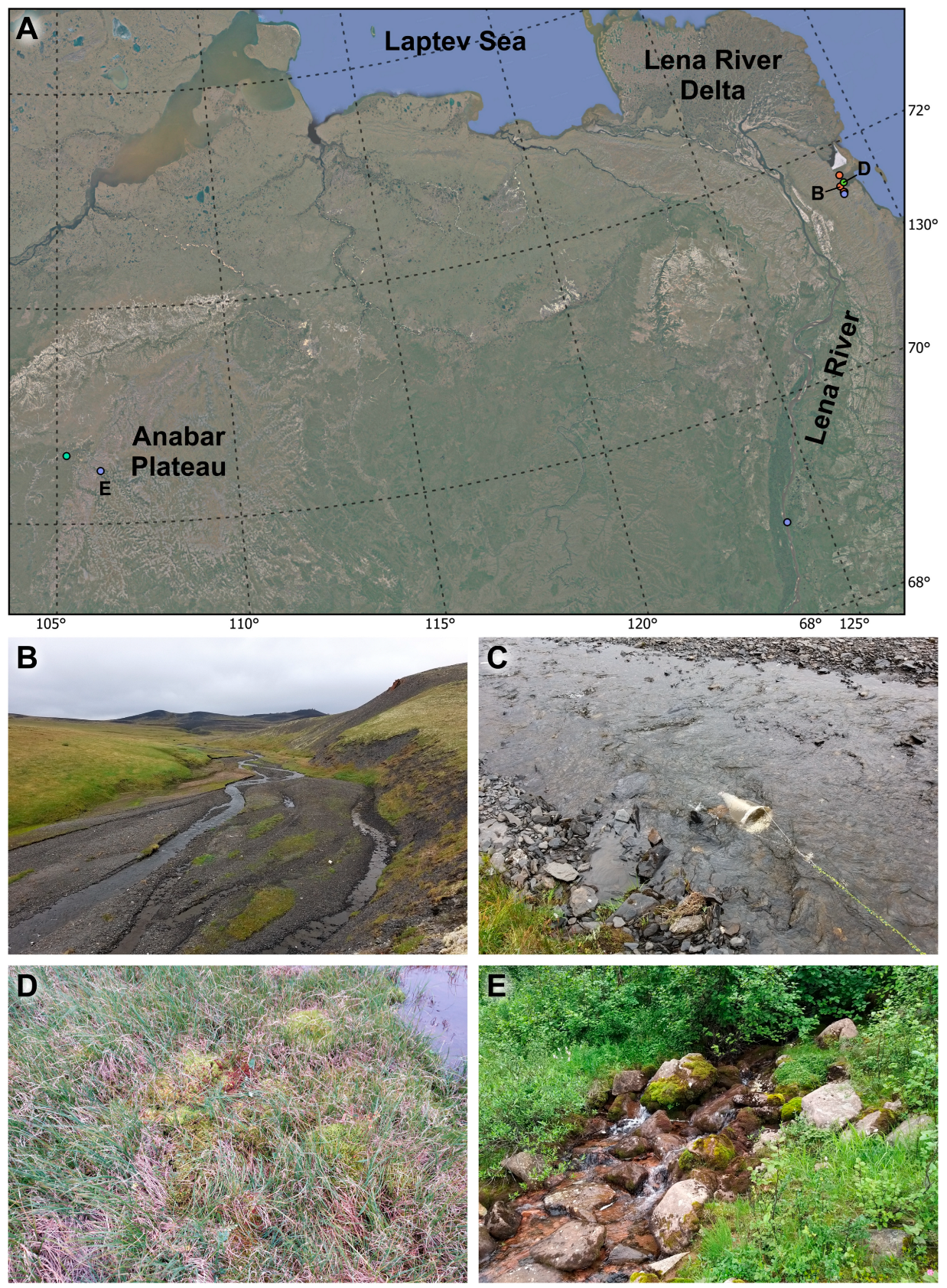
2. Materials and Methods

Studies were carried out during the summer seasons of 2019–2023 in water bodies and water sources in several parts of the Palearctic (North Yakutia, Anabar Plateau, Tatarstan Republic and Abkhazia) (Figure 1). Samples were collected using a small plankton net with a mesh size of 100 μm. Basically, for collection, a net was placed in the course of a river or stream for 30 min to collect drifting organisms (Figure 1C). Also, some of the samples were collected from small puddles and wet moss (Figure 1D). Samples from Tatarstan and Abkhazia were collected using the Karaman–Chappuis method [15,16]. To achieve this, in the immediate vicinity of the watercourse, a pit 30–70 cm deep was dug with a shovel, which was gradually filled with water. Then, using a small plankton net, water was filtered from the pit. Samples were fixed in 4% formalin or 96% ethanol.
Specimens were dissected under a stereomicroscope, with each element being placed under a separate cover slip. Rough drawings were generated on printed photographs of elements, and the final drawings were prepared using the program CorelDraw.
Congo red staining and fluorescence microscopy were used to image the habitus. To stain the specimens, they were first washed in distilled water, and the specimens were kept in Congo red (1.5 mg per 1 mL) for 24 h. Next, the crustaceans were transferred to glycerol and covered with a coverslip. For observations, we used a ZEISS Axio Imager M2 microscope with the following filter settings: excitation BP 460–490 nm, beamsplitter FT 495 nm, emission BP 500–600 nm. With this filter, Congo red glows yellow and chitin glows green. Photographs were taken using Z-stacking.
Figure 1. (A) Map of the distribution of Diacyclops dyabdar sp. nov. Points B, D and E correspond to the photographs below; (BE) Types of water bodies in which Diacyclops dyabdar sp. nov. is collected; (B) Type locality of Diacyclops dyabdar sp. nov. Nameless river on the Tiksi area (North Yakutia); (C) The process of collecting drifting organisms in type locality; (D) Wet moss on the Tiksi area; (E) Stream on the Anabar Plateau.
Diversity 16 00208 g001
For morphometric analysis, we used both individuals from our materials and figures by other authors (Table 1). In total, we used 13 relative measurements regarding P4 and caudal rami:
  • Ratio of the length of the coxal seta to the distance between the base of this seta and the end of the inner spine of the P4 basis;
  • Ratio of the length to the width of the distal segment of the P4 endopod;
  • Ratio of the length of the outer seta to the length of the distal segment of the P4 endopod;
  • Ratio of the length of the inner apical spine to the length of the outer spine of the distal segment of the P4 endopod;
  • Ratio of the length of the distal inner seta to the length of the proximal inner seta of the distal segment of the P4 endopod;
  • Ratio of the length of the inner apical spine to the length of the distal segment of the P4 endopod;
  • Ratio of the sum of the lengths of the inner setae of the distal segment of the P4 endopod to the length of the distal segment of the P4 endopod;
  • Ratio of the length to the width of caudal rami;
  • Ratio of the distance from the base of the caudal ramus to seta II to the length of the caudal ramus;
  • Ratio of the length of the outer spine (seta III) to the length of the caudal ramus;
  • Ratio of the length of the inner seta (seta VI) to the length of the outer spine (seta III);
  • Ratio of the length of the inner seta (seta VI) to the width of the caudal ramus;
  • Ratio of the length of the dorsal seta (seta VII) to the length of the caudal ramus.
To compare morphometric indices, we used two multivariate analyses in the PAST4 program: principal coordinates analysis (PCoA) and clustering with Gower’s distance [17].
Nomenclature and descriptive terminology follows Huys and Boxshall [18]. The armature formulae of swimming legs are given according to Sewell [19]. Terminology and homology of maxillary structures follow Ferrari and Ivanenko [20]. The numbering of the groups of spinules on the posterior side of the P4 coxa is given according to Einsle [21].
Abbreviations used in the text are as follows: ae = aesthetasc; P1–P6 = legs 1–6; Enp1–3 = first–third endopodal segment; and Exp1–3 = first–third exopodal segment.
All material was deposited in the Zoological Museum of Kazan Federal University.
Table 1. List of females used for morphometric analysis. The upper part of the table is individuals from our materials, the lower part is morphometry based on figures from various references. Abbreviations: d—dyabdar, c—crassicaudis, Y—Yakutia, A—Anabar Plateau, T—Tatarstan, R—individual from reference.
SpecimenSpeciesSample
d_Holotype
d_Paratype1
d_Paratype2
D. dyabdar sp. nov. type locality
d_Y1, d_Y2D. dyabdar sp. nov. North Yakutia, Tiksi area
71.529281° N, 128.770956° E
d_A1D. dyabdar sp. nov. Anabar plateau
70.635118° N, 105.266465° E
c_Y1, c_Y2D. crassicaudisNorth Yakutia, Tiksi area
71.529281° N, 128.770956° E
c_T1, c_T2, c_T3, c_T4, c_T5D. crassicaudisTatarstan Republic
55.767969° N, 48.846781° E
specimenspecies/subspeciesreference
c_c_R4D. crassicaudis s. str.[22]
c_c_R1D. crassicaudis s. str.[23]
c_c_R3D. crassicaudis s. str.[24]
c_c_R5D. crassicaudis s. str.[25]
c_c_R2D. crassicaudis s. str.[26]
c_brachycer._R1D. crassicaudis brachycercus[22]
c_cretensis_R3D. crassicaudis cretensis[27]
c_cretensis_R1, D. crassicaudis cretensis[28]
c_cretensis_R2D. crassicaudis cf. cretensis[28]
c_lagrecai_R1D. crassicaudis lagrecai[13]
c_taipehen._R1D. crassicaudis taipehensis[29]
c_trinacriae_R1D. crassicaudis trinacriae[13]
fontinalis_R1D. fontinalis[30]
iranicus_R1D. iranicus[31]
antrincola_R1D. antrincola[32]
antrincola_R3D. antrincola[33]
antrincola_R2D. antrincola[34]
c_cosana_R1D. crassicaudis var. cosana[35]
ruffoi_R1D. ruffoi[12]
skopljensis_R1D. skopljensis[36]
c_s.lat._R1D. crassicaudis s. lat. [37]
karamani_R1D. karamani[36]

3. Molecular Analysis

For molecular analysis, we used one female of D. dyabdar sp. nov., collected from Anabar Plateau (Anabar Plateau, stream flowing into the Kotuykan River; 70.492244° N, 106.210403° E; 23 July 2023), fixed in 96% ethanol and stored at −20 °C. Molecular analysis was carried out at the Instrumentation Center “Electronic Microscopy” of the Collective Instrumental Center “Ultramicroanalysis” (LIN SB RAS).Total DNA was extracted from somatic tissue using Proteinase K according to the protocol described by Mayor et al. [38]. A gene fragment of cytochrome c oxidase of mtDNA (COI), the first and second internal transcribed spacers (ITS1, ITS2) and a gene fragment of 18S rRNA of nuclear DNA were analyzed. PCR was performed using universal primers LCO-1490 and HCO-2198 to amplify the COI [39], KP2 (5′-AAAAAGCTTCCGTAGGTGAACCTGCG-3′) and 5.8S (5′-AGCTTGGTGCGTTCTTCATCGA-3′), ITS4 (5′-TCCTCCGCTTATTGATATGC-3′) and ITS5 (5′-GGAAGTAAAAGTCGTAACAAGG-3′) to amplify ITS1 and ITS2 [40,41] and 18sI (5′-AACTYAAAGGAATTGACGG-3′) and 18s329 (5′-TAATGATCCTTCCGCAGGTT-3′) to amplify 18S rRNA [42]. The PCR (10 µL) contained 5–10 ng of total DNA, 1x Encyclo PCR buffer, 3.5 mM magnesium, 0.5 µM of each primer, 0.2 mM of each dNTP and 0.5 units of Encyclo DNA polymerase. Amplification conditions consisted of 4 min at 95 °C, and then 15 s at 94 °C, 20 s at 48 °C or 57 °C (for COI and ITS1, ITS2 and 18S rRNA, respectively), 1 min at 72 °C for 35–40 cycles, and the final elongation stage for 4 min at 72 °C. The amplicons were extracted for sequencing from the 0.6% agarose gel following the previously described protocol [38]. The nucleotide sequences of the target fragments were bi-directional sequencing using the ABI PRISM BigDye Terminator v. 3.1 sequencing kit in a Nanophor 05 genetic analyzer (Sintol, Russia). Sequences were manually edited and assembled into a consensus using Unipro UGENE 49.1 [43]. The obtained consensus sequences were deposited in the GenBank. The nucleotide sequences were aligned and p-distances were calculated using the MegaX program [44]. Additional sequences of Cyclopoida were obtained from GenBank and used for phylogenetic analysis. Their accession numbers are shown on the phylogenetic trees. Substitution saturation was assessed using DAMBE [45,46]. Maximum Likelihood trees were constructed using IQ-TREE2 2.2.2.6 software [47]. The trees were visualized and edited using Interactive Tree Of Life (iTOL) version 6.8.1 (https://itol.embl.de, accessed on 9 February 2024) [48]. The Basic Local Alignment Search Tool (BLAST) and BOLD Systems were used to search for sequence similarity [48,49].

4. Results

Subclass Copepoda H. Milne Edwards, 1840
Order Cyclopoida Burmeister, 1834
Family Cyclopidae Rafinesque, 1815
Genus Diacyclops Kiefer, 1927

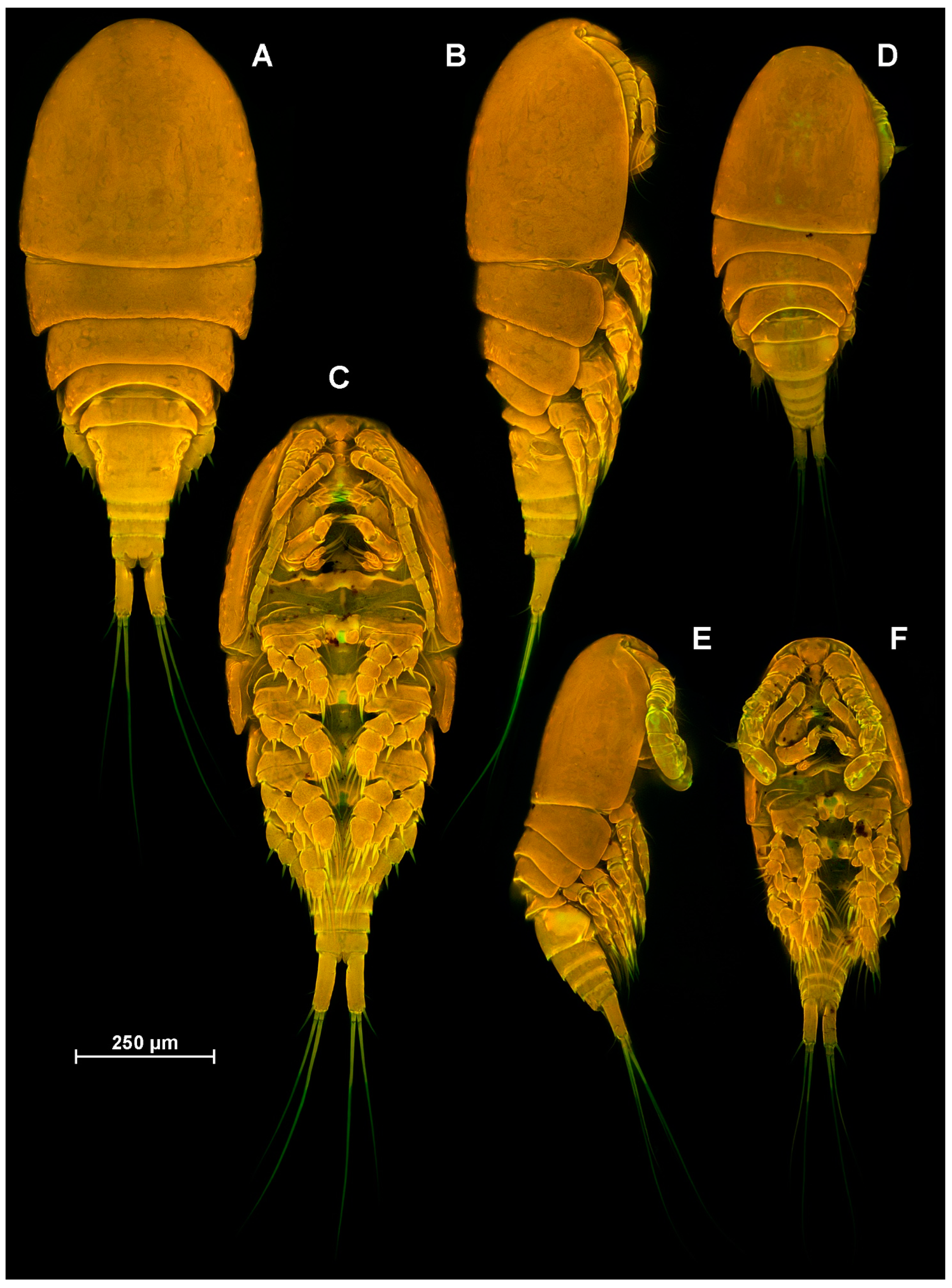
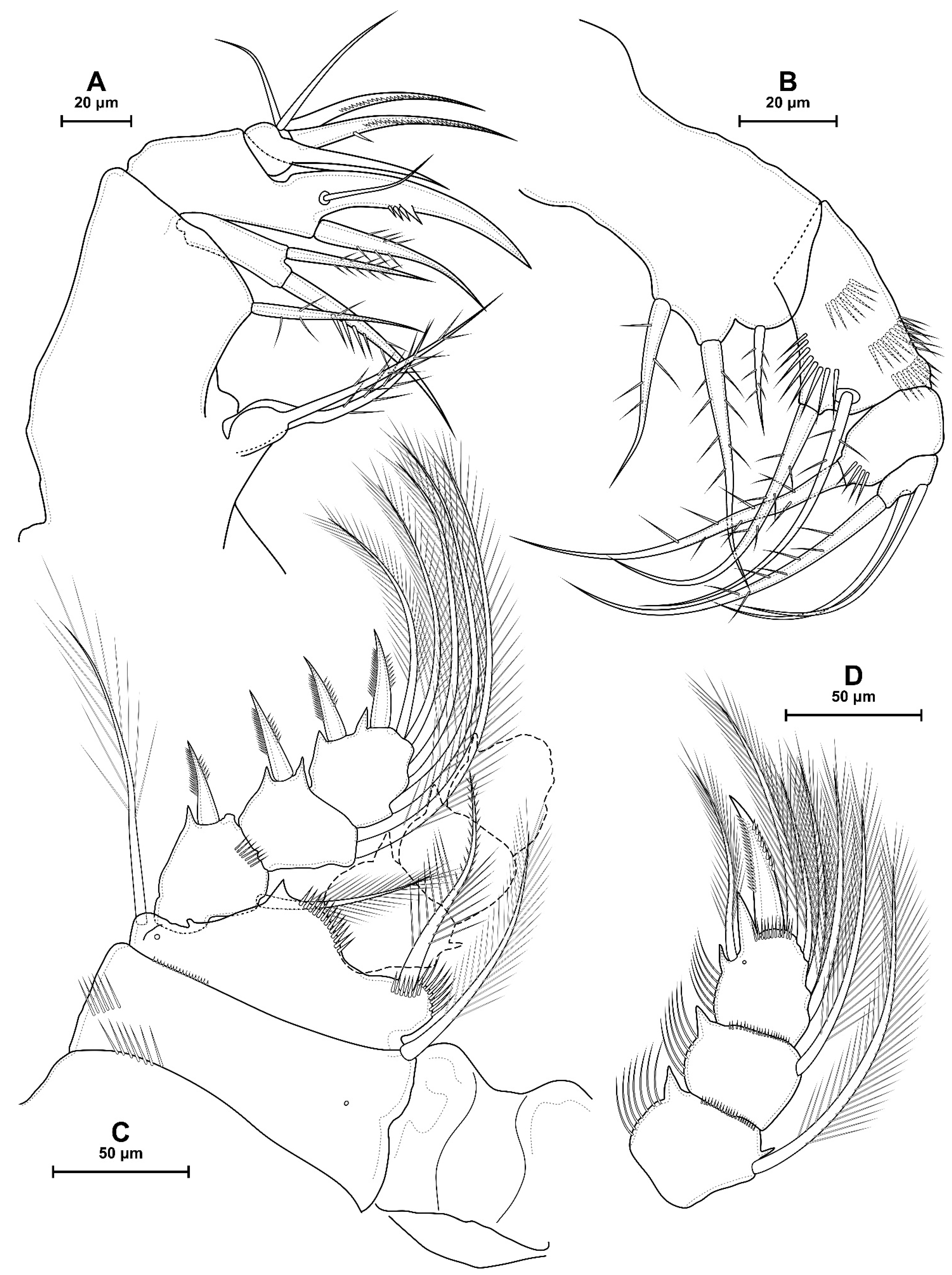
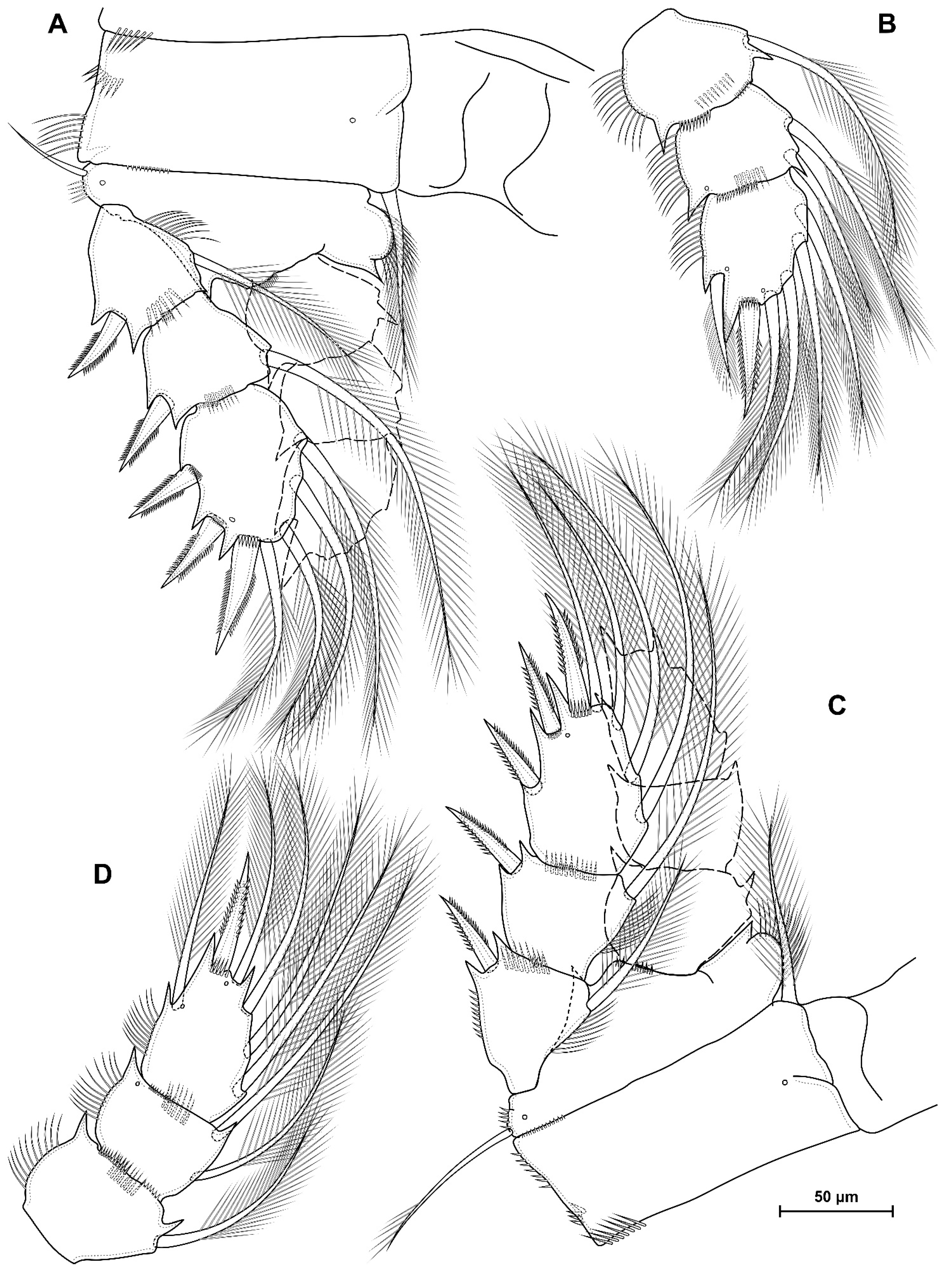
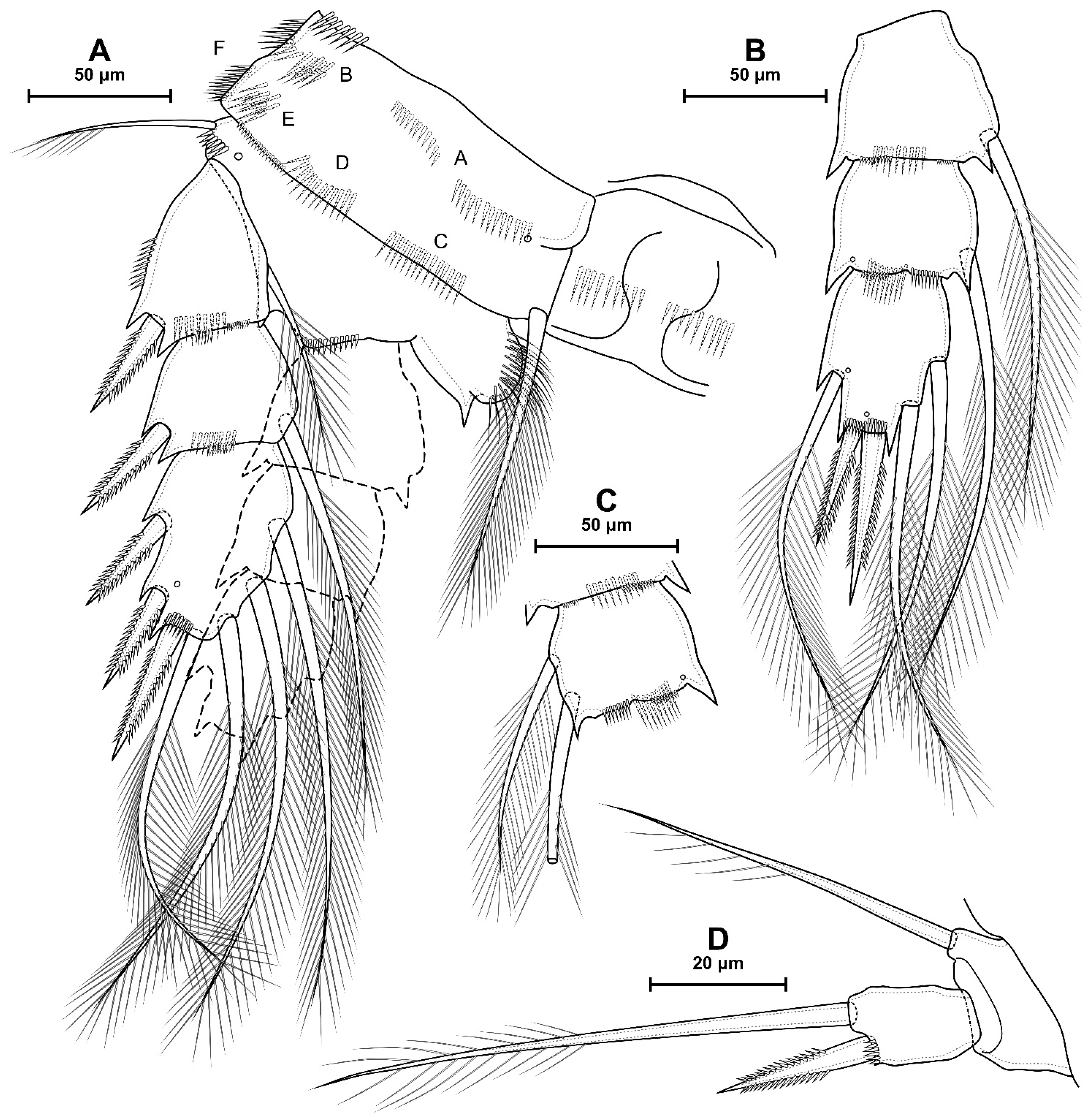
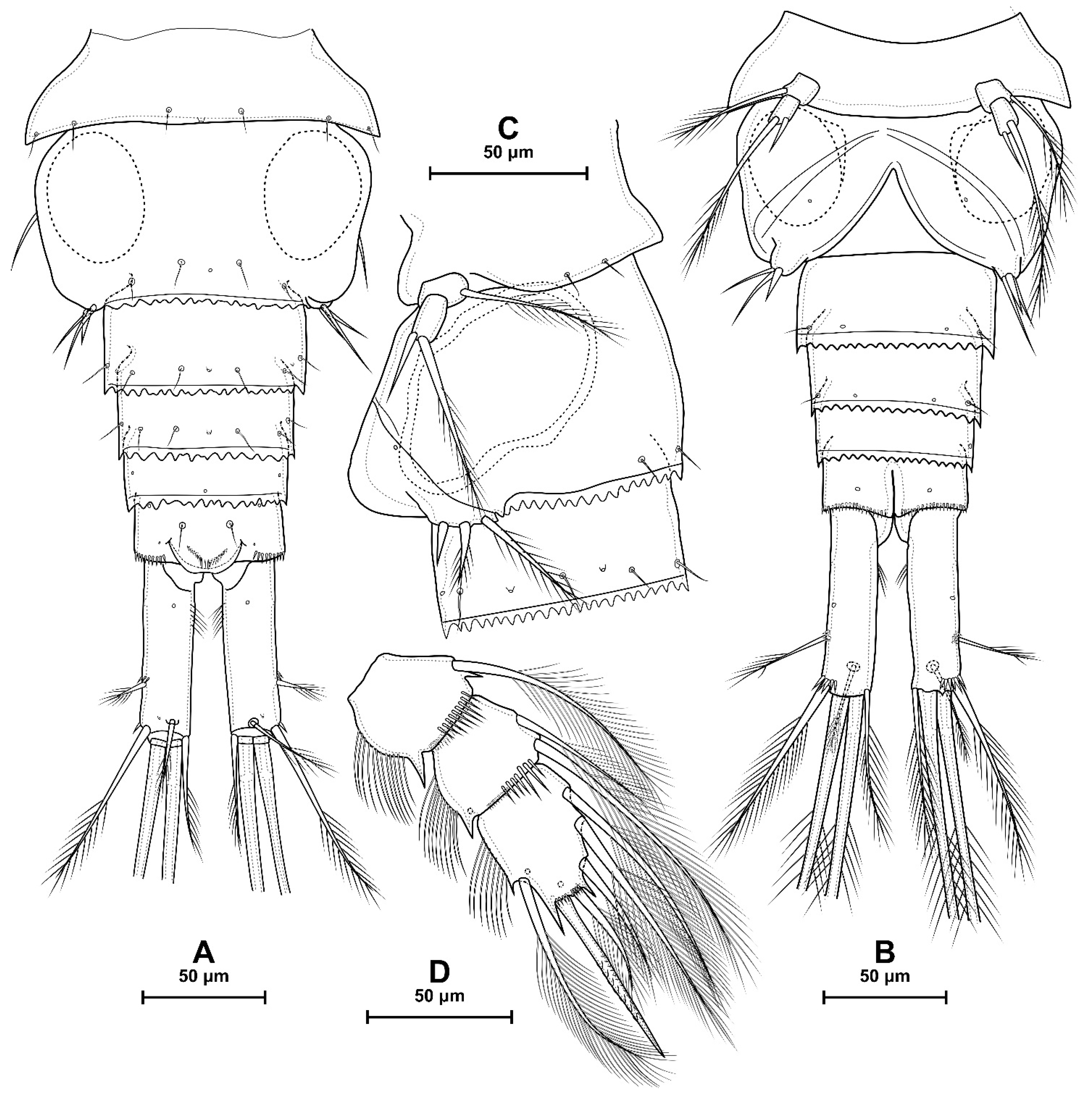
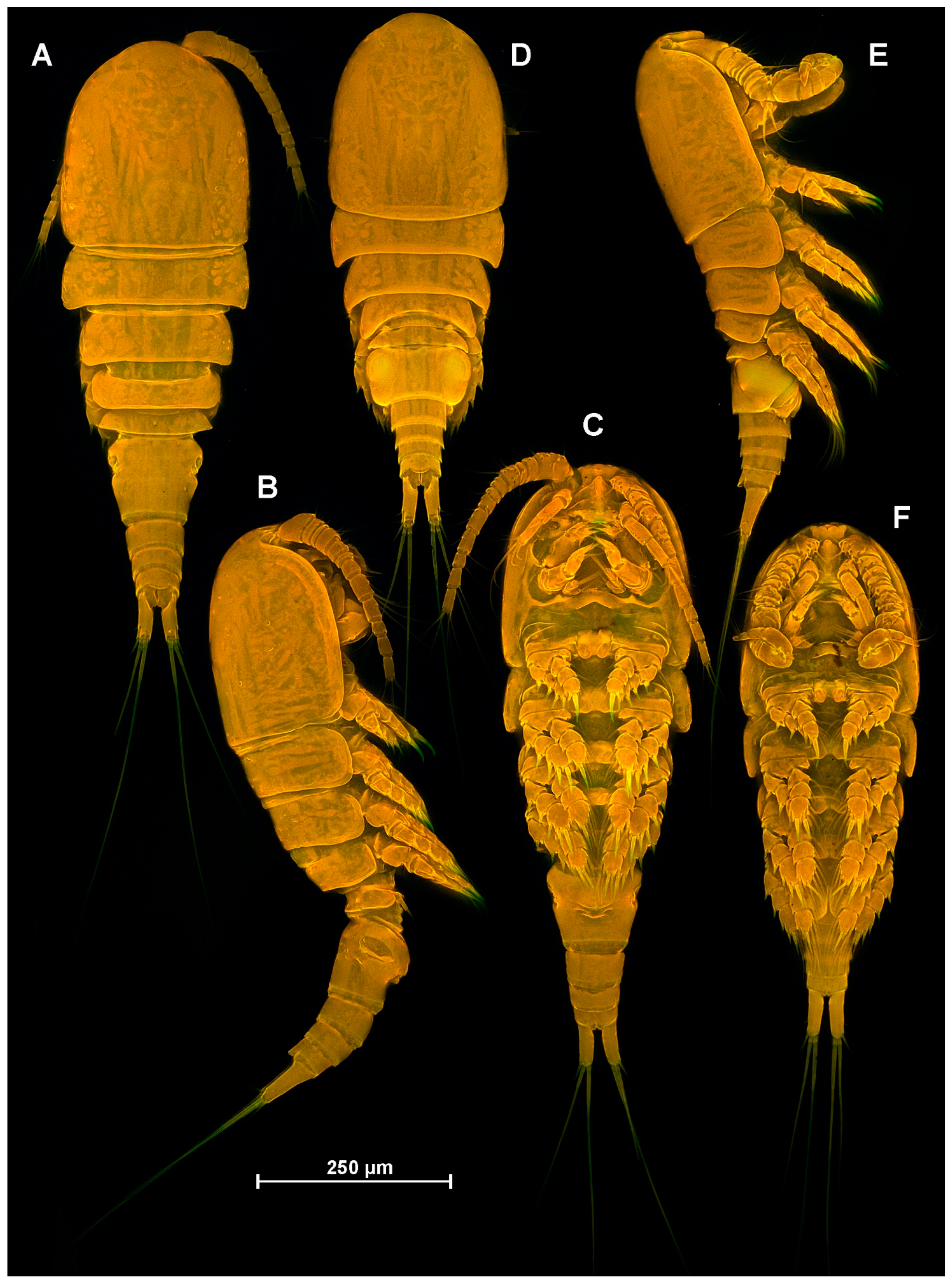
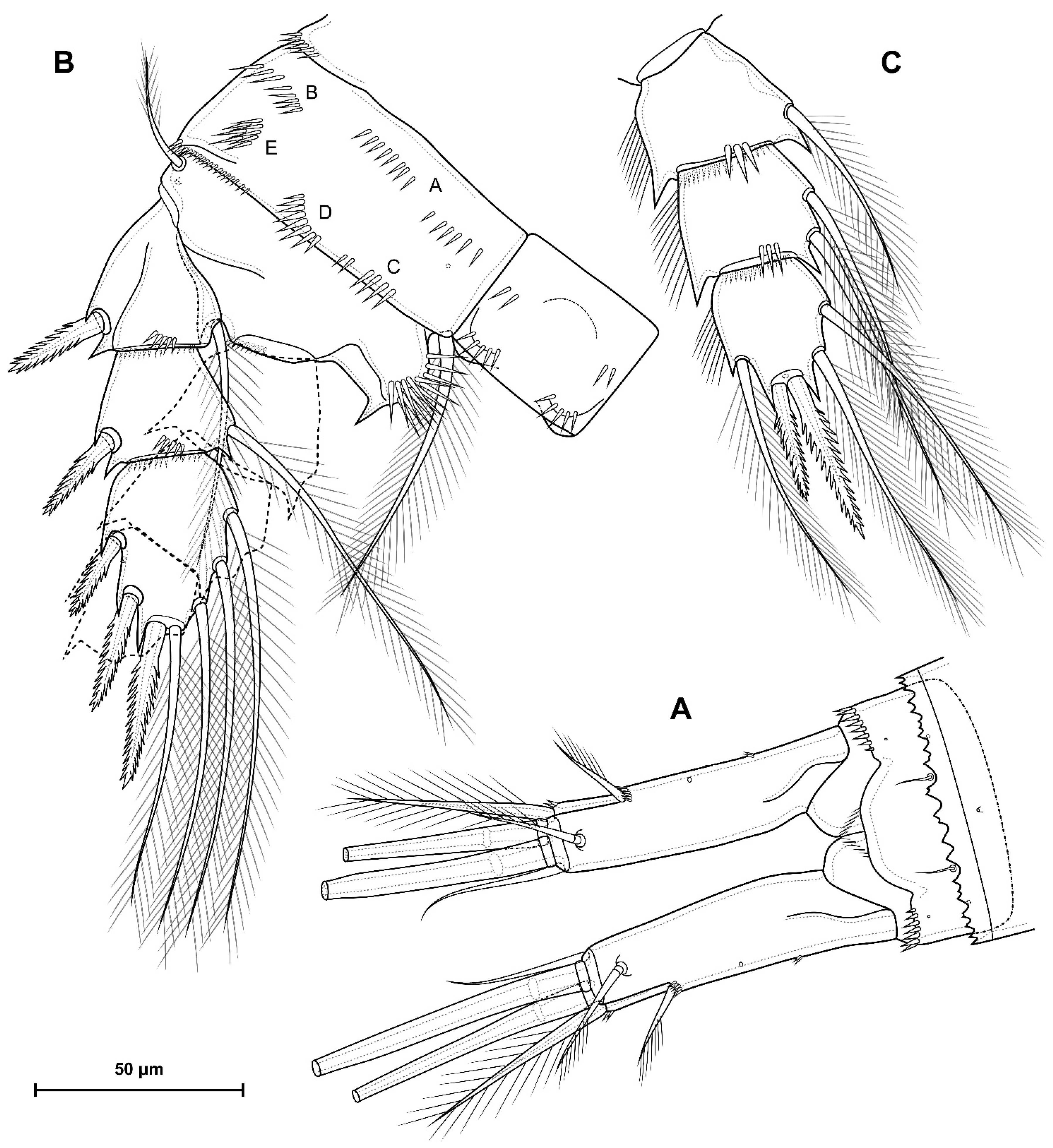
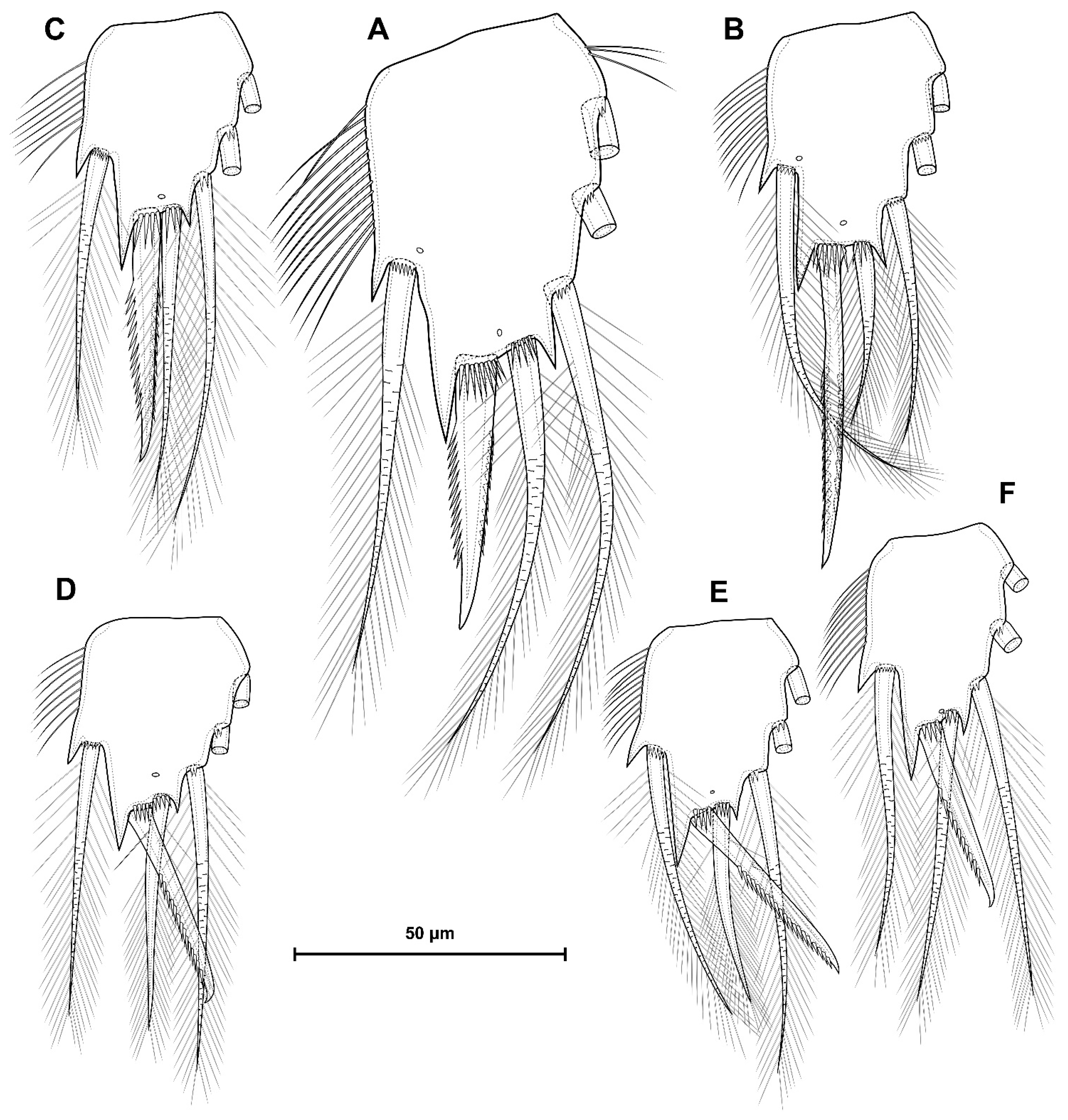
4.1. Species Diacyclops dyabdar sp. nov.

urn:lsid:zoobank.org:act:7008FC60-E385-4D76-8645-2395CFDA6309
Synonymy.
Diacyclops sp. 1—Novikov et al. (2023) [2], Table 1.
Etymology. The species is named after a mythical creature from the mythology of the Evenki, the indigenous people of the territory in which the new species lives. The name “dyabdar” is the latinized word from the Evenki “Дябдар”, and it is a noun in the nominative singular standing in apposition. Dyabdar is a huge mythical snake that lives underground, and with its body it creates river beds. The new species lives mainly in rivers and streams.
Material. Holotype: RUSSIA • ♀ dissected on two slides; North Yakutia, Tiksi area, nameless river, flow; 71.609229° N, 128.752738° E; 14 August 2022; A. Novikov leg; VH EFZK 22/1, VH EFZK 22/2. Allotype: RUSSIA •♂ dissected on one slide; collection data completed as for the holotype; VH EFZK 22/3. Paratypes: RUSSIA • Two ♀ and one ♂ undissected, preserved in 4% formalin, two ♀ dissected on one slide; collection data completed as for the holotype; VH EFZK 22/4, VH EFZK 22/5.
Additional material. RUSSIA • One ♂ undissected; North Yakutia, stream flowing into the Lena River; 68.889394° N, 124.044170° E; 27 June 2019; A. Novikov leg; retained in the collection of the first author.
RUSSIA • One ♀ and one ♂ undissected; North Yakutia, Tiksi area, Sevastyan–Yuryage River, flow; 71.546744° N, 128.801305° E; 2 August 2021; A. Novikov leg; retained in the collection of the first author.
RUSSIA • One ♀ undissected; North Yakutia, Tiksi area, Snezhinka River, flow; 71.707836° N, 128.862999° E; 12 August 2022; A. Novikov leg; retained in the collection of the first author.
RUSSIA • Two ♀ and one ♂ undissected; North Yakutia, Tiksi area, Sevastyan–Yuryage River, flow; 71.549866° N, 128.782585° E; 16 August 2022; A. Novikov leg; retained in the collection of the first author.
RUSSIA • One ♀ undissected and two ♀ dissected on two slides; North Yakutia, Tiksi area, stream flowing into the Kobchik Lake; 71.529281° N, 128.770956° E; 16 August 2022; A. Novikov leg; retained in the collection of the first author.
RUSSIA • One ♂ undissected; North Yakutia, Tiksi area, wet moss; 71.631423° N, 128.894520° E; 22 August 2022; A. Novikov leg; retained in the collection of the first author.
RUSSIA • One ♀ undissected; Anabar Plateau, stream flowing into the Kotuykan River; 70.492244° N, 106.210403° E; 23 July 2023; A. Novikov leg; lost during molecular genetic analysis.
RUSSIA • One ♀ dissected on one slide; Anabar Plateau, small puddles with moss; 70.635118° N, 105.266465° E; 28 July 2023; A. Novikov leg; retained in the collection of the first author.
Description. Female. Body robust (Figure 2A–C). Total body length from anterior margin of rostrum to posterior margin of caudal rami 1072 µm (n = 1). Cephalothorax wider than first and second free somites, largest width 435 µm (n = 1), and surface with pores and sensillae. Naupliar eye not discovered. Rostrum fused with cephalothorax. Posterior margin of cephalothorax and all pedigerous somites smooth.
Figure 2. Diacyclops dyabdar sp. nov. Female paratype, habitus: (A) dorsal; (B) lateral; and (C) ventral. Male paratype, habitus: (D) dorsal; (E) lateral; (F) ventral.
Diversity 16 00208 g002
Abdomen (Figure 3) consists of genital-double somite, two free abdominal somites, and anal somite with caudal rami. Genital, second, and third abdominal somites on posterior margin serrated. Genital-double somite consists of last thoracic somite and first abdominal somite, wider than long; anterior part with two dorsal pairs of sensillae and dorsal unpaired pore; and posterior part with four pairs of sensillae, three pairs of pores, and dorsal unpaired pore.
Figure 3. Diacyclops dyabdar sp. nov. Female holotype, abdomen: (A) dorsal; (B) ventral; (C) lateral. II–VII—number of caudal setae.
Diversity 16 00208 g003
P6 (Figure 3C) lateral, fused with somite; with one long seta, one short seta and small spiniform process. Genital field (Figure 3B) small, bean-shaped. The egg sac not observed.
Second abdominal somite (Figure 3) with four pairs of sensillae, three pairs of pores and dorsal unpaired pore. Third abdominal somite (Figure 3) with dorsal unpaired pore and two pairs of lateral pores. Anal somite with one dorsal pair of sensillae, two dorsal and one ventral pairs of pores and ventro-lateral rows of small spinules; proctodeum with rows of setules. Anal operculum square.
Caudal rami (Figure 3): length/width ratio of 2.96–3.79 (mean 3.35, n = 6). Ramus has one ventral, two lateral, and one dorsal pores; group of fine setules on proximal part of inner side; and spinules at base of setae II and III. Seta I absent. Seta II pinnate and located laterally. Seta III strong and bipinnate. Apical setae IV and V long and bipinnate with fracture plane; length ratio of 1 and 1.33, respectively. Seta VI slightly shorter than seta III (ratio of length of seta VI to seta III is 0.57–1.02, mean of 0.81, n = 6). Seta VII biarticulated.
Antennule (Figure 4A) 12-segmented, slightly curved along posterior margin, with short row of spinules on first segment, with one short aesthetasc on ninth segment, with one slender aesthetasc on eleventh segment and one long aesthetasc on twelfth segment. Armature formula as follows: 1-(8), 2-(4), 3-(2), 4-(6), 5-(4), 6-(2), 7-(2), 8-(3), 9-(2 + ae), 10-(2), 11-(2 + ae), 12-(7 + ae). One seta on sixth segment spiniform and short; other setae slender and most setae naked, only nine setae plumose or pinnate at their distal parts. One apical seta on 12th segment fused basally to aesthetasc.
Figure 4. Diacyclops dyabdar sp. nov. Female holotype: (A) antennule; (B) antenna, anterior side; (C) mandible; (D) mandibular gnathobase; (E) maxillule.
Diversity 16 00208 g004
Antenna (Figure 4B) four-segmented include short coxabasis and three-segmented endopod. Coxobasis rectangular, with two spinular rows on posterior side, two rows anterior side, one row on outer and one row on inner margin; distally with three setae, one of these exopodal long pinnate seta; exopodal seta reaching end of distal endopodal segment. First endopodal segment with one naked seta and one row spinules on posterio-lateral side. Second endopodal segment cylindrical, witn nine naked setae on inner margin, these setae longer from proximal to distal, with one row of spinules along outer margin. Third endopodal segment cylindrical, same length as second segment; with seven naked apical setae with claw-like ends, with two spinular rows on outer margin.
Labrum trapezoidal. Anterior surface with two groups of long spinules. Distal margin with small lateral denticles and 13 medial blunt teeth.
Mandible (Figure 4C) composed of coxa and small palp. Coxal gnathobase (Figure 4D) with 14 distal elements: one serrate seta; eight strong teeth; five slender teeth, three of these fused in base; gnathobase subdistaly with spinular row. Palp with three apical setae: two long and pinnate and one short and naked.
Maxillule (Figure 4E) composed of syncoxa and two-segmented palp: one-segmented basis and one-segmented endopod. Praecoxal arthrite medially with three distal strong teeth, distal pinnate seta, two strong medial spines, two small medial setae, one small proximal seta and one long robust proximal seta. Basis distally with two setae and one spine. Exopod represented by one seta on proximal part of basis. Endopod with three setae.
Maxilla (Figure 5A) five-segmented, composed of syncoxa, basis and three-segmented endopod. Syncoxa with endite with two long pinnate setae. Basis with two endites: proximal with one pinnate seta; distal with two pinnate setae. First endopodal segment with slender seta, strong subdistal pinnate seta and strong claw with several spinule. Second endopodal segment with two strong setae, one of this serrate. Third endopodal segment with two naked setae and one strong serrate seta.
Maxilliped (Figure 5B) four-segmented, composed of syncoxa, basis and two-segmented endopod. Syncoxa elongated with three setae. Basis with three outer rows of spinules, row of spinules on anterior side; with two pinnate setae. First endopodal segment with row of spinules and long pinnate seta. Second endopodal segment with long pinnate and two naked setae.
P1–P4 well developed, setae/spine armature presented in Table 2.
P1 (Figure 5C,D). Praecoxa with row of long spinules. Coxa rectangular, with antero-lateral and distal spinular rows; with one pore; with long inner seta. Intercoxal sclerite wide with large lateral lobes, without ornamentation. Basis with proximal pore, spinular rows at base of endopod and inner seta, inner row of setules; with outer long pinnate seta and inner thick pinnate seta. All exopodal and endopodal setae pinnate. Exopod three-segmented; first segment with distal posterior spinular row, outer spinulose spine and inner seta; second segment with inner seta and outer spinulose spine; third exopodal segment with two outer spinulose spines, two apical and two inner long setae. Endopod three-segmented; first and second endopodal segments with distal row of small spinules, outer row of setules and one inner seta; third endopodal segment with outer seta, apical strong curve spinulose spine, apical seta, three inner setae, with outer row of setules and row of spinules near base of apical spine.
P2 (Figure 6A,B). Praecoxa with row of long spinules. Coxa rectangular, with two antero-lateral rows of long spinules and distal row of small spinules; with one pore; with long inner seta. Intercoxal sclerite square, without ornamentation. Basis with proximal pore, spinular rows at base of endopod and outer seta, inner row of setules; with outer naked seta. All exopodal and endopodal setae pinnate. Exopod three-segmented; first segment with distal posterior and anterior spinular rows, outer spinulose spine and inner seta; second segment with distal posterior and anterior spinular rows, with inner seta and outer spinulose spine; third exopodal segment with one pore, two outer spinulose spines, apical spinulose spine, one apical and three inner setae. Endopod three-segmented; first endopodal segment with distal anterior and posterior rows of spinules, outer row of setules and one inner seta; second endopodal segment with distolateral anterior pore, distal anterior and posterior rows of spinules, outer and inner rows of setules and two inner setae; third endopodal segment with outer seta, apical spinulose spine, apical seta, three inner setae, with outer row of setules, row of spinules near base of apical spine and two pores near base of outer and apical seta.
Figure 5. Diacyclops dyabdar sp. nov. Female holotype: (A) maxilla; (B) maxilliped; (C) P1, anterior side; (D) P1 endopod.
Diversity 16 00208 g005
Figure 6. Diacyclops dyabdar sp. nov. Female holotype: (A) P2, anterior side; (B) P2 endopod; (C) P3, anterior side; (D) P3 endopod.
Diversity 16 00208 g006
P3 (Figure 6C,D and Figure 14A) similar to P2. First exopodal segment with lateral row of spinules.
P4 (Figure 7A–C). Praecoxa with row of long spinules. Coxa rectangular, with one pore; with long inner seta; anterior side with distal group of small spinules; posterior side with groups of spinules A, B, C, D, E, F; group C and D separated; group B composed of two different group; group A with gap in middle part. Intercoxal sclerite square, without outstanding humps, with posterior row of spinules. Basis with proximal pore, spinular rows at base of endopod and outer seta, inner row of setules; with outer naked seta; inner side of basis with two protrusions: spiniform and large lobe-shaped process. Exopod similar to that of P3, but first and third segments without inner setules. Endopod three-segmented. First and second endopodal segments similar to that of P2 and P3, but without inner setules on second segment. Third endopodal segment with outer seta, two apical spinulose spines, two inner setae, with outer row of setules, row of spinules near base of apical spines and two pores near base of outer and apical spines; ratio between inner to outer apical spines 1.43–1.53 (mean 1.47, n = 6).
P5 (Figure 7D) two-segmented, composed of coxobasis and exopod, without intercoxal sclerite. Coxobasis small with outer pinnate seta. Exopod with apical pinnate seta and subapical serrate spine; with spinules near base of spine; spine longer than exopodal segment.
Figure 7. Diacyclops dyabdar sp. nov. Female holotype: (A) P4, anterior side; (B) P4 endopod; (C), P4 endopodal segment from the other side; (D) P5. Numbers of coxal spinular groups (A–F) are given according to Einsle [21].
Diversity 16 00208 g007
Male. Significantly smaller than female (Figure 2D–F). Total body length from anterior margin of rostrum to posterior margin of caudal rami: 746 µm (n = 1), largest width of cephalothorax 300 µm (n = 1). Sexual dimorphism expressed in the antennule, P3 endopod, genital segmentation and ornamentation, P6. Cephalothorax and thoracic somites as in female. P1-P5, except P3 endopod, identical to this in female, differences only in relative length of segments, setae and spines.
Genital somite (Figure 8A–C) free; with two spermatophores visible inside; with P6. Spermatophores (Figure 8A–C) rounded with corner on inner side. P6 (Figure 8C) presented by two large flaps fused to the somite; with ventral pores; with outer pinnate long seta, middle pinnate short seta and inner spinulose spine; inner part of flaps protruded to ventral side.
Figure 8. Diacyclops dyabdar sp. nov. Male allotype: (A) abdomen, dorsal; (B) abdomen, ventral; (C) first-third urosomites, lateral; (D) P3 endopod, posterior side.
Diversity 16 00208 g008
Antennule (Figure 9A–C) 17-segmented, haplocer with geniculation between segments 14 and 15; with seven aesthetascs. Segment 1 long, with spinular row, eight setae and three aesthetascs. Segment 2 with four setae. Segments 3, 5, 6, 7, 8 similar, short, each with two setae. Segments 4 and 9 with two setae and aesthetasc. Segments 10 and 11 widened, each with two setae. Segment 12 with one small and one spiniform robust seta. Segment 13 with two small setae and one aesthetasc. Segment 14 elongate, with two small setae, one modified flattened seta and one small papillary process, which probably modified seta. Segment 15 elongate with one long and two modified flattened setae. Segments 16 and 17 partially fused. Segment 16 with four setae. Segment 17 triangular, with seven setae, one of which fused in base with one aesthetasc. Armature formula is as follows: 1-(8 + 3ae), 2-(4), 3-(2), 4-(2 + ae), 5-(2), 6-(2), 7-(2), 8-(2), 9-(2 + ae), 10-(2), 11-(2), 12-(2), 13-(2 + ae), 14-(2 + 2 modified), 15-(1 + 2 modified), 16-(4), 17-(6 + (1 + ae)).
Figure 9. Diacyclops dyabdar sp. nov. Male allotype: (A) antennule; (B) 10–14 segments of antennule; (C) 15–17 segments of antennule; (D) P4, posterior side; (E) P4 endopod.
Diversity 16 00208 g009
P3 endopod (Figure 8D and Figure 14B) with dimorphic third segment. Apical spine long, slightly widened in middle, ratio of length of spine to length of endopodal segment 1.5. Apical seta and distal inner setae shorter than spine.
P4 (Figure 9D,E) similar to this in female, differences only in relative length of segments, setae and spines.
Variability. Female holotype has an asymmetrical P4: Enp2 has one seta on one side (Figure 7B) and two setae on other (Figure 7C). Other individuals have two setae on this segment, so this could be considered a morphological abnormality. Structure of genital field is also variable, with different sizes in different females.
Ecology and distribution. Diacyclops dyabdar sp. nov. is most often found in watercourses. Out of nine finds, four were in small rivers (Figure 1B,C), three in streams (Figure 1E) and two in wet mosses (Figure 1D). At the same time, other Copepoda species are also found in watercourses, but it is D. dyabdar and Moraria sp. that are almost always characteristic of these types of water bodies [2]. For two samples with this species from the Anabar Plateau, pH values (6.16 and 6.31) and total dissolved solids (59 and 158 ppm) are known.
At present, the range of the species is rather narrow and includes Northern Yakutia and the Anabar Plateau (Figure 1A). However, the difficulty of detection is probably due to the fact that watercourses are rarely surveyed for Copepoda. Thus, we can expect the species’ range to expand at least within northern Siberia.

4.2. Morphometric Analysis

Cluster analysis (Figure 10A) and the method of principal coordinates (Figure 10B) show similar data. All studied individuals form three main clusters. The first includes only one species D. karamani (Kiefer, 1932), which is well-characterized by elongated caudal rami. The second cluster brings together D. ruffoi Kiefer, 1981, D. skopljensis Kiefer, 1932, D. antrincola Kiefer, 1968, D. crassicaudis cosana Stella & Salvadori, 1957 and D. crassicaudis s. lat sensu Pesce, 1992. A common feature of these species is the long caudal seta VI, which is at least 1.5 times longer than the caudal ramus.
The third cluster includes D. crassicaudis s. str. and subspecies, D. dyabdar sp. nov., D. fontinalis Naidenow, 1969 and D. iranicus Pesce & Maggi, 1982. Most of the different subspecies finds are not grouped into subclusters. The same is true for the individuals we measured. Diacyclops crassicaudis from Tatarstan fell into two different subclusters. D. dyabdar sp. nov. also fell into two different subclusters, one of which is formed only by this species, and the second includes D. crassicaudis from Tatarstan. Diacyclops iranicus and D. fontinalis do not form their own subclusters; hence, they have no significant morphometric differences from subspecies of D. crassicaudis.
Figure 10. Morphometric analysis of individuals from species of D. crassicaudis group: (A) clustering tree based on Gower’s distances; (B) principal coordinates analysis based on Gower’s distances. Filled circles—our individuals; open circles—individuals from references (Table 1). Red—D. crassicaudis and close related species; blue—D. dyabdar sp. nov.
Diversity 16 00208 g010

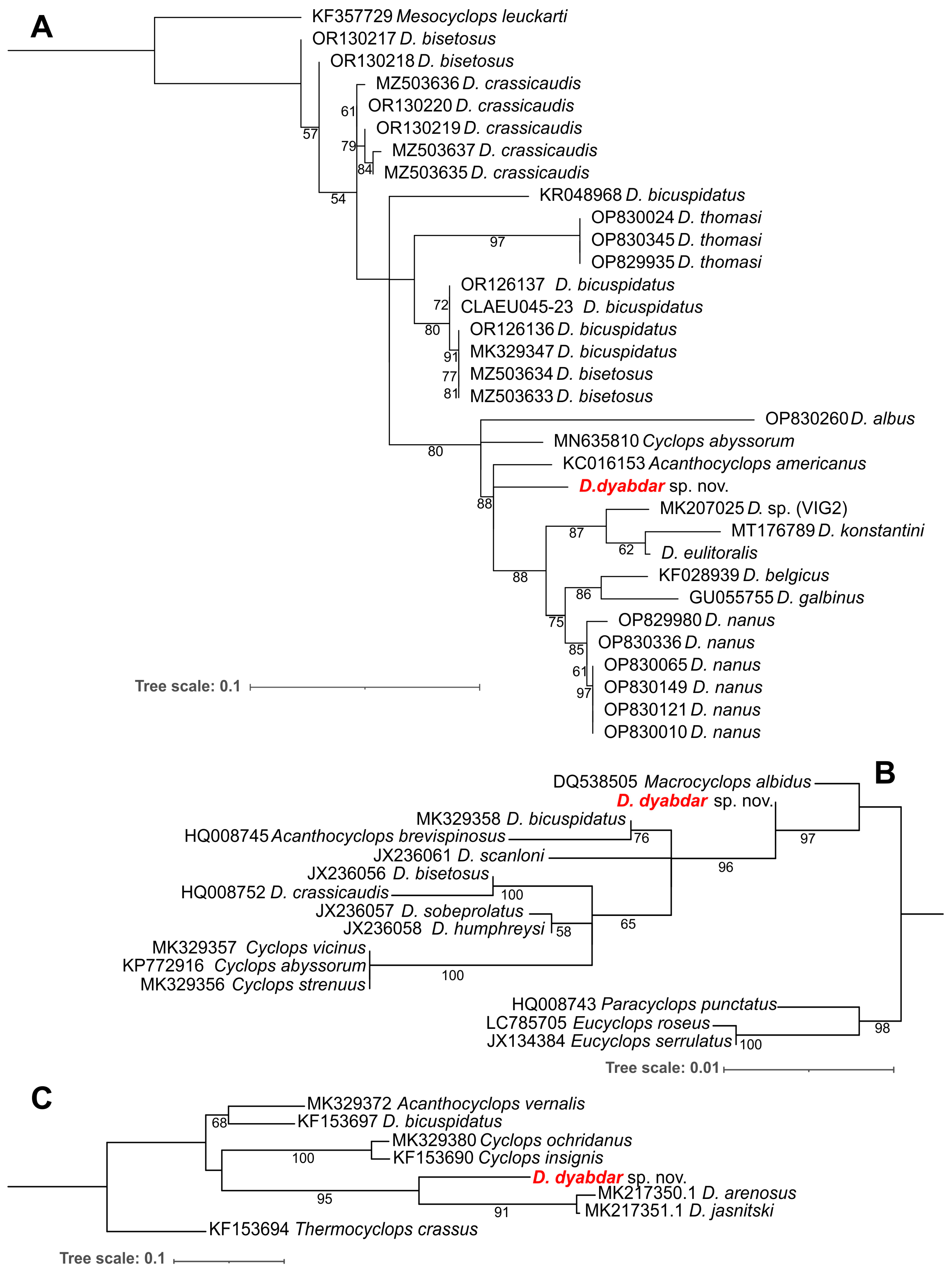
4.3. Molecular Analysis

Four nucleotide sequences of COI (547 bp), ITS1 (197 bp), 5.8S-ITS2 (347 bp) and 18S rRNA (547 bp) were obtained for a single female of D. dyabdar sp. nov. The COI, ITS1 and 18S sequences were assembled from forward and reverse sequence reads, while the ITS2 sequences remain unassembled sequence reads. These sequences were deposited in GenBank under accession numbers PP254237, PP326082, PP488223 and PP254238. BLAST analysis revealed that the sequences of D. dyabdar sp. nov. are similar to those of A. americanus (Marsh, 1893) (KC016153) with 81.63% identity and 99% query coverage and D. nanus (Sars, 1863) (OP830336) with 80.41% identity and 97% query coverage by COI; Macrocyclops albidus Jurine, 1820 (KY421594), and D. bicuspidatus (Claus, 1857) (MK329358) with 98.72% and 98.54% identity and 100% query coverage, respectively, by 18S rRNA; D. jasnitskii (Mazepova, 1950) (MK217351) with 85.75% identity and 99% query coverage by ITS2; and D. sp. with 71.01% identity and 82% query coverage by ITS1 in GenBank. A search in the BOLD database using COI revealed the top 100 matches for D. dyabdar sp. nov. with a similarity range of 81.78–82.68% to Acanthocyclops americanus, A. eduardoi Mercado-Salas & Álvarez-Silva, 2013, A. robustus (Sars, 1863), and Cyclops abyssorum Sars, 1863. The substitution saturation was evaluated for ITS data using Xia’s method. We obtained an Iss = 0.1466, which was much smaller than Iss.c (=0.8215 assuming a symmetrical topology and 0.7267 assuming an asymmetrical topology). The sequences have experienced little substitution saturation and are useful for phylogeny. On the ITS2 (ML, GTR) phylogenetic tree, D. dyabdar sp. nov. reliably clusters with representatives of the same genera, including two endemic species, D. arenosus (Mazepova, 1950) and D. jasnitski, from Lake Baikal (Figure 11C). However, D. dyabdar sp. nov. is genetically distant from D. bicuspidatus. Substitution saturation was evaluated for the third nucleotide position of the COI sequences using Xia’s method. The obtained was Iss = 0.634, which was not significantly smaller than Iss.c (=0.712) assuming a symmetrical topology, and much larger than Iss.c (=0.419) assuming an asymmetrical topology. The COI data are substantially saturated. Steel’s method of substitution saturation measuring revealed that the D. dyabdar sp. nov. COI sequence might be problematic. Therefore, the COI (ML, TN + F + I + G4) tree was constructed based only on the first and second nucleotide positions. In both the COI and 18S rRNA trees (ML, K2P + I), D. dyabdar sp. nov. is positioned separately and distantly from D. crassicaudis (Figure 11A,B). On the COI tree D. dyabdar sp. nov. clusters with A. americanus, endemic Diacyclops species from Lake Baikal, stygobitic D. belgicus and D. nanus. The genetic interspecies COI p-distances, taking into account only the first and second nucleotide positions, range from 3.3% to 6.9%. The p-distances between D. dyabdar and D. crassicaudis were 4.8%. The minimal p-distances were calculated between D. dyabdar and D. nanus, A. americanus and C. abyssorum (3.3%) (Table S1).
Figure 11. (A) Phylogenetic tree constructed on the base of the COI (ML, TN + F + I); (B) phylogenetic tree constructed on the base of the 18S (ML, K2P + I); (C) phylogenetic tree constructed on the base of the ITS2 (ML, GTR). The number in the node is the bootstrap value of the branching node support. Red color—new species.
Diversity 16 00208 g011

4.4. Diacyclops crassicaudis Morphological Species Group

Diagnosis. Medium-sized Diacyclops with robust body. Antennule of female 12-segmented. P1–P4 with three-segmented rami. P1–P4 Exp3 with two, three, three, and three spines, respectively, and four setae. P1 Enp2 with only one seta. Inner apical spine of P4 Enp3 longer than outer one.
Species: D. antrincola Kiefer, 1968, D. crassicaudis (Sars, 1863), D. dyabdar sp. nov., D. karamani Kiefer, 1932, D. ruffoi Kiefer, 1981 and D. skopljensis Kiefer, 1932.
Species inquirendae: Diacyclops crassicaudis cosana Stella & Salvadori, 1953 and Diacyclops crassicaudis s. lat. sensu Pesce (1992)

4.5. Diacyclops crassicaudis (Sars, 1863)

Major synonyms.
Cyclops crassicaudis—Sars (1863) [50], 236; Sars (1913) [23], 49, pl. XXIX; Gurney (1933) [26], 232, Figures 1686–1696; Coker (1938) [51], 77, Figures 1–13.
Diacyclops crassicaudis—Ito (1957), 12, Figures 24–34; Monchenko (1974), 290; Karpowicz et al. (2021) [52], 7, Figures 6–7.
Diacyclops crassicaudis crassicaudis—Monchenko (1974) [22], 291, Figure 100.
Cyclops brucei—Scott (1899) [53], 93, pl. 6, Figures 1–6.
Cyclops bissextilis—Willey (1925) [54], 141, Figure 3.
Cyclops (Diacyclops) crassicaudis brachycercus syn. nov.—Kiefer (1927) [55], 262, Figures 1–2.
Cyclops crassicaudis brachycercus syn. nov.—Yeatman (1944) [56], 60, Figures 114–121.
Diacyclops crassicaudis brachycercus syn. nov.—Monchenko (1974) [22], 294, Figures 101–102.
Cyclops crassicaudis cretensis syn. nov.—Kiefer (1928) [57], 245, Figures 1–3.
Diacyclops crassicaudis cretensis syn. nov.—Pesce & Maggi (1981) [28], 170, Figure 3a,c,e,f,g; Kim & Chang (1989) [27], 242, Figure 9.
Diacyclops crassicaudis cf. cretensis—Pesce & Maggi (1981) [28], 171, Figure 3b,d.
Cyclops (Diacyclops) crassicaudis taipehensis syn. nov.—Harada (1931) [29], 158, Figures 17–18.
Acanthocyclops (Diacyclops) fontinalis syn. nov.—Naidenow (1969) [30], 1063, Figures a–e.
Diacyclops iranicus syn. nov.—Pesce & Maggi (1982) [31], 177, Figure 1.
Diacyclops crassicaudis lagrecai syn. nov.—Pesce & Galassi (1987) [13], 209, Figures 19–24.
Diacyclops crassicaudis trinacriae syn. nov.—Pesce & Galassi (1987) [13], 211, Figures 25–31.
Material examined. RUSSIA • One ♀ dissected on one slide; North Yakutia, small river flowing into the Lena River; 72.118771° N, 126.987816° E; 22 July 2021; A. Novikov leg; retained in the collection of the first author.
RUSSIA • One ♂ dissected on one slide, one ♀ undissected; North Yakutia, Tiksi area, nameless river, flow; 71.609229° N, 128.752738° E; 14 August 2022; A. Novikov leg; retained in the collection of the first author.
RUSSIA • One ♀ undissected and one ♀ dissected on one slide; North Yakutia, Tiksi area, stream flowing into the Kobchik Lake; 71.529281° N, 128.770956° E; 16 August 2022; A. Novikov leg; retained in the collection of the first author.
RUSSIA • Five ♀ dissected on three slides, ten ♀ and three ♂ undissected; Tatarstan Republic, Morkvashinka River, Karaman-Chappuis pit; 55.767969° N, 48.846781° E; 17 June 2020; A. Novikov leg; retained in the collection of the first author.
RUSSIA • One ♂ dissected on one slide, three ♀ and one ♂ undissected; Tatarstan Republic, stream flowing into the Morkvashinka River, Karaman-Chappuis pit; 55.765465° N, 48.836648° E; 30 August 2022; A. Novikov and D. Sharafutdinova leg; retained in the collection of the first author.
Short description. Female. Body relatively slender (Figure 12A–C). Total body length from anterior margin of rostrum to posterior margin of caudal rami 765 µm (n = 1). Cephalothorax not wider than first free somite, with largest width 242 µm (n = 1).
Figure 12. Diacyclops crassicaudis from Tatarstan (European part of Russia). Female, habitus: (A) dorsal; (B) lateral; (C) ventral. Male, habitus: (D) dorsal; (E) lateral; (F) ventral.
Diversity 16 00208 g012
Anal somite (Figure 13A) with one dorsal pair of sensillae, two dorsal and one ventral pair of pores, and ventro-lateral rows of small spinules; proctodeum with rows of setules. Anal operculum trapezoidal. Caudal rami (Figure 13A) length/width ratio: 3.67–4.57 (mean of 4.16, n = 7). Ramus with one ventral and two lateral pores; with spinules at base of lateral pore; setae II and III; and a ridge between bases of seta II and seta IV. Seta I absent. Seta II pinnate and located laterally. Seta III strong and bipinnate. Apical setae IV and V long and bipinnate and with fracture plane. Seta VI shorter than seta III (ratio of length of seta VI to seta III is 0.61–0.88, with a mean of 0.72, n = 7). Seta VII biarticulated.
Figure 13. Diacyclops crassicaudis from Tatarstan (European part of Russia). Female: (A) caudal rami, dorsal; (B) P4, posterior side; (C) P4 endopod. Numbers of coxal spinular groups (A–E) are given according to Einsle [21].
Diversity 16 00208 g013
P4 (Figure 13B,C). Praecoxa with row of long spinules. Coxa rectangular, with one pore; with long inner seta; anterior side with distal group of small spinules; posterior side with groups of spinules A, B, C, D, E; groups C and D separated; group A with gap in middle part. Intercoxal sclerite square, with outstanding humps; with or without posterior rows of spinules on middle part and with posterior rows of spinules on distal part. Basis with proximal pore, spinular row at base of endopod, inner group of long spinules; with outer seta; inner side of basis with spiniform process and rounded inner lobe. Exopod three-segmented. First and second segments with distal posterior and anterior spinular rows, outer spinulose spine and inner seta. Third exopodal segment with one pore, displaced to lateral side of segment; two outer spinulose spines, apical spinulose spine, one apical and three inner setae. Endopod three-segmented. First endopodal segment with distal anterior and posterior rows of spinules, outer row of setules and one inner seta. Second endopodal segment with distal anterior and posterior rows of spinules, outer and inner rows of setules and two inner setae. Third endopodal segment with outer seta, two apical spinulose spines, two inner setae, with outer row of setules, row of spinules near base of apical spines and pore near base of apical spines; ratio between inner to outer apical spines1.40–1.80 (mean of 1.55, n = 6).
Male. Body almost the same size as the female (Figure 12D–F). Total body length from anterior margin of rostrum to posterior margin of caudal rami: 652 µm (n = 1), largest width of cephalothorax 230 µm (n = 1).
P3 Endopod (Figure 14D,E) with dimorphic third segment. Apical spine long, slightly widened in middle, ratio of length of spine to length of endopodal segment 1.5. Apical seta widened, spiniform. Apical seta and distal inner setae shorter or equal than spine.

4.6. Diacyclops cf. crassicaudis (Sars, 1863)

Material examined. GEORGIA/ABKHAZIA• One ♂ dissected on one slide; Pshandra River, Karaman–Chappuis pit; 43.091355° N, 40.591239° E; 20 June 2022; A. Novikov and D. Sharafutdinova leg; retained in the collection of the first author.
Differences. It is similar to D. crassicaudis, but slightly different in the structure of P3 and P4: the P3 Enp3 has an unmodified apical seta (Figure 14F) and the P4 coxa is without spinular group E. Unfortunately, only one male was found, so the taxonomic position of this individual is questionable.
Figure 14. Distal segment of P3 endopod of studied species of Diacyclops, anterior side: (A) D. dyabdar sp. nov, female; (B) D. dyabdar sp. nov., male; (C) D. crassicaudis, female (Tatarstan); (D) D. crassicaudis, male (Tiksi area, Yakutia); (E) D. crassicaudis, male (Tatarstan); (F) D. cf. crassicaudis, male (Abkhazia).
Diversity 16 00208 g014

5. Discussion

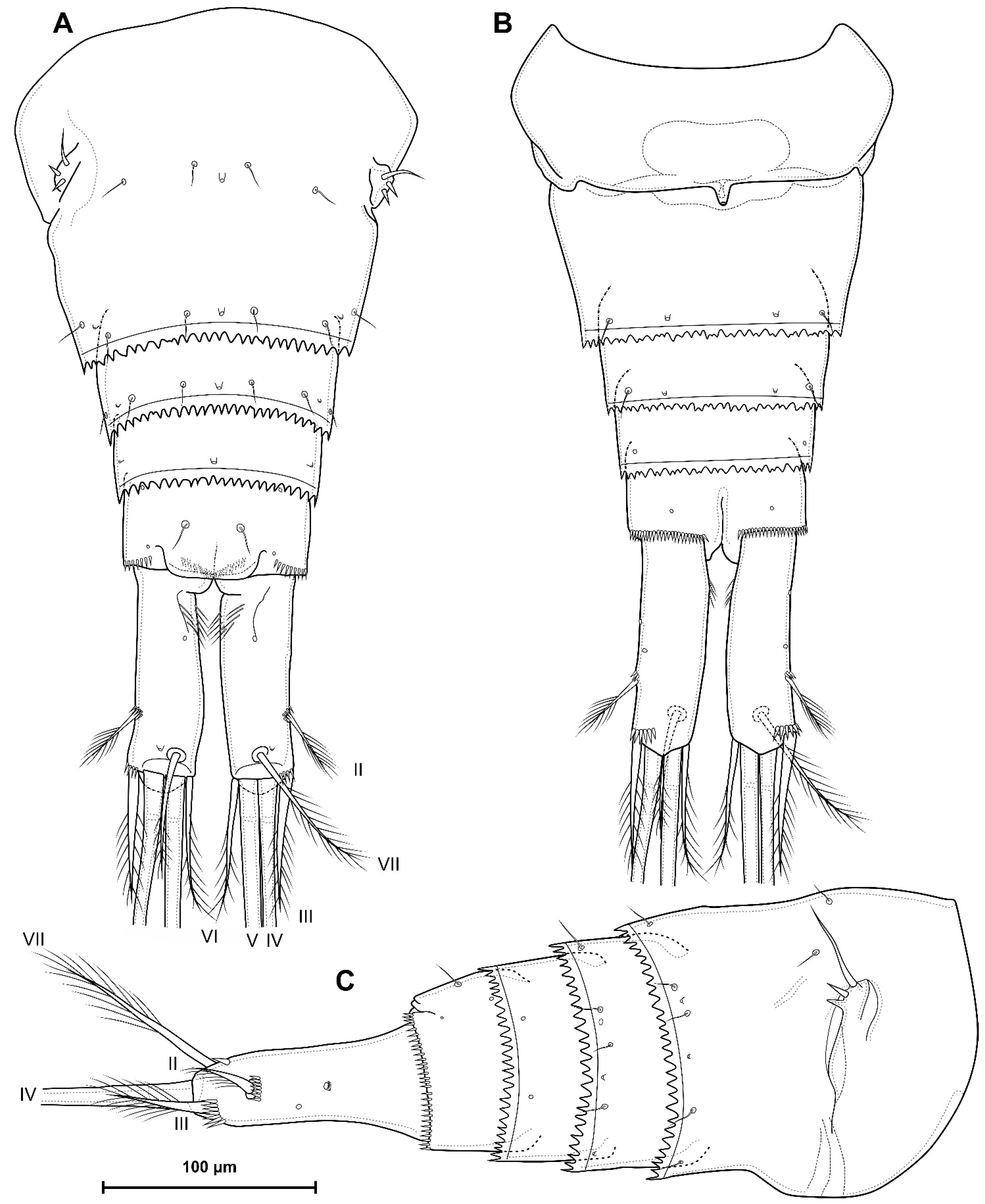
5.1. Difference between D. crassicaudis and D. dyabdar sp. nov.

Diacyclops crassicaudis and D. dyabdar sp. nov. belong to the same species group, are similar in habitus, armature and segmentation of appendages, and are often found together in samples from northern Siberia. Also, these species are quite similar in morphometric indices, but differ well in the following morphological characters:
  • Females of D. dyabdar sp. nov. are significantly larger than females of D. crassicaudis.
  • The caudal rami of D. dyabdar sp. nov. have a group of thin inner setules at the base.
  • The caudal rami of D. crassicaudis distally after seta II have a well-marked ridge, which is absent in D. dyabdar sp. nov.
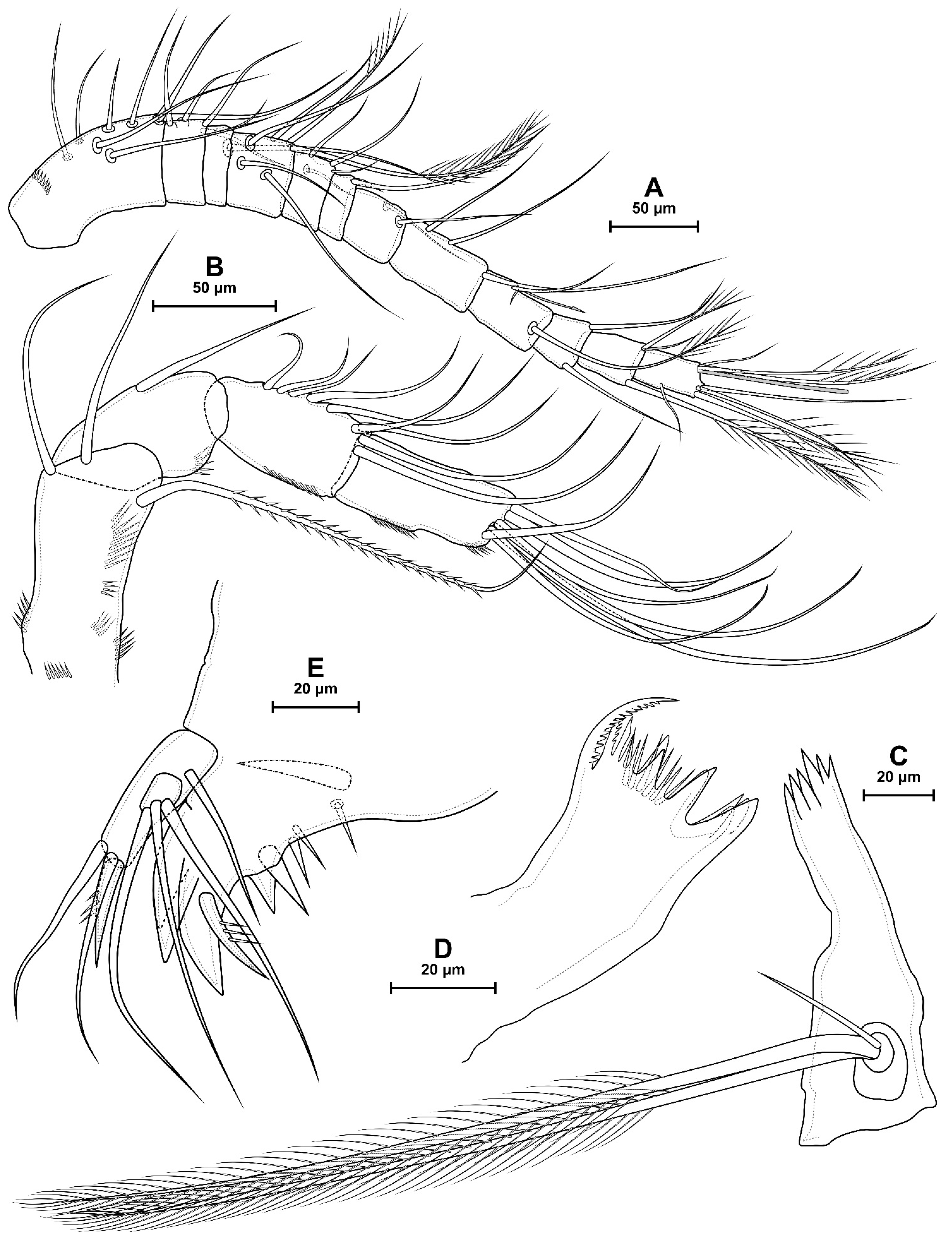
  • P4 coxa in D. dyabdar sp. nov. have spinular group F.
  • The first segments of exopods P3 and P4 in D. dyabdar sp. nov. have outer groups of well-marked spinules.
  • The intercoxal sclerite of P4 on the posterior side has one row of spinules in D. dyabdar sp. nov. and two rows of spinules in D. crassicaudis.
  • The intercoxal sclerite of P4 in D. crassicaudis has outstanding humps and D. dyabdar sp. nov. are without them.
  • P4 basis has a group of outer spinules in D. dyabdar sp. nov. but not in D. crassicaudis.
  • The inner lobe of the P4 basis is strongly protruded in D. dyabdar sp. nov., but in D. crassicaudis, this lobe forms only a semicircle.
  • The pore on P4 Exp 3 of D. crassicaudis is displaced to the outer side.
  • P4 Enp3 has two pores in D. dyabdar sp. nov. and one in D. crassicaudis.

5.2. Diacyclops crassicaudis Species Group

According to Pesce and De Laurentiis [11], the D. crassicaudis species group includes 15 species and subspecies. Later, Karanovic [10] also included in the group the Acanthocyclops rupestris Mazepova, 1950, D. conversus Reid, 2004 and several described taxa closely related to D. humphreysi Pesce & De Laurentiis, 1996. In this article, we consider this species group in a narrower sense considering the characters given in the species group diagnosis. With this diagnosis, the group includes only six species together with D. dyabdar sp. nov. Taking into account molecular genetic data (Figure 11), the group is most likely not monophyletic. However, the species have similar morphology and the identical segmentation and armature of antennules and appendages. Therefore, we use the concept of species group only from a morphological point of view for ease of identification. And, first of all, we excluded species that are close to other groups from the species list of the group.
Diacyclops conversus, like other species of the group D. nearcticus (Kiefer, 1934), has only one seta on P4 Enp2 and no exopodal seta of antenna [58]. This species is similar to D. crassicaudis in having a 12-segmented antennule, but in other species of the D. nearcticus group, the antennule may also be 17-segmented, as in D. nearcticus or D. jeanneli (Chappuis, 1929) [25], so this could be considered a convergent similarity.
Acanthocyclops rupestris (D. rupestris in Karanovic 2006) should belong to the genus Acanthocyclops Kiefer, 1927 by a number of characters and is included in the group A. venustus (Norman & Scott, 1906). Diacyclops talievi (Mazepova 1970) was originally described as a member of the genus Acanthocyclops [7]. However, later, together with a number of other endemic species of Lake Baikal, it was transferred to the genus Diacyclops on the basis of the long inner spine of the P5 distal segment [59]. Diacyclops talievi has a highly modified morphology related to its lifestyle in the abyssal zone of the Baikal Lake. It has elongated segments and setae on the segments of the swimming legs. It is probable the inner spine has undergone the same modification. We believe that this species belongs to the genus Acanthocyclops and is included in the A. rupestris species flock according to Boxshall and Evstigneeva [6]. The large number of spines and setules on caudal rami, the large size and the shape of the P4 endopod bring it close to these species. Mazepova [7] also points out the closeness of this species to A. rupestris and A. signifier Mazepova, 1952.
Diacyclops alticola Kiefer, 1935 and D. longifurcus Shen & Sung, 1963 have two setae on P1Enp2 and a longer outer spine P4 Enp3 compared to the inner one [60,61], which brings this species closer to D. bicuspidatus. In the D. bicuspidatus species group, the segmentation of the female antennule is rather variable: 17 segments in D. bicuspidatus and 14 in D. odessanus (Schmankevitsch, 1875) [22]. So, this does not contradict the assignment of this species to the same group.
Diacyclops humphreysi and closely related species may be closely related to the D. crassicaudis group, but have a number of differences, such as the longer outer spine of P4 Enp3 compared to the inner one and the long caudal seta VII [10].

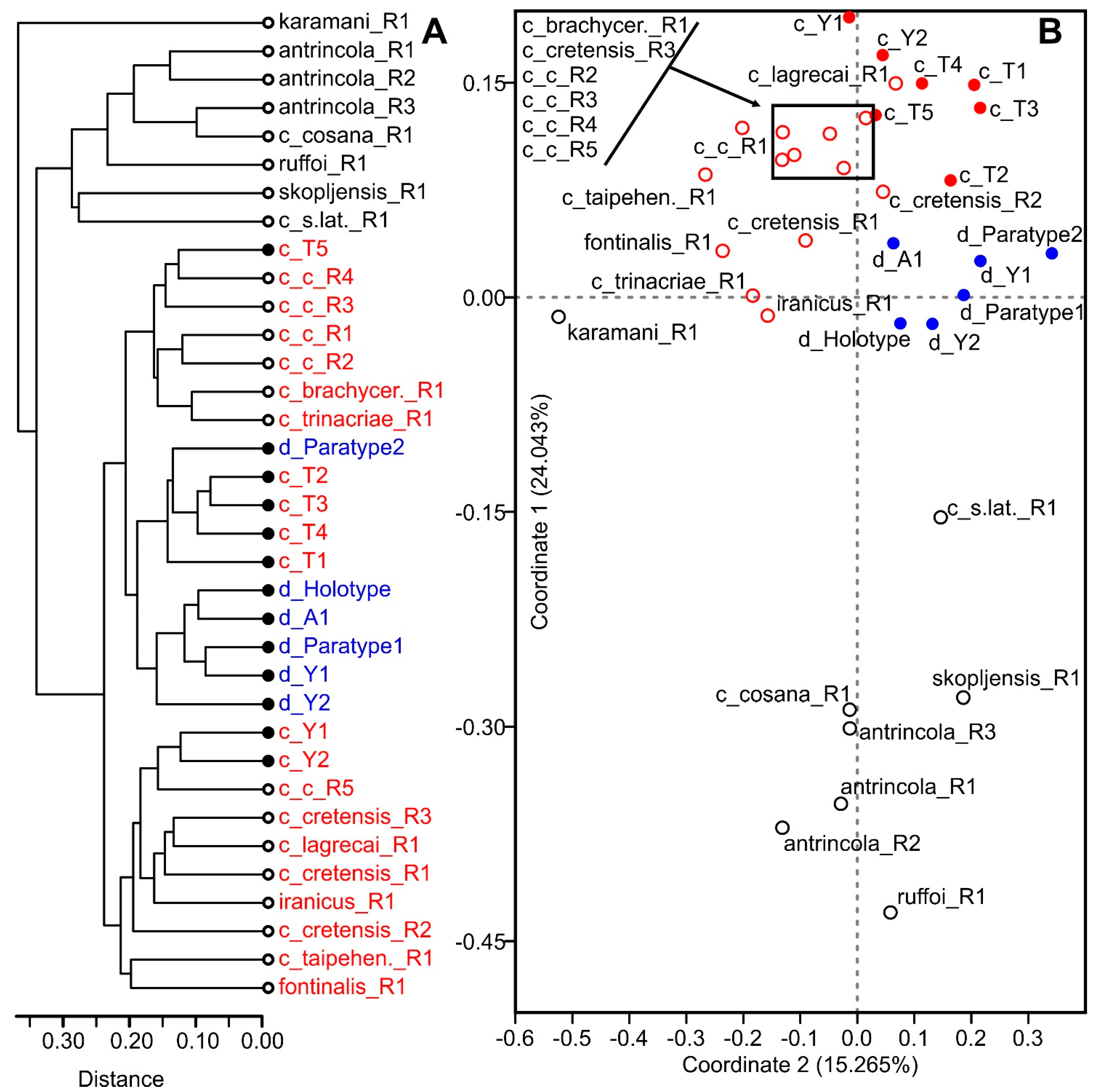
5.3. Morphometric Indices in Taxonomy of Diacyclops crassicaudis Species Group

According to the morphometric analysis performed, it is hardly possible to use classical morphometry (morphometric indices) to separate subspecies or species within the D. crassicaudis group. The specimens of D. crassicaudis from regions thousands of kilometers away showed no differences in obvious morphological characters. At the same time, two different species (D. crassicaudis and D. dyabdar sp. nov.) morphometrically appeared sometimes more similar than different representatives of the same subspecies of D. crassicaudis, for example, D. crassicaudis cretensis [13].
In the reference data, we also do not see any particular differences between individuals from distant regions and different subspecies. In works, individuals with different morphometry often appear under the same name; for example, the ratio of the length of the caudal rami in D. crassicaudis cretensis is indicated by authors in different ranges as 4.0–4.5 [27], 5.2–5.5 [28] and 4.0–6.0 [13]. Conversely, for example, in Monchenko [22], D. crassicaudis s. str. and D. crassicaudis brachycercus are almost identical in all characters except for the relative length of the caudal rami (4.0–5.5 and 3.2–3.8, respectively).
It is also important to note the sympatric habitat of different subspecies, for example in D. crassicaudis s. str. and D. crassicaudis brachycercus [22] or D. crassicaudis lagrecai and D. crassicaudis trinacriae [13], which contradicts the concept of the subspecies. It is worth noting the high variability of the species even within one sample, as in D. crassicaudis from Tatarstan, in which the relative length of the caudal rami ranges from 3.6 to 4.6. Actually, we reduce all subspecies to one species for the reason that there are no clear boundaries between them.
Without a doubt, this species is polymorphic. This is partially visible in the variability of male P3 Enp3 (Figure 14D–F). It cannot be excluded that it is genetically heterogeneous and represents a complex of species. However, differences should be sought in obvious morphological characters, certainly related to morphometry, but not based on it. But, this is difficult to achieve without the use of molecular systematics or very extensive collections from the entire range of D. crassicaudis.

5.4. About Dubious Taxa within the Genus Diacyclops

Mahoon and Zia [62] described a new species Cyclops (Diacyclops) landei Mahoon & Zia, 1985 from Pakistan. The figures obviously depict the copepodite stage of CIV. First, the number of abdominal somites is three (the first somite indicated as abdominal is a P5-bearing somite). Second, the antennule of this species is 10-segmented. According to Schutze et al. [63], the 10-segmented antennule are present in most Cyclopidae at stage CIV. Given the shape and length of the caudal rami and the structure of P5, it is likely that this species is a copepodite stage species in the genus Mesocyclops Sars, 1914.
The next doubtful species is Cyclops (Diacyclops) landei richardi Parveen, Mahoon & Saleem, 1988. The description of this species in the figures [64] probably depicts a male Mesocyclops based on the following characters: shape P5 with a long inner exopodal spine, strongly elongated distal segment of the P4 endopod, short caudal rami and a long inner seta VI and seta II close to the middle of the rami.
Cyclops (Diacyclops) jurenei Parveen, Mahoon & Saleem, 1988 is described in the same article. Like the first species, it has only three abdominal somites and a 10-segmented antennule [64], so, that too is the copepodite stage CIV. This species is much more similar to Diacyclops in the shape of the caudal rami and length of the caudal setae, but the structure of P5 is also closest to Mesocyclops. The pattern of P4 with four outer spines and five setae of the distal exopodal segment and three inner setae of the distal endopodal segment also looks rather strange. It is most likely that P3 is depicted in this figure.
Diacyclops bicuspidatus jurenei Najam-un-Nisa, Mahoon & Irfan Khan, 1987 is unclear as Thermocyclops Kiefer, 1927 or Mesocyclops, and was probably described from an adult specimen. However, the female lacks one abdominal somite and P4 [65].
Given the quality of the descriptions and the fact that some of them are described from juvenile individuals, we believe that all these species cannot be accurately identified. It is also important to note that holotypes and type series of all these species are not assigned and probably absent (there is no mention of collections in the articles); we consider all these taxa as invalid and summarize them as nomina dubia: D. landei nomen dubium, D. landei richardi nomen dubium, D. jurenei nomen dubium and D. bicuspidatus jurenei nomen dubium.

Supplementary Materials

The following supporting information can be downloaded at: https://www.mdpi.com/article/10.3390/d16040208/s1, Table S1: COI p-distances (%) between D. dyabdar and the analysed Cyclopoida species, taking into account the 1 and 2 codon positions.

Author Contributions

Conceptualization A.A.N.; methodology A.A.N., E.S.C., T.Y.M. and D.N.S.; visualization A.A.N., T.Y.M. and D.N.S.; writing A.A.N., E.S.C. and T.Y.M.; revision A.A.N., E.S.C., T.Y.M. and D.N.S. All authors have read and agreed to the published version of the manuscript.

Funding

The study was supported by the Russian Science Foundation (grant No. 23-24-00054).

Institutional Review Board Statement

Not applicable.

Informed Consent Statement

Not applicable.

Data Availability Statement

Data are contained within the article and Supplementary Materials.

Acknowledgments

We would like to thank Ekaterina Abramova for their assistance in collecting samples in the Tiksi area. Thanks to Russell Shiel for editing English.

Conflicts of Interest

The authors declare no conflicts of interest. The funders had no role in the design of the study; in the collection, analyses, or interpretation of data; in the writing of the manuscript; or in the decision to publish the results.

References

  1. Chertoprud, E.S.; Novichkova, A.A. Crustaceans in the Meiobenthos and Plankton of the Thermokarst Lakes and Polygonal Ponds in the Lena River Delta (Northern Yakutia, Russia): Species Composition and Factors Regulating Assemblage Structures. Water 2021, 13, 1936. [Google Scholar] [CrossRef]
  2. Novikov, A.; Abramova, E.; Novichkova, A.; Chertoprud, E. Unveiling Copepod Diversity and Faunal Patterns in Middle Siberia: Insights from Tiksi Settlement Vicinity. Acta Biol. Sib. 2023, 9, 683–708. [Google Scholar]
  3. Novikov, A.; Sharafutdinova, D.; Chertoprud, E. Two New Species of Bryocamptus (Copepoda, Harpacticoida, Canthocamptidae) from the Russian Arctic and Comparison with Bryocamptus Minutus (Claus, 1863). ZooKeys 2023, 1138, 89–141. [Google Scholar] [CrossRef]
  4. Chertoprud, E.S.; Novichkova, A.A.; Novikov, A.A.; Fefilova, E.B.; Vorobjeva, L.V.; Pechenkin, D.S.; Glubokov, A.I. Assemblages of Meiobenthic and Planktonic Microcrustaceans (Cladocera and Copepoda) from Small Water Bodies of Mountain Subarctic (Putorana Plateau, Middle Siberia). Diversity 2022, 14, 492. [Google Scholar] [CrossRef]
  5. Walter, T.C.; Boxshall, G.A. World of Copepods Database. Diacyclops Kiefer, 1927. Available online: https://www.Marinespecies.Org/Copepoda/Aphia.Php?P=taxdetails&id=149783 (accessed on 4 February 2024).
  6. Boxshall, G.A.; Evstigneeva, T.D. The Evolution of Species Flocks of Copepods in Lake Baikal: A Preliminary Analysis. Ergeb. Limnol. 1994, 44, 235–246. [Google Scholar]
  7. Mazepova, G.F. A new species of cyclops (Crustacea, Copepoda) from the deep zone of Baikal. Zool. Zhurnal 1970, 49, 354–361. [Google Scholar]
  8. Stoch, F. How Many Species of Diacyclops? New Taxonomic Characters and Species Richness in a Freshwater Cyclopid Genus (Copepoda, Cyclopoida). Hydrobiologia 2001, 453, 525–531. [Google Scholar] [CrossRef]
  9. Karanovic, T.; Grygier, M.J.; Lee, W. Endemism of Subterranean Diacyclops in Korea and Japan, with Descriptions of Seven New Species of the Languidoides-Group and Redescriptions of D. Brevifurcus Ishida, 2006 and D. Suoensis Ito, 1954 (Crustacea, Copepoda, Cyclopoida). ZooKeys 2013, 267, 1–76. [Google Scholar] [CrossRef]
  10. Karanovic, T. Subterranean Copepods (Crustacea, Copepoda) from the Pilbara Region in Western Australia. Rec. West. Aust. Mus. Suppl. 2006, 70, 1. [Google Scholar] [CrossRef]
  11. Pesce, G.L.; De Laurentiis, P. Copepods from Ground Waters of Western Australia. III. Diacyclops Humphreysi n. Sp., and Comments on the Diacyclops Crassicaudis-Complex (Copepoda, Cyclopidae). Crustaceana 1996, 69, 524–531. [Google Scholar]
  12. Kiefer, F. Ruderfusskrebse (Crustacea Copepoda) Aus Dem Interstitial Einiger Norditalienischen Flusse. Boll. Mus. Civ. Stor. Nat. Verona 1981, 8, 275–285. [Google Scholar]
  13. Pesce, G.L.; Galassi, D.P. Copepodi Di Acque Sotterranee Della Sicilia. Animalia 1987, 14, 193–235. [Google Scholar]
  14. Rylov, V.M. Fauna USSR. Crustaceana. Cyclopoida of Freshwater; Moscow-Leningrad: Moscow, Russia, 1948. [Google Scholar]
  15. Chappuis, P.A. Eine Neue Methode Zur Untersuchung Der Grundwasserfauna. Acta Scientifica Mathematisch. Naturwissenschaftlichen Univ. Fr. Josephinae Kolozsvar. 1942, 6, 1–7. [Google Scholar]
  16. Karaman, S. Die Fauna Der Unterirdischen Gewässer Jugoslaviens: Mit 5 Abbildungen. Int. Ver. Für Theor. Und Angew. Limnol. Verhandlungen 1935, 7, 46–73. [Google Scholar] [CrossRef]
  17. Gower, J.C. A General Coefficient of Similarity and Some of Its Properties. Biometrics 1971, 27, 857–871. [Google Scholar] [CrossRef]
  18. Huys, R.; Boxshall, G.A. Copepod Evolution; The Ray Society: London, UK, 1991. [Google Scholar]
  19. Sewell, R.B.S. The Littoral and Semi-Parasitic Cyclopoida, Monstrilloida and Notodelphyoida. John Murray Exped 1933–34 Sci. Rep. 1949, 9, 17–199. [Google Scholar]
  20. Ferrari, F.D.; Ivanenko, V.N. The Identity of Protopodal Segments and the Ramus of Maxilla 2 of Copepods (Copepoda). Crustaceana 2008, 81, 823–835. [Google Scholar] [CrossRef]
  21. Einsle, U. A Further Criterion for the Identification of Species in the Genus Cyclops s. Str.(Copepoda, Cyclopoida). Crustaceana 1985, 49, 299–309. [Google Scholar] [CrossRef]
  22. Monchenko, V.I. Fauna of Ukraine. Cyclopidae; Naukova Dumka: Kiev, Ukraine, 1974. [Google Scholar]
  23. Sars, G.O. An Account of the Crustacea of Norway Vol. VI. Copepoda. Cyclopoida. Parts III and IV. Cyclopidae; Bergen Museum: Bergen, Norway, 1913. [Google Scholar]
  24. Ito, T. Groundwater Copepods from South-Western Japan. Hydrobiologia 1957, 11, 1–28. [Google Scholar] [CrossRef]
  25. Reid, J.W. Redescription of Diacyclops Nearcticus (Kiefer, 1934) and Description of Four Similar New Congeners from North America, with Comments on D. Crassicaudis (G. 0. Sars, 1863) and D. Crassicaudis Var. Brachycercus (Kiefer, 1927) (Crustacea: Copepoda). Can. J. Zool. 1992, 70, 1445–1469. [Google Scholar] [CrossRef]
  26. Gurney, R. British Fresh-Water Copepoda. Volume III; The Ray Society: London, UK, 1933. [Google Scholar]
  27. Kim, H.S.; Chang, C.Y. Freshwater Cyclopoid Copepods (Cyclopoida, Cyclopidae) of Korea. Korean J. Syst. Zool. 1989, 5, 225–256. [Google Scholar]
  28. Pesce, G.L.; Maggi, D. Cyclopides et Calanoïdes Des Eaux Phréatiques de La Grèce Méridionale et Insulaire (Crutacea: Copepoda). Ecol. Mediterr. 1981, 7, 163–182. [Google Scholar] [CrossRef]
  29. Harada, I. Studien Über Die Süsswasserfauna Formosas, IV. Susswasser-Cyclopiden Aus Formosa. Süsswasser-Cyclopiden Formosa. Annot. Zool. Jpn. 1931, 13, 149–168. [Google Scholar]
  30. Naidenow, W. Eine Neue Acanthocyclops (Diacyclops)-Art Aus Bulgarischen Grundgewassern. Comptes Rendus L’académie Bulg. Sci. 1969, 22, 1063–1066. [Google Scholar]
  31. Pesce, G.; Maggi, D. Diacyclops Iranicus n. Sp., a Phreatic Cyclopoid from Subterranean Waters of Iran (Crustacea: Copepoda). Rev. Suisse Zool. 1982, 89, 177–181. [Google Scholar] [CrossRef]
  32. Kiefer, F. Ein Neuer Cyclopide (Crustacea-Copepoda) Aus Einer Hohle in Mittelitalien. Riv. Idroniologia 1967, 6, 133–138. [Google Scholar]
  33. Pesce, G.L. The Occurrence of Diacyclops Antrincola Kiefer (Crustaces, Copepoda) in Subterranean Waters of Turkey, and Remarks on Its Variability and Distribution. Commun. Fac. Sci. L’université D’ankara 1980, 24, 1–6. [Google Scholar]
  34. Pesce, G.L.; Fusacchia, G.; Maggi, D.; Tete, P. Ricerche Faunistiche in Acque Freatiche Del Salento. Thalass. Salentina 1978, 8, 3–51. [Google Scholar]
  35. Stella, E.; Salvadori, F.B. La Fauna Acquatica Della Grotta “Di Punta Degli Stretti” (Monte Argentario). Arch. Zool. Ital. 1953, 38, 441–483. [Google Scholar]
  36. Kiefer, F. Neue Süsswassercopepoden Aus Jugoslawien. I. Cyclopiden. Zool. Anz. 1932, 101, 49–60. [Google Scholar]
  37. Pesce, G.L. Cyclopids from Ground Waters of Turkey and Secription of Diacyclops Languidoides Anatolicus n. Ssp. Fragm. Entomol. 1992, 24, 1–12. [Google Scholar]
  38. Mayor, T.Y.; Sheveleva, N.G.; Sukhanova, L.V.; Timoshkin, O.A.; Kiril’chik, S.V. Molecular-Phylogenetic Analysis of Cyclopoids (Copepoda: Cyclopoida) from Lake Baikal and Its Water Catchment Basin. Russ. J. Genet. 2010, 46, 1373–1380. [Google Scholar] [CrossRef]
  39. Folmer, O.; Black, M.; Hoeh, W.; Lutz, R.; Vrijenhoek, R. DNA Primers for Amplification of Mitochondrial Cytochrome c Oxidase Subunit I from Diverse Metazoan Invertebrates. Mol. Mar. Biol. Biotechnol. 1994, 3, 294–299. [Google Scholar] [PubMed]
  40. Phillips, R.B.; Matsuoka, M.P.; Konon, I.; Reed, K.M. Phylogenetic Analysis of Mitochondrial and Nuclear Sequences Supports Inclusion of Acantholingua Ohridana in the Genus Salmo. Copeia 2000, 2000, 546–550. [Google Scholar] [CrossRef]
  41. White, T.J. Amplification and Direct Sequencing of Fungal Ribosomal RNA Genes for Phylogenetics. PCR Protoc. A Guide Methods Appl. 1990, 18, 315–322. [Google Scholar]
  42. Spears, T.; Abele, L.G.; Kim, W. The Monophyly of Brachyuran Crabs: A Phylogenetic Study Based on 18S rRNÅ. Syst. Biol. 1992, 41, 446–461. [Google Scholar] [CrossRef]
  43. Okonechnikov, K.; Golosova, O.; Fursov, M.; Team, U. Unipro UGENE: A Unified Bioinformatics Toolkit. Bioinformatics 2012, 28, 1166–1167. [Google Scholar] [CrossRef]
  44. Kumar, S.; Stecher, G.; Li, M.; Knyaz, C.; Tamura, K. MEGA X: Molecular Evolutionary Genetics Analysis across Computing Platforms. Mol. Biol. Evol. 2018, 35, 1547. [Google Scholar] [CrossRef]
  45. Steel, M.A.; Lockhart, P.J.; Penny, D. Confidence in Evolutionary Trees from Biological Sequence Data. Nature 1993, 364, 440–442. [Google Scholar] [CrossRef]
  46. Xia, X.; Xie, Z.; Salemi, M.; Chen, L.; Wang, Y. An Index of Substitution Saturation and Its Application. Mol. Phylogenetics Evol. 2003, 26, 1–7. [Google Scholar] [CrossRef]
  47. Minh, B.Q.; Schmidt, H.A.; Chernomor, O.; Schrempf, D.; Woodhams, M.D.; Von Haeseler, A.; Lanfear, R. IQ-TREE 2: New Models and Efficient Methods for Phylogenetic Inference in the Genomic Era. Mol. Biol. Evol. 2020, 37, 1530–1534. [Google Scholar] [CrossRef] [PubMed]
  48. Letunic, I.; Bork, P. Interactive Tree Of Life (iTOL) v5: An Online Tool for Phylogenetic Tree Display and Annotation. Nucleic Acids Res. 2021, 49, W293–W296. [Google Scholar] [CrossRef] [PubMed]
  49. Ratnasingham, S.; Hebert, P.D.N. BOLD: The Barcode of Life Data System (http://www.barcodinglife.org). Mol. Ecol. Notes 2007, 7, 355–364. [Google Scholar] [CrossRef] [PubMed]
  50. Sars, G.O. Oversigt Af de Indenlandske Ferskvandscopepoder. Forh. Vidensk.-Selsk. Christ. 1863, 1862, 212–262. [Google Scholar]
  51. Coker, R.E. Anomalies of Crustacean Distribution in the Carolinas with List of Cyclopoids of the General Region of Chapel Hill, N.C. J. Elisha Mitchell Sci. Soc. 1938, 54, 76–87. [Google Scholar]
  52. Karpowicz, M.; Smolska, S.; Świsłocka, M.; Moroz, J. First Insight into Groundwater Copepods of the Polish Lowland. Water 2021, 13, 2086. [Google Scholar] [CrossRef]
  53. Scott, T. Report on the Marine and Freshwater Crustacea from Franz-Josef Land, Collected by Mr. William S. Bruce, of the Jackson-Harmsworth Expedition. J. Linn. Soc. Zool. 1899, 27, 60–126. [Google Scholar] [CrossRef]
  54. Willey, A. Northern Cyclopidae and Canthocamptidae. Trans. R. Soc. Can. 1925, 5, 137–158. [Google Scholar]
  55. Kiefer, F. Freilebende Susswasser-Copepoden Aus Nordamerika. I. Cyclopiden. Zool. Anz. 1927, 72, 262–268. [Google Scholar]
  56. Yeatman, H.C. American Cyclopoid Copepods of the Viridis-Vernalis Group, (Including a Description of Cyclops Carolinianus n. Sp.). Am. Midl. Nat. 1944, 32, 1–90. [Google Scholar] [CrossRef]
  57. Kiefer, F. Beiträge Zur Copepodenkunde (XI). 27. Eine Neue Unterart Des Cyclops Crassicaudis Sars. 28. Ein Paracyclops Fimbriatus Mit Aussergewöhnlich Gebautem Fünften Fusspaar. 29. Die Gattungsnamen Paracyclops Claus, Ectocyclops Brady Und Platycyclops Sars. Zool. Anz. 1928, 79, 244–250. [Google Scholar]
  58. Reid, J.W. New Records and New Species of the Genus Diacyclops (Crustacea: Copepoda) from Subterranean Habitats in Southern Indiana, USA. Jeffersoniana. 2004, 12, 1–65. [Google Scholar]
  59. Boxshall, G.A.; Evstigneeva, T.D.; Clark, P.F. A New Interstitial Cyclopoid Copepod from a Sandy Beach on the Western Shore of Lake Baikal, Siberia. Hydrobiologia 1993, 268, 99–107. [Google Scholar] [CrossRef]
  60. Mirabdullayev, I.; Rustamova, N. Redescription of Diacyclops Alticola Kiefer, 1935 (Copepoda, Cyclopoida) from the Pamirs (Tajikistan). Turk. J. Zool. 2007, 31, 411–417. [Google Scholar]
  61. Shen, C.J.; Sung, T.H. Notes on Copepoda Collected from Shigatze and Gyangtse Regions in Tibet, China. Acta Zool. Sin. 1963, 15, 79–97. [Google Scholar]
  62. Mahoon, M.S.; Zia, Z. Taxonomic Studies in Copepoda (Calanoida and Cyclopoida). Biologia 1985, 31, 251–292. [Google Scholar]
  63. Schutze, M.L.M.; da Rocha, C.E.F.; Boxshall, G.A. Antennulary Development during the Copepodid Phase in the Family Cyclopidae (Copepoda, Cyclopoida). Zoosystema 2000, 22, 749–806. [Google Scholar]
  64. Parveen, K.; Mahoon, M.S.; Saleem, P.M. An Addition to the Copepod Fauna of the Punjab. Biologia 1988, 34, 49–78. [Google Scholar]
  65. Najam-Un-Nisa; Mahoon, M.S.; Khan, M.I. Copepods (Cyclopoida) of the Punjab (Pakistan). Biologia 1987, 33, 121–138. [Google Scholar]
Table 2. P1–P4 armature of Diacyclops dyabdar sp. nov.
CoxaBasisExopodEndopod
P10-11-1I-1; I-1: II, 2, 20-1; 0-1; 1, I + 1; 3
P20-11-0I-1; I-1: II, I + 1, 30-1; 0-2; 1, I + 1; 3
P30-11-0I-1; I-1: II, I + 1, 30-1; 0-2; 1, I + 1; 3
P40-11-0I-1; I-1: II, I + 1, 30-1; 0-2; 1, II; 2
Disclaimer/Publisher’s Note: The statements, opinions and data contained in all publications are solely those of the individual author(s) and contributor(s) and not of MDPI and/or the editor(s). MDPI and/or the editor(s) disclaim responsibility for any injury to people or property resulting from any ideas, methods, instructions or products referred to in the content.

