
Solid State Structure and Hydrogen Bonding of Some Cyclic NH Carboximides

EaStCHEM School of Chemistry, University of St Andrews, North Haugh, St Andrews, Fife KY16 9ST, UK
*
Author to whom correspondence should be addressed.
Crystals 2023, 13(1), 150; https://doi.org/10.3390/cryst13010150
Submission received: 29 December 2022
/
Revised: 6 January 2023
/
Accepted: 11 January 2023
/
Published: 15 January 2023
(This article belongs to the Special Issue Feature Papers in Crystal Engineering in 2022)
Abstract
:Thirteen new crystal structures of cyclic NH carboximides have been determined and are compared with respect to the mode of intermolecular hydrogen bonding observed in the crystal. The structures include a new cyclobutane-fused succinimide, seven new simple bi- and tricyclic succinimides derived from Diels–Alder reactions of maleimide, three methylated glutarimides, a morpholinedione and adipimide, the first seven-membered ring NH carboximide to be structurally characterised. Overall, seven of the compounds adopt a ribbon structure, five show centrosymmetric dimers, and one has bonding between NH and a remote bridging ether oxygen. Halogen bonding was also detected in one case.
1. Introduction
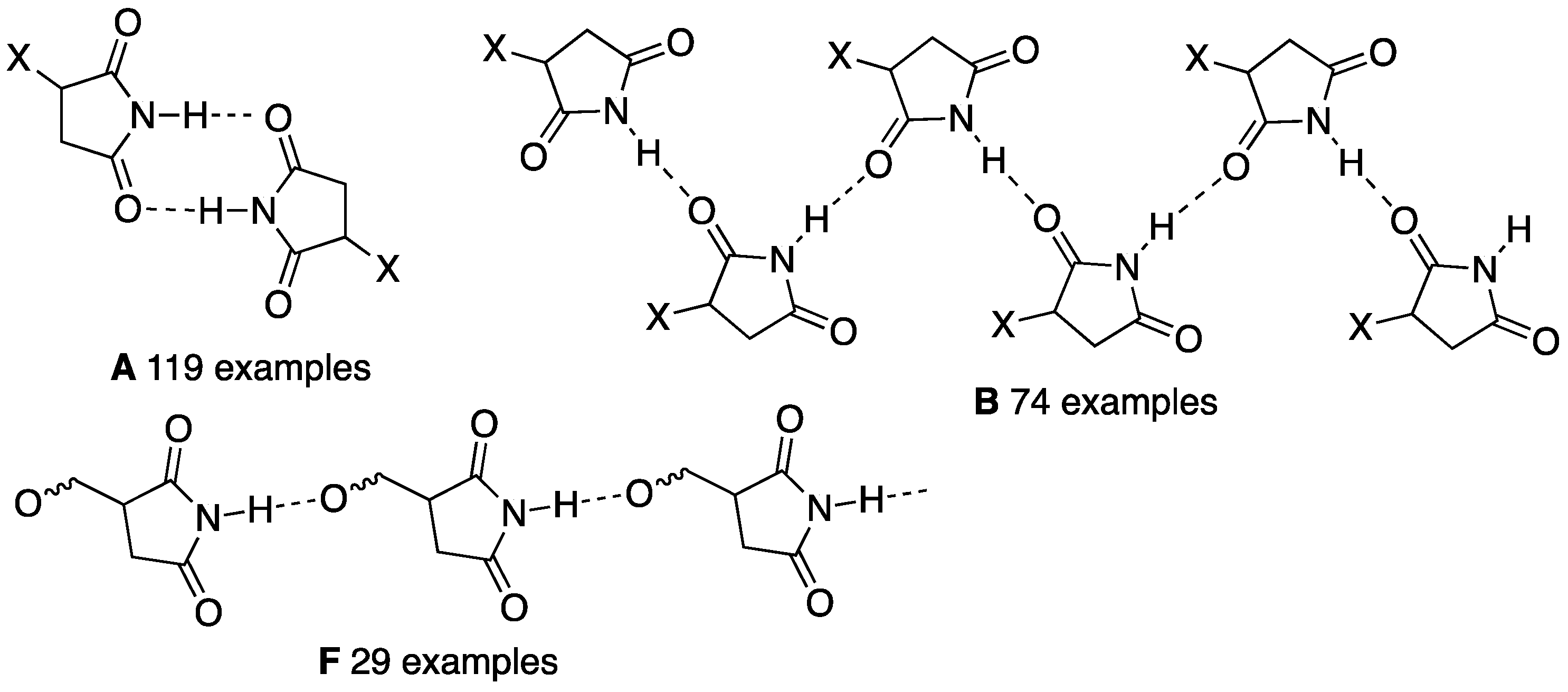
In a recent review in this journal [1] we surveyed the published crystal structures of over 300 cyclic NH carboximides containing the CO–NH–CO functional group and this revealed some important trends. In particular, two hydrogen bonded structures, namely, a centrosymmetric dimer A and a linear ribbon structure B involving two parallel rows of molecules exemplified for a simple monosubstituted succinimide (Figure 1) were by far the most common, accounting respectively for 119 and 74 examples among the total of 311 analysed. A further less common pattern F was one involving a chain formed by hydrogen bonding of NH to a remote non-imide oxygen elsewhere in the structure (29 examples). There were also less common modes of bonding such as hydrogen bonding of NH to a remote non-imide nitrogen (11 examples), dimers formed by bonding of imide NH to a remote oxygen (9 examples) or nitrogen (2 examples), hydrogen bonding of NH to imide CO in four molecules leading to a square shape (2 examples) and the equivalent to the centrosymmetric dimer but with bis imides leading to a doubly linked chain (11 examples). In addition, a host of more complex structures involving additional hydrogen bonding interactions with other functional groups were described.
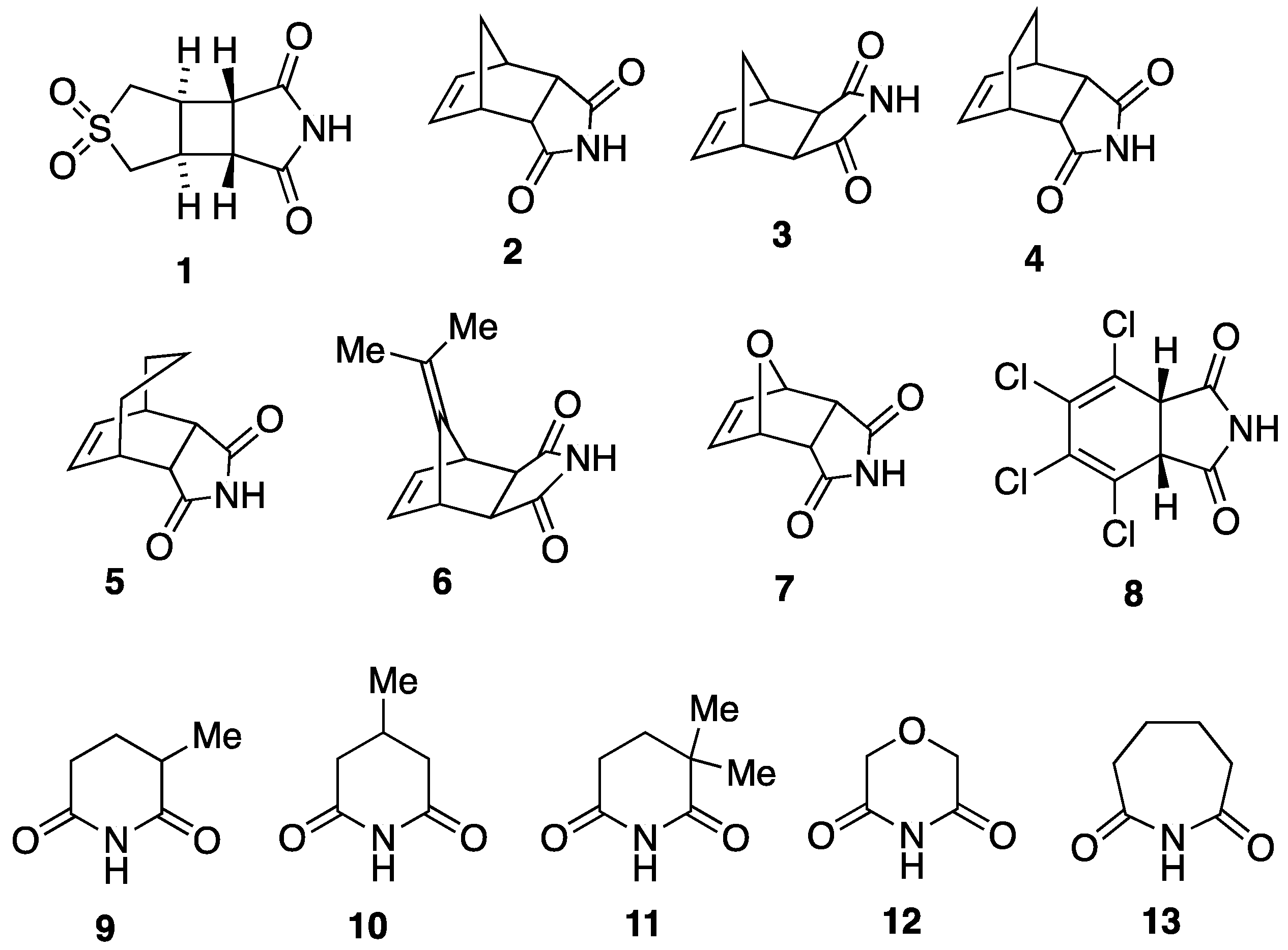
The review also highlighted some notable gaps in previous structural studies. In this paper, we describe the determination of 13 new cyclic NH imide crystal structures and analysis of their hydrogen bonding patterns, including those of seven new simple maleimide Diels–Alder adducts, three methylated glutarimides, and the first seven-membered ring imide adipimide (hexahydroazepine-2,7-dione). The structures 1–13 involved are summarised in Figure 2.
2. Materials and Methods
2.1. Synthesis of Imides 1–13
The cyclic imides, all previously known compounds, were prepared by reported methods and had physical and spectroscopic data in agreement with literature values. Specifically, 1 was prepared by photochemical [2 + 2]-cycloaddition between 3-sulfolene and maleimide in acetone [2], compounds 2, 4, 5, 6, 7 and 8 were prepared by Diels–Alder reaction of maleimide with, respectively, cyclopentadiene [3], 1,3-cyclohexadiene [4], 1,3-cycloheptadiene [5], 6,6-dimethylfulvene [6,7], furan [8], and tetrachlorothiophene 1,1-dioxide [9]. In the case of the furan adduct 7 it was essential to avoid undue heating since that resulted in isomerisation to the exo isomer whose structure has already been determined (see below). The exo-isomer 3 was prepared by reaction of the corresponding exo-anhydride with ammonium acetate in acetic acid [10]. Imides 9–12 were prepared by treatment of the corresponding dicarboxylic acids with ammonia; specifically, 9 was prepared by treatment of 2-methylglutaric acid with aqueous ammonia followed by evaporation to dryness and distillation [11], 10 was formed by reaction of 3-methylglutaric acid with thionyl chloride then aqueous ammonia followed by evaporation to dryness and distillation [12], 11 was prepared by reaction of 2,2-dimethylglutaric acid with urea at 160 °C [13], and 12 was prepared by treating diglycolic acid with aqueous ammonia followed by evaporation to dryness and distillation [14]. Imide 13 was prepared by treatment of ε-caprolactam with ozone in CCl4 [15].
2.2. Crystallography
Data were collected on either a Rigaku XtaLAB P200 diffractometer (Tokyo, Japan) using graphite-monochromated Mo (1, 3, 6, 7, 8, 10, 11, 12) or Cu (4, 5) radiation or a Rigaku XtaLAB P100 diffractometer using Cu radiation (2, 9, 13). Structures were solved by direct methods and refined by full-matrix least squares against F2 (SHELXL version 2018/3 [16]). The crystallographic data are summarised in detail in Table 1, Table 2 and Table 3. The cif files are available in the Supplementary Materials.
3. Results and Discussion
3.1. Cyclobutane-Fused Succimimide 1
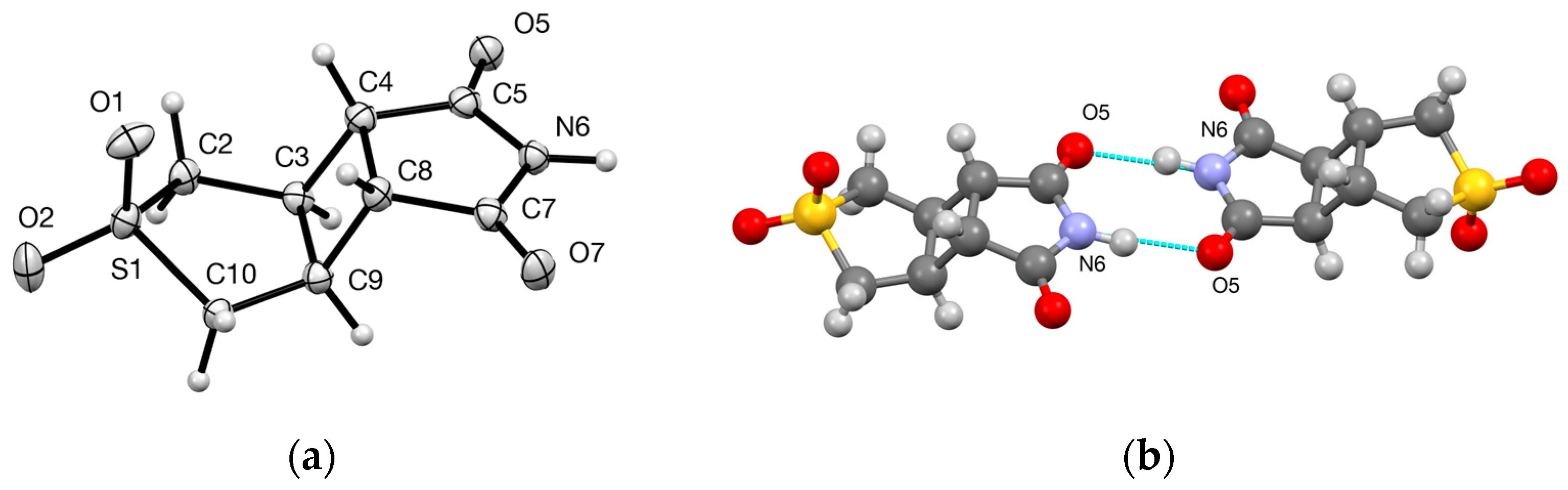
The molecular structure of 1 (Figure 3a) clearly confirms the anti-arrangement of the two five-membered rings with respect to the central cyclobutane. This is also consistent with that of the corresponding anhydride 14 (Figure 4) whose crystal structure was reported recently [17]. In the crystal the molecules are arranged in centrosymmetric hydrogen bonded dimers (Figure 3b) with parameters within the normal ranges (Table 4). None of the previously reported seven structures for cyclobutane-fused succinimides show this structure, which using the graph set notation developed by Etter and Bernstein [18,19] would be denoted R22(8).
3.2. Bi- and Tricyclic Diels–Alder Adducts of Maleimide 2–8
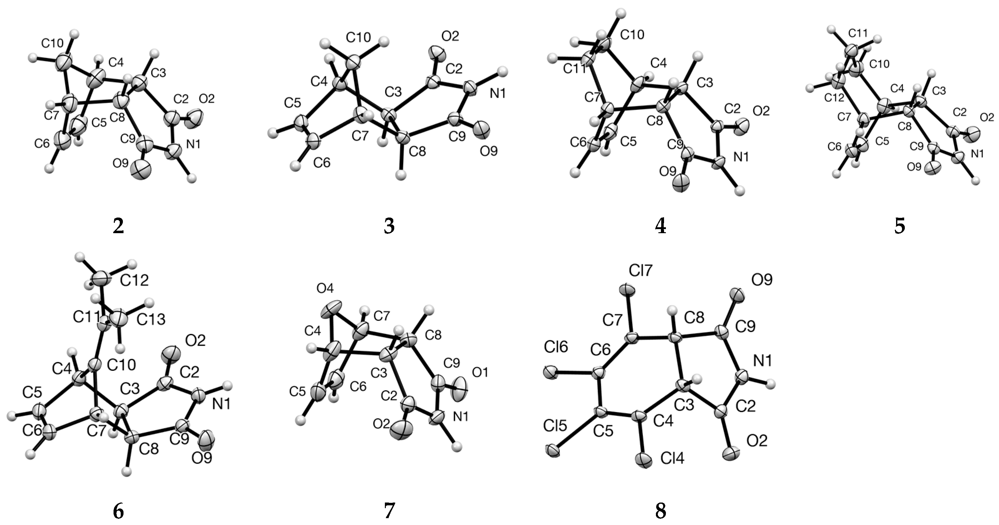
The Diels–Alder reaction of maleimide has been well investigated and at least 50 previous bicyclo[2.2.1] and [2.2.2] adducts derived in this way have been crystallographically characterised [1]. However, it is notable that these do not include many of the very simplest examples, such as the endo cyclopentadiene adduct 2 first reported as early as 1944 [20]. We report here the structure of both endo and exo cyclopentadiene adducts 2 and 3, the endo adducts from 1,3-cyclohexadiene and 1,3-cycloheptadiene 4 and 5, the exo dimethylfulvene adduct 6, the endo furan adduct 7 and the cis tetrachlorocyclohexadiene-fused compound 8 formed by cycloaddition with tetrachlorothiophene 1,1-dioxide followed by extrusion of SO2. The molecular structures (Figure 5) confirm the exo/endo configuration in each case. It is perhaps surprising that the C(11) bridge in 5 is oriented towards the apparently more hindered imide function rather than the double bond. It should be noted here that in a previous publication [21], the structure of a 2:1 adduct of dimethylfulvene with maleimide 15 was determined (Figure 4) which clearly results from a further diene addition onto the initial exo adduct 6. Despite this, the 1:1 adduct also obtained was shown with the endo configuration [21] which is clearly an error as now confirmed by the structure of 6.
All the molecular structures 2–8 show fairly conventional bond lengths and angles but when we come to the mode of hydrogen bonding an interesting pattern emerges. Only the endo cyclopentadiene adduct 2 forms the centrosymmetric R22(8) structure (Figure 6) while its exo isomer 3 as well as the higher homologues 4 and 5 and compounds 6 and 8 all form the C(4) ribbon structure (Figure 7). The hydrogen bonding parameters (Table 4) are standard. It should be noted that for compound 3 there are two independent molecules and these alternate in the hydrogen bonding pattern.
In our review [1], fifty Diels–Alder derived tricyclic succinimides were covered and fifteen of these show the centrosymmetric R22(8) dimer structure A while twenty show the linear C(4) ribbon structure B. The factors that favour one of these as opposed to the other are subtle and there seems to be a fine balance. The endo furan adduct 7 was of particular interest since the exo isomer 16 (Figure 4) was previously found to form the centrosymmetric dimer structure [22] but, by way of contrast, compound 7 adopts the linear C(6) structure (Figure 7) with NH hydrogen bonding to the bridging oxygen of the next molecule. It is worth noting that this is the structure also observed for several simple substituted derivatives such as 17 in the exo series [22] and 18 in the endo series [23] (Figure 4).
A further element of interest was the detection of a strong intermolecular halogen bonding interaction [24] in the structure of 8 between one ring C–Cl bond and the same imide carbonyl as is involved in hydrogen bonding to NH. Since this forms additional chains in a direction perpendicular to those formed by the hydrogen bonding interaction, the overall structure is quite complex and for simplicity the halogen bonding is shown separately (Figure 8). Examples in which a C–Cl halogen bonds to C=O seem to be rather uncommon but a comparable example is provided by sydnone-containing hydroximoyl chloride compound 23 [25].
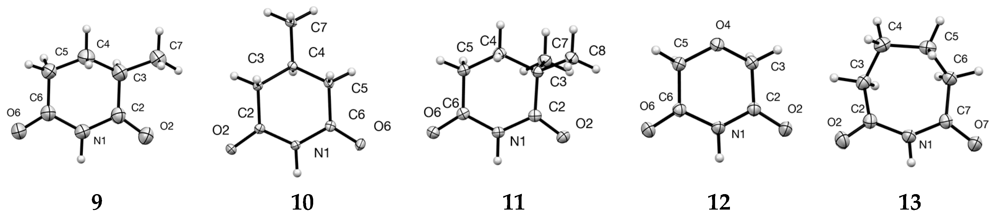
3.3. Ring-Methylated Glutarimides 9–11 and Morpholine Analogue 12
The structure of glutarimide (piperidine-2,6-dione) was determined at an early stage and it exhibits the C(4) ribbon structure B [26]. Very few simple derivatives have been structurally characterised so far and we describe here the structures of the 3- and 4-methyl derivatives 9 and 10 as well as the 3,3-dimethyl compound 11 (Figure 9). The morpholine-3,5-dione structure 12 is also included.
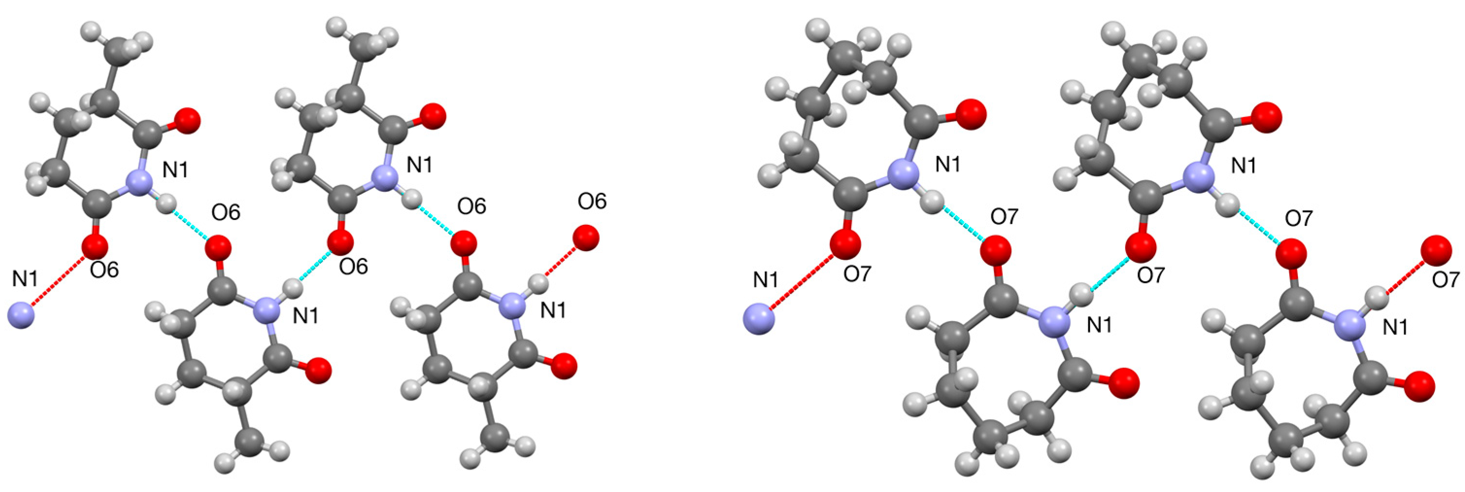
Among these four compounds only one, the 3-methylglutarimide 9, shows a C(4) ribbon structure (Figure 10) while the remaining three all adopt the centrosymmetric R22(8) dimer structure (Figure 6). Comparison with the few similar structures reported previously (Figure 4) shows that while the 4-ethyl-4-methyl compound 19 [27] and the 3,3,5,5-tetramethyl compound 21 [28] both have dimeric structures similar to 10 and 11, the 4,4-dimethyl compound 20 [29] forms a C(4) ribbon similar to 9. The dimer observed for 12 also contrasts with the ribbon structure reported for the thiomorpholine analogue 22 [30].
3.4. Adipimide 13
Although X-ray structures of five- and six-membered cyclic NH imides are very common and there a few structures of eight- and nine-membered rings [1], no previous examples of a seven-membered ring have been reported as far as we are aware. The molecular structure of the first such example, adipimide or hexahydroazepine-2,7-dione 13, showed a bent structure with C(4) and C(5) above the plane formed by the approximately coplanar remaining five ring atoms (Figure 9). This compound again adopted a C(4) ribbon structure (Figure 10).
4. Conclusions
In this paper we have filled in obvious gaps in the structural characterisation of cyclic NH carboximides. The observed hydrogen bonding patterns among the 13 structures fall mainly into the two commonest types for this class of compounds with seven adopting a C(4) ribbon structure and five exhibiting R22(8) centrosymmetric dimers. The remaining example has a less common C(6) structure with NH bonding to the bridging ether oxygen of the next molecule. Overall it is clear that there is a fine balance between the different possible hydrogen binding modes with closely similar compounds often giving different results.
Supplementary Materials
The following supporting information can be downloaded at: https://www.mdpi.com/article/10.3390/cryst13010150/s1, cif and checkcif files for 1–13.
Author Contributions
D.K.S. and A.J.B.N. prepared the compounds; A.M.Z.S. collected the X-ray diffraction data and solved the structures; R.A.A. devised the study, supervised the work, analysed the data and wrote the paper. All authors have read and agreed to the published version of the manuscript.
Funding
This research received no external funding.
Data Availability Statement
Conflicts of Interest
The authors declare no conflict of interest.
References
- Aitken, R.A.; Sonecha, D.K. The solid state structures of cyclic NH carboximides. Crystals 2020, 10, 606. [Google Scholar] [CrossRef]
- Shaikhrazieva, V.S.; Tolstikov, G.A.; Enikeev, R.S. Photoinitiated addition of maleic anhydride and its derivatives to 3-sulfolene. Zh. Org. Khim 1972, 8, 377–382. [Google Scholar]
- Michaelis, S.; Blechert, S. Ring-opening cross-metathesis (ROCM) as a novel tool for the ligation of peptides. Chem. Eur. J. 2007, 13, 2358–2368. [Google Scholar] [CrossRef]
- Fujimoto, M.; Okabe, K. Nouvelle méthode de synthèse des dérivés d’ethano-4,7-polyhydroisoindoline. Chem. Pharm. Bull. 1962, 10, 714–719. [Google Scholar] [CrossRef] [Green Version]
- Abou-Gharbia, M.; Patel, U.R.; Webb, M.B.; Moyer, J.A.; Andree, T.H.; Muth, E.A. Polycyclic aryl- and heteroarylpiperazinyl imides as 5-HT1A receptor ligands and potential anxiolytic agents: Synthesis and structure–activity relationship studies. J. Med. Chem. 1988, 31, 1382–1392. [Google Scholar] [CrossRef] [PubMed]
- Poos, G.I.; Lehman, M.M.; Landis, E.B.; Rosenau, J.D. Bicyclic bases. IV. Aryl substituted bridged hydroisoindolines. J. Med. Chem. 1962, 5, 883–896. [Google Scholar] [CrossRef] [PubMed]
- Graham, B.; Fayter, A.E.R.; Houston, J.E.; Evans, R.C.; Gibson, M.I. Facially amphipathic glycopolymers inhibit ice recrystallzation. J. Am. Chem. Soc. 2018, 140, 5682–5685. [Google Scholar] [CrossRef] [Green Version]
- Kwart, H.; Burchuk, I. Isomerism and adduct stability in the Diels-Alder reaction. I. The adducts of furan and maleimide. J. Am. Chem. Soc. 1952, 74, 3094–3097. [Google Scholar] [CrossRef]
- Raasch, M.S. Annelations with tetrachlorothiophene 1,1-dioxide. J. Org. Chem. 1980, 45, 856–867. [Google Scholar] [CrossRef]
- Kreituss, I.; Chen, K.-Y.; Eitel, S.H.; Adam, J.-M.; Wuitschik, G.; Fettes, A.; Bode, J.W. A robust, recyclable resin for decagram scale resolution of (±)-mefloquine and other chiral N-heterocycles. Angew. Chem. Int. Ed. 2016, 55, 1553–1556. [Google Scholar] [CrossRef] [Green Version]
- Aitken, R.A.; Farrell, D.M.M.; Kirton, E.H.M. Synthesis and pyrolysis of tetrahydro-1,4-oxazine-3,5-diones and tetrahydro-1,4-thiazine-3,5-diones. Chem. Heterocycl. Compd. 2001, 37, 1526–1531. [Google Scholar] [CrossRef]
- Sircar, S.S.G. The influence of groups and associated rings on the stability of certain heterocyclic ring systems. Part 1. The substituted glutarimides. J. Chem. Soc. 1927, 600–605. [Google Scholar] [CrossRef]
- Dabrowski, Z.; Cybulski, J. Structure and stereochemistry of lactams and cyclic imides. V. Mass spectrometry of substituted glutarimides. Bull. Acad. Pol. Sci.-Chim. 1981, 29, 11–16. [Google Scholar]
- Mizuno, H.; Manabe, A. Pyrimidine Compounds and Pests Controlling Composition Containing the Same. WO2004/99160 A1, 18 November 2004. [Google Scholar]
- Alekseeva, O.; Konstantinova, M.; Rasumovskii, S. Efficient method of cyclic imides synthesis under ozone influence by the example of ε-caprolactam oxidation reaction. Heteroatom Chem. 2008, 19, 661–666. [Google Scholar] [CrossRef]
- Sheldrick, G.M. A short history of SHELXL. Acta Crystallogr. Sect. A 2008, 64, 112–122. [Google Scholar] [CrossRef] [Green Version]
- Chen, T.-G.; Barton, L.M.; Lin, Y.; Tsien, J.; Kossler, D.; Bastida, I.; Asai, S.; Bi, C.; Chen, J.S.; Shan, M.; et al. Building C(sp3)-rich complexity by combining cycloaddition and C–C cross-coupling reactions. Nature 2018, 560, 350–354. [Google Scholar] [CrossRef]
- Etter, M.C. Encoding and decoding hydrogen-bond patterns of organic compounds. Acc. Chem. Res. 1990, 23, 120–126. [Google Scholar] [CrossRef]
- Etter, M.C.; MacDonald, J.C.; Bernstein, J. Graph-set analysis of hydrogen-bond patterns in organic crystals. Acta Crystallogr. Sect. B 1990, 46, 256–262. [Google Scholar] [CrossRef]
- Morgan, M.S.; Tipson, R.S.; Lowy, A.; Baldwin, W.E. Some derivatives of cis-3,6-endomethylene-Δ4-tetrahydrophthalic acid. J. Am. Chem. Soc. 1944, 66, 404–407. [Google Scholar] [CrossRef]
- Hong, B.-C.; Shr, Y.-J.; Liao, J.-H. Unprecedented microwave effects on the cycloaddition of fulvenes. A new approach to the construction of polycyclic ring systems. Org. Lett. 2002, 4, 663–666. [Google Scholar] [CrossRef]
- Struga, M.; Miroslaw, B.; Pakosinska-Parys, M.; Drzewiecka, A.; Borowski, P.; Kossakowski, J.; Koziol, A.E. Synthesis, characterization and supramolecular synthons in crystals of new derivatives of 10-oxa-4-azatricyclo[5.2.1.02,6]dec-8-ene-3,5-dione. J. Mol. Struct. 2010, 965, 23–30. [Google Scholar] [CrossRef]
- Bennett, G.D.; Spengler, A.O.; Wheeler, K.A. CSD Communication; Cambridge Crystallographic Data Centre: Cambridge, UK, 2016; BAMSUG. [Google Scholar]
- Cavallo, G.; Metrangolo, P.; Milani, R.; Pilati, T.; Priimagi, A.; Resnati, G.; Terraneo, G. The halogen bond. Chem. Rev. 2016, 116, 2478–2601. [Google Scholar] [CrossRef] [PubMed]
- Shih, M.-H.; Yeh, M.-Y.; Lee, M.-J.; Su, Y.-S. Efficient syntheses of 3-(3-arylsydnon-4-yl)triazole derivatives. Synthesis 2004, 2877–2885. [Google Scholar] [CrossRef]
- Petersen, C.S. The crystal structure of glutarimide. Acta Chem. Scand. 1971, 25, 379. [Google Scholar] [CrossRef]
- Tutughamiarso, M.; Bolte, M. 4-Ethyl-4-methyl piperidine-2,6-dione. Acta Crystallogr. Sect. E 2007, 63, o4743. [Google Scholar] [CrossRef]
- Maurin, J.K.; Czarnocki, Z.; Paluchowska, B.; Winnicka-Maurin, M. Conformation-related reaction efficiency of glutarimides with phenyllithium. Structures of 3,3,5,5-tetramethylglutarimide and 2-hydroxy-2-phenyl-3,3,5,5-tetramethyl-6-piperidone. X-ray and theoretical study. Acta Crystallogr. Sect. B 1997, 53, 719–725. [Google Scholar] [CrossRef]
- Bocelli, G.; Grenier-Loustalot, M.F. The structure of 4,4-dimethylazacyclohexane-2,6-dione (4,4-dimethyl-2,6-piperidinedione). Acta Crystallogr. Sect. B 1981, 37, 1302–1304. [Google Scholar] [CrossRef]
- Aitken, R.A.; Slawin, A.M.Z.; Yeh, P.-P. Tetrahydro-1,4-thiazine-3,5-dione. Molbank 2018, 2018, M1036. [Google Scholar] [CrossRef]
Figure 1.
Most common previously reported hydrogen bonding patterns.

Figure 2.
Cyclic NH imides for which structures were determined.

Figure 3.
(a) Molecular structure of 1 with numbering system used (50% thermal ellipsoids), (b) hydrogen bonding pattern for 1.
Figure 3.
(a) Molecular structure of 1 with numbering system used (50% thermal ellipsoids), (b) hydrogen bonding pattern for 1.

Figure 4.
Structure of crystallographically characterised reference compounds 14–22 with CCDC Ref. Codes and literature references.
Figure 4.
Structure of crystallographically characterised reference compounds 14–22 with CCDC Ref. Codes and literature references.

Figure 5.
Molecular structures for Diels–Alder derived compounds 2–8 showing numbering systems used (50% probability ellipsoids).
Figure 5.
Molecular structures for Diels–Alder derived compounds 2–8 showing numbering systems used (50% probability ellipsoids).

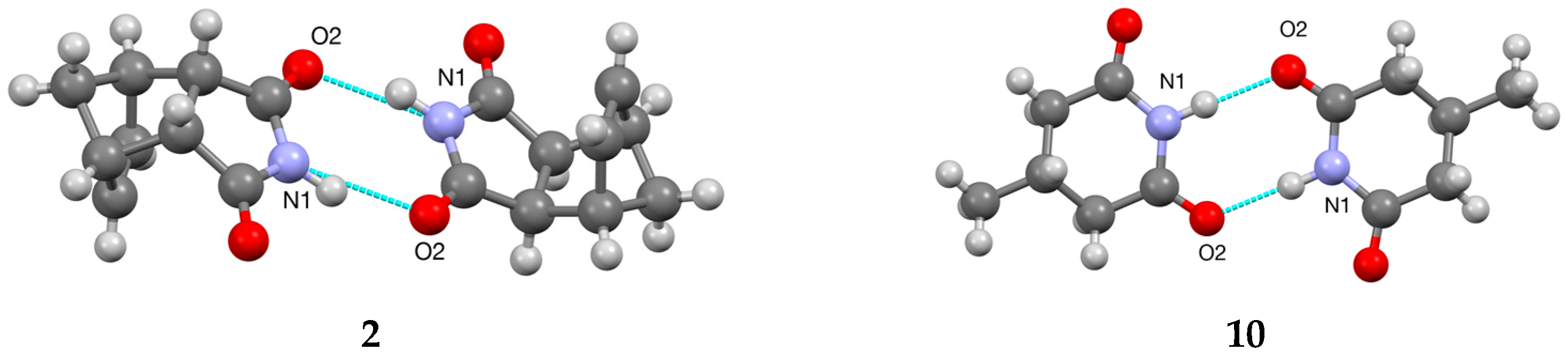
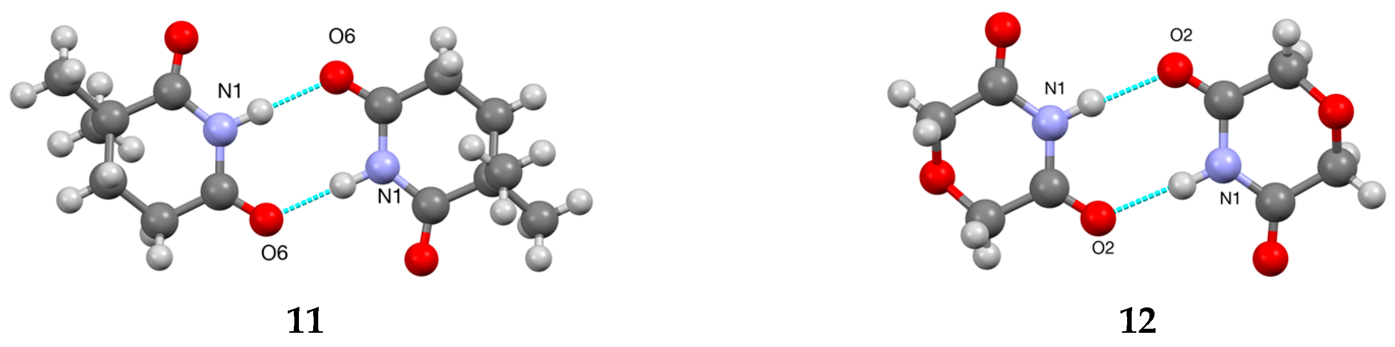
Figure 6.
Hydrogen bonded R22(8) dimer structures for 2, 10, 11 and 12.


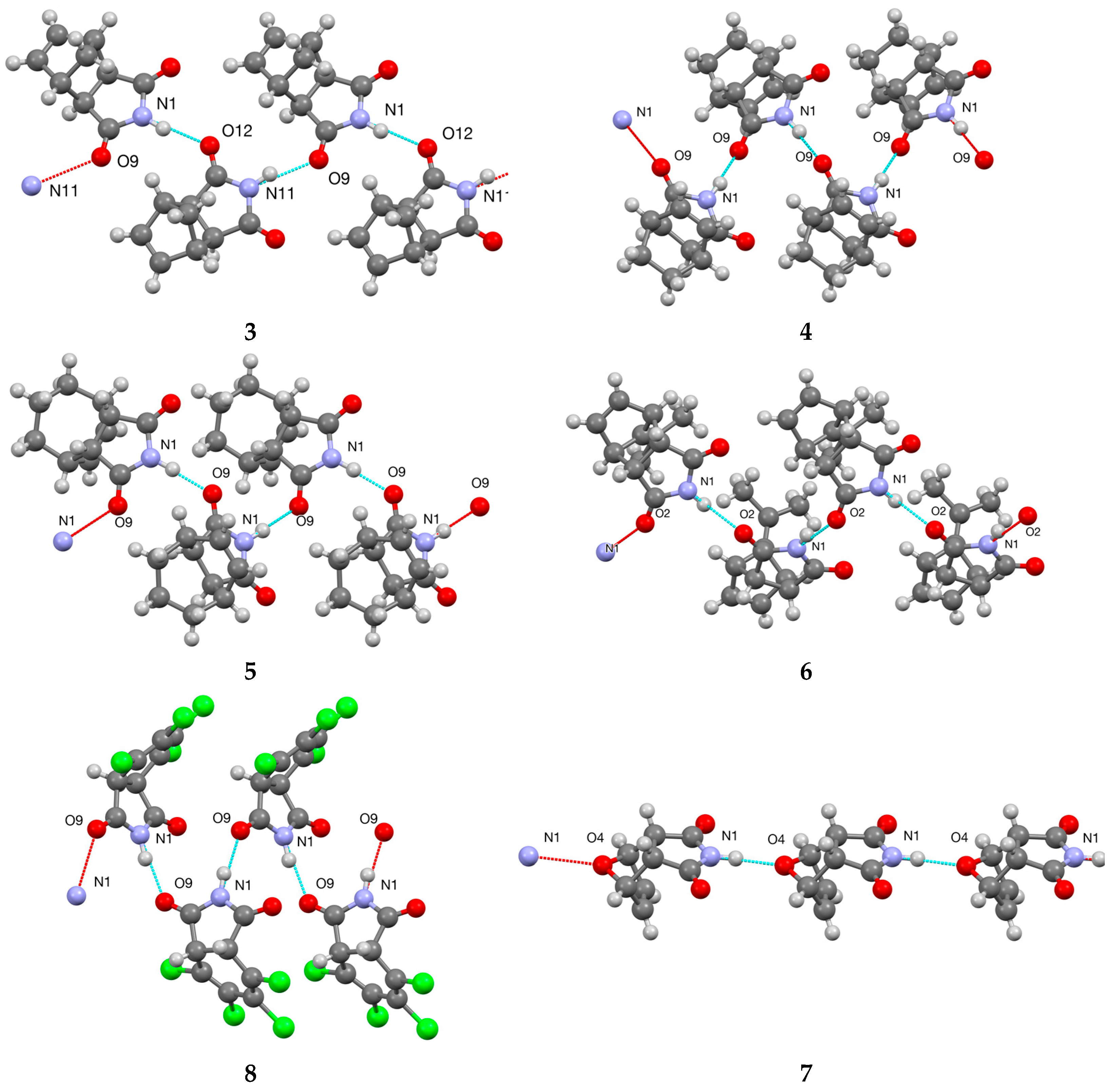
Figure 7.
Hydrogen bonded C(4) structures for 3, 4, 5, 6 and 8 and C(6) structure for 7.

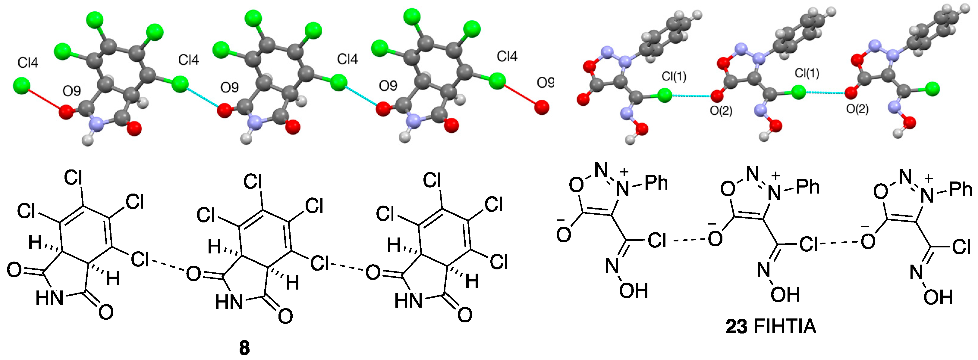
Figure 8.
Halogen bonding interaction in structure of 8 [C–Cl…O parameters: C(4)–Cl(4) 1.722(2), Cl(4)…O(9) 2.940(1), C(4)–O(9) 4.569(2) Å, angle 156.26(6)°] and for comparison 23 [C–Cl…O parameters: C(9)–Cl(1) 1.717(2), Cl(1)…O(2) 3.150(2), C(9)–O(2) 4.861(3) Å, angle 173.57(9)°].
Figure 8.
Halogen bonding interaction in structure of 8 [C–Cl…O parameters: C(4)–Cl(4) 1.722(2), Cl(4)…O(9) 2.940(1), C(4)–O(9) 4.569(2) Å, angle 156.26(6)°] and for comparison 23 [C–Cl…O parameters: C(9)–Cl(1) 1.717(2), Cl(1)…O(2) 3.150(2), C(9)–O(2) 4.861(3) Å, angle 173.57(9)°].

Figure 9.
Molecular structures for glutarimides 9–11, morpholine analogue 12 and adipamide 13 showing numbering systems used (50% probability ellipsoids).
Figure 9.
Molecular structures for glutarimides 9–11, morpholine analogue 12 and adipamide 13 showing numbering systems used (50% probability ellipsoids).

Figure 10.
Hydrogen bonded C(4) structures for 9 and 13.


Table 1.
Summary of crystallographic data obtained for compounds 1–4.
| Compound | 1 | 2 | 3 | 4 |
|---|---|---|---|---|
| CCDC deposit no. | 2225715 | 2225716 | 2225713 | 2225707 |
| Empirical formula | C8H9NO4S | C9H9NO2 | C9H9NO2 | C10H11NO2 |
| Crystal system | monoclinic | monoclinic | monoclinic | monoclinic |
| Space group | P21/n (No. 14) | P21/c (No. 14) | P21/c (No. 14) | P21/c (No. 14) |
| Temperature (K) | 93 | 173 | 93 | 125 |
| Crystal form | colourless prism | colourless prism | colourless prism | colourless prism |
| Size (mm) | 0.10 × 0.10 × 0.03 | 0.12 × 0.03 × 0.03 | 0.20 × 0.10 × 0.10 | 0.12 × 0.05 × 0.02 |
| Unit cell | a = 6.0139 (3) | a = 11.1131 (3) | a = 8.394 (2) Å | a = 10.3980 (2) |
| dimensions (Å, °) | b = 14.2212 (6) | b = 6.07791 (12) | b = 16.964 (5) | b = 6.53350 (10) |
| c = 10.2142 (5) | c = 12.2001 (3) | c = 10.681 (3) | c = 12.8770 (2) | |
| β = 100.804 (5) | β = 100.690 (3) | β = 90.818 (7) | β = 105.469 (2) | |
| Volume (Å3) | 858.08 (7) | 770.90 (4) | 1520.8 (7) | 843.11 (3) |
| Z | 4 | 4 | 8 | 4 |
| Dc (g cm−3) | 1.666 | 1.406 | 1.425 | 1.396 |
| Absorption coefficient | 0.363 mm−1 | 0.830 mm−1 | 0.102 mm−1 | 0.802 mm−1 |
| Radiation type, wavelength | Mo Kα, 0.71073 Å | Cu Kα, 1.54184 Å | Mo Kα, 0.71075 Å | Cu Kα, 1.54184 Å |
| F(000) | 448.00 | 344.00 | 688.00 | 376.00 |
| θ range | 2.484–28.235° | 4.253–68.319° | 2.253–25.355° | 4.412–75.133° |
| Limiting indices | –7 ≤ h ≤ 7, –18 ≤ k ≤ 18, –12 ≤ l ≤ 13 | –13 ≤ h ≤ 11, –6 ≤ k ≤ 7, –14 ≤ l ≤ 14 | –10 ≤ h ≤ 8, –14 ≤ k ≤ 20, –12 ≤ l ≤ 12 | –12 ≤ h ≤ 12, –8 ≤ k ≤ 8, –16 ≤ l ≤ 15 |
| Reflns collected/unique | 12460/1899 | 7229/1409 | 7224/2745 | 9087/1705 |
| Rint | 0.0799 | 0.0216 | 0.0281 | 0.0159 |
| Data/restraints/parameters | 1899/1/131 | 1409/1/114 | 2745/2/226 | 1705/1/122 |
| Data with I > 2σ (I) | 1659 | 1385 | 2584 | 1672 |
| Goodness of fit on F2 | 1.175 | 1.068 | 1.048 | 1.094 |
| R1, wR2 (data I > 2σ (I)) | 0.0505, 0.1563 | 0.0781, 0.1697 | 0.0393, 0.1062 | 0.0389, 0.1039 |
| R1, wR2 (all data) | 0.0552, 0.1584 | 0.0783, 0.1703 | 0.0410, 0.1075 | 0.0394, 0.1044 |
| Largest diff. peak/hole (e Å2) | 0.77 and −0.55 | 0.42 and −0.68 | 0.48 and −0.26 | 0.28 and −0.24 |
Table 2.
Summary of crystallographic data obtained for compounds 5–8.
| Compound | 5 | 6 | 7 | 8 |
|---|---|---|---|---|
| CCDC deposit no. | 2225709 | 2225714 | 2225712 | 2225711 |
| Empirical formula | C11H13NO2 | C12H13NO2 | C8H7NO3 | C8H3Cl4NO2 |
| Crystal system | monoclinic | monoclinic | orthorhombic | monoclinic |
| Space group | P21/c (No. 14) | P21/c (No. 14) | P212121 (No. 19) | P21/c (No. 14) |
| Temperature (K) | 125 | 173 | 173 | 93 |
| Crystal form | colourless plate | colourless prism | colourless prism | colourless plate |
| Size (mm) | 0.12 × 0.04 × 0.01 | 0.12 × 0.10 × 0.03 | 0.15 × 0.15 × 0.08 | 0.10 × 0.10 × 0.02 |
| Unit cell | a = 11.3039 (4) | a = 10.978 (3) | a = 7.0068 (7) | a = 6.4439 (4) |
| dimensions (Å, °) | b = 7.28194 (18) | b = 11.441 (3) | b = 8.1073 (8) | b = 5.8690 (3) |
| c = 12.3790 (4) | c = 8.122 (2) | c = 12.7199 (13) | c = 26.8276 (19) | |
| β = 113.542 (2) | β = 92.417 (7) | — | β = 95.696 (6) | |
| Volume (Å3) | 934.16 (6) | 1019.2 (5) | 722.57 (13) | 1009.59 (11) |
| Z | 4 | 4 | 4 | 4 |
| Dc (g cm−3) | 1.360 | 1.324 | 1.518 | 1.888 |
| Absorption coefficient | 0.763 mm−1 | 0.090 mm−1 | 0.118 mm−1 | 1.143 mm−1 |
| Radiation type, wavelength | Cu Kα, 1.54184 Å | Mo Kα, 0.71075 Å | Mo Kα, 0.71075 Å | Mo Kα, 0.71075 Å |
| F(000) | 408.00 | 432.00 | 344.00 | 568.00 |
| θ range | 4.266–75.347° | 1.857–25.361° | 2.980–25.375° | 3.052–28.139° |
| Limiting indices | −13 ≤ h ≤ 14, −8 ≤ k ≤ 9, −15 ≤ l ≤ 15 | −13 ≤ h ≤ 13, −13 ≤ k ≤ 13, –9 ≤ l ≤ 9 | −8 ≤ h ≤ 7, −9 ≤ k ≤ 9, −15 ≤ l≤ 14 | −8 ≤ h ≤ 8, −7 ≤ k ≤ 6, −32 ≤ l ≤ 32 |
| Reflns collected/unique | 10,206/1886 | 11,923/1856 | 8213/1307 | 9882/2148 |
| Rint | 0.0172 | 0.0286 | 0.0295 | 0.0365 |
| Data/restraints/parameters | 1886/1/131 | 1856/0/142 | 1307/1/113 | 2148/1/140 |
| Data with I > 2σ (I) | 1845 | 1743 | 1296 | 1972 |
| Goodness of fit on F2 | 1.041 | 1.090 | 1.022 | 1.006 |
| R1, wR2 (data I > 2σ (I)) | 0.0360, 0.0967 | 0.0370, 0.0969 | 0.0267, 0.0815 | 0.0303, 0.0848 |
| R1, wR2 (all data) | 0.0364, 0.0970 | 0.0385, 0.0981 | 0.0269, 0.0818 | 0.0324, 0.0862 |
| Largest diff. peak/hole (e Å2) | 0.25 and −0.19 | 0.21 and −0.20 | 0.15 and −0.18 | 0.58 and −0.28 |
Table 3.
Summary of crystallographic data obtained for compounds 9–13.
| Compound | 9 | 10 | 11 | 12 | 13 |
|---|---|---|---|---|---|
| CCDC deposit no. | 2225705 | 2225704 | 2225708 | 2225710 | 2225706 |
| Empirical formula | C6H9NO2 | C6H9NO2 | C7H11NO2 | C4H5NO3 | C6H9NO2 |
| Crystal system | monoclinic | monoclinic | monoclinic | triclinic | monoclinic |
| Space group | P21/c (No. 14) | P21/c (No. 14) | P21/n (No. 14) | P–1 (No. 2) | P21/n (No. 14) |
| Temperature (K) | 173 | 93 | 93 | 173 | 173 |
| Crystal form | colourless platelet | colourless prism | colourless prism | colourless prism | colourless prism |
| Size (mm) | 0.10 × 0.10 × 0.01 | 0.10 × 0.10 × 0.03 | 0.20 × 0.03 × 0.03 | 0.15 × 0.05 × 0.05 | 0.15 × 0.12 × 0.03 |
| Unit cell | a = 11.795 (8) | a = 10.231 (4) | a = 15.276 (3) | a = 3.92406 (12) | a = 10.9883 (2) |
| dimensions | b = 7.040 (4) | b = 5.949 (2) | b = 6.3447 (11) | b = 6.67474 (18) | b = 7.18070 (11) |
| (Å, °) | c = 7.721 (5) | c = 10.412 (4) | c = 15.358 (3) | c = 9.7328 (3) | c = 8.21778 (18) |
| α = 76.716 (2) | |||||
| β = 103.355 (8) | β = 95.784 (8) | β = 90.108 (5) | β = 82.516 (2) | β = 109.066 (2) | |
| γ = 77.665 (2) | |||||
| Volume (Å3) | 623.8 (7) | 630.5 (4) | 1488.5 (5) | 241.489 (13) | 612.84 (2) |
| Z | 4 | 4 | 8 | 2 | 4 |
| Dc (g cm−3) | 1.354 | 1.339 | 1.260 | 1.583 | 1.378 |
| Absorp’n coefficient | 0.853 mm−1 | 0.101 mm−1 | 0.092 mm−1 | 0.137 mm−1 | 0.868 mm−1 |
| Radiation type, wavelength | Cu Kα, 1.54187 Å | Mo Kα, 0.71075 Å | Mo Kα, 0.71075 Å | Mo Kα, 0.71075 Å | Cu Kα, 1.54184 Å |
| F(000) | 272.00 | 272.00 | 608.00 | 120.00 | 272.00 |
| θ range | 3.852–67.810° | 2.001–25.339° | 2.653–25.335° | 2.158–28.247° | 4.257–68.071° |
| Limiting indices | −13 ≤ h ≤ 14, −7 ≤ k ≤ 8, −8 ≤ l ≤ 9 | −10 ≤ h ≤ 12, −5 ≤ k ≤ 7, −11 ≤ l ≤ 12 | −18 ≤ h ≤ 18, −7 ≤ k≤ 7, −18 ≤ l ≤ 18 | −5 ≤ h ≤ 4, −8 ≤ k ≤ 8, −12 ≤ l ≤ 12 | −13 ≤ h ≤ 12, −5 ≤ k ≤ 8, −9 ≤ l ≤ 9 |
| Refln total/unique | 6167/1122 | 3953/1145 | 15821/2699 | 7485/1052 | 5849/1111 |
| Rint | 0.0670 | 0.0300 | 0.0399 | 0.0254 | 0.0174 |
| Data/restraints /parameters | 1122/1/88 | 1145/1/87 | 2699/2/194 | 1052/1/77 | 1111/187 |
| Data I > 2σ (I) | 1061 | 1067 | 2490 | 965 | 1100 |
| Goodness of fit F2 | 1.081 | 1.090 | 1.196 | 1.076 | 1.273 |
| R1, wR2 (I > 2σ (I)) | 0.0643, 0.1706 | 0.0461, 0.1488 | 0.0452, 0.1592 | 0.0292, 0.0838 | 0.0664, 0.1431 |
| R1, wR2 (all data) | 0.0657, 0.1726 | 0.0615, 0.2137 | 0.0486, 0.1618 | 0.0318, 0.0853 | 0.0666, 0.1433 |
| Largest diff. peak/hole (e Å2) | 0.62 and −0.29 | 0.49 and −0.62 | 0.27 and −0.17 | 0.32 and −0.21 | 0.40 and −0.65 |
Table 4.
Hydrogen bonding parameters (Å, °).
| Compound | D–H…A | D–A | D–H | H…A | Angle DHA |
|---|---|---|---|---|---|
| 1 | N(6)–H(6)…O(5) | 2.906(2) | 0.97(2) | 1.94(2) | 170(2) |
| 2 | N(1)–H(1)…O(2) | 2.8438(15) | 0.977(15) | 1.902(15) | 160.9(14) |
| 3 | N(1)–H(1)…O(12) | 2.9057(16) | 0.975(13) | 1.946(12) | 167.6(14) |
| N(11)–H(11)…O(9) | 2.9036(16) | 0.972(13) | 1.963(12) | 162.1(13) | |
| 4 | N(1)–H(1)…O(9) | 2.8432(12) | 0.975(14) | 1.878(13) | 169.8(13) |
| 5 | N(1)–H(1)…O(9) | 2.7991(11) | 0.973(11) | 1.832(11) | 172.4(17) |
| 6 | N(1)–H(1)…O(2) | 2.8616(15) | 0.880(17) | 2.020(17) | 159.8(15) |
| 7 | N(1)–H(1)…O(4) | 2.8735(19) | 0.975(8) | 1.909(8) | 170.0(19) |
| 8 | N(1)–H(1)…O(9) | 2.8576(18) | 0.975(16) | 1.897(18) | 168(2) |
| 9 | N(1)–H(1)…O(6) | 2.908(2) | 0.978(17) | 1.935(18) | 173(2) |
| 10 | N(1)–H(1)…O(2) | 2.9300(17) | 0.973(13) | 1.957(13) | 177.0(16) |
| 11 | N(1)–H(1)…O(6) | 2.849(2) | 0.977(17) | 1.871(17) | 179.1(18) |
| N(11)–H(11)…O(16) | 2.883(2) | 0.977(19) | 1.907(19) | 178(2) | |
| 12 | N(1)–H(1)…O(2) | 2.8872(11) | 0.971(11) | 1.920(11) | 173.3(13) |
| 13 | N(1)–H(1)…O(7) | 2.9278(17) | 0.975(17) | 1.957(17) | 173.1(14) |
Disclaimer/Publisher’s Note: The statements, opinions and data contained in all publications are solely those of the individual author(s) and contributor(s) and not of MDPI and/or the editor(s). MDPI and/or the editor(s) disclaim responsibility for any injury to people or property resulting from any ideas, methods, instructions or products referred to in the content. |
© 2023 by the authors. Licensee MDPI, Basel, Switzerland. This article is an open access article distributed under the terms and conditions of the Creative Commons Attribution (CC BY) license (https://creativecommons.org/licenses/by/4.0/).
Share and Cite
MDPI and ACS Style
Aitken, R.A.; Nelson, A.J.B.; Slawin, A.M.Z.; Sonecha, D.K. Solid State Structure and Hydrogen Bonding of Some Cyclic NH Carboximides. Crystals 2023, 13, 150. https://doi.org/10.3390/cryst13010150
AMA Style
Aitken RA, Nelson AJB, Slawin AMZ, Sonecha DK. Solid State Structure and Hydrogen Bonding of Some Cyclic NH Carboximides. Crystals. 2023; 13(1):150. https://doi.org/10.3390/cryst13010150
Chicago/Turabian StyleAitken, R. Alan, Alexander J. B. Nelson, Alexandra M. Z. Slawin, and Dheirya K. Sonecha. 2023. "Solid State Structure and Hydrogen Bonding of Some Cyclic NH Carboximides" Crystals 13, no. 1: 150. https://doi.org/10.3390/cryst13010150
Note that from the first issue of 2016, this journal uses article numbers instead of page numbers. See further details here.