Review

The Future of Regenerative Medicine: Cell Therapy Using Pluripotent Stem Cells and Acellular Therapies Based on Extracellular Vesicles

by
Margot Jarrige
1,2,3,†,
Elie Frank
1,2,3,†,
Elise Herardot
1,2,†,
Sabrina Martineau
1,2,
Annabelle Darle
1,2,3,
Manon Benabides
1,2,3,
Sophie Domingues
1,2,3,
Olivier Chose
1,2,3,
Walter Habeler
1,2,3,
Judith Lorant
1,2,
Christine Baldeschi
1,2,
Cécile Martinat
1,2,
Christelle Monville
1,2,
Lise Morizur
1,2,3,*,‡ and
Karim Ben M’Barek
1,2,3,*,‡
1
INSERM U861, I-Stem, AFM, Institute for Stem Cell Therapy and Exploration of Monogenic Diseases, 91100 Corbeil-Essonnes, France
2
Université Paris-Saclay, Université d’Evry, U861, 91100 Corbeil-Essonnes, France
3
Centre d’Etude des Cellules Souches, 91100 Corbeil-Essonnes, France
*
Authors to whom correspondence should be addressed.
Equal contribution.
These authors share equal senior authorship.
Cells 2021, 10(2), 240; https://doi.org/10.3390/cells10020240
Submission received: 30 November 2020 / Revised: 13 January 2021 / Accepted: 23 January 2021 / Published: 27 January 2021
(This article belongs to the Special Issue Feature Papers in Stem Cells)

Abstract

:
The rapid progress in the field of stem cell research has laid strong foundations for their use in regenerative medicine applications of injured or diseased tissues. Growing evidences indicate that some observed therapeutic outcomes of stem cell-based therapy are due to paracrine effects rather than long-term engraftment and survival of transplanted cells. Given their ability to cross biological barriers and mediate intercellular information transfer of bioactive molecules, extracellular vesicles are being explored as potential cell-free therapeutic agents. In this review, we first discuss the state of the art of regenerative medicine and its current limitations and challenges, with particular attention on pluripotent stem cell-derived products to repair organs like the eye, heart, skeletal muscle and skin. We then focus on emerging beneficial roles of extracellular vesicles to alleviate these pathological conditions and address hurdles and operational issues of this acellular strategy. Finally, we discuss future directions and examine how careful integration of different approaches presented in this review could help to potentiate therapeutic results in preclinical models and their good manufacturing practice (GMP) implementation for future clinical trials.

1. Introduction

In the background of many diseases, the degeneration and/or dysfunction of a particular cell type affects tissues and organs, which will deteriorate over time and lose their functions. In this context, regenerative medicine offers new perspectives for pathologies that otherwise remain untreated [1,2]. Hundreds of clinical trials have been initiated in the last few years for a large panel of indications. Strategies are more and more refined to comply with large-scale productions and identify the most efficient formulation to maximize therapeutic effects [3]. Tissue engineering has recently emerged and constitutes the second generation of cell-based regenerative therapy by incorporating three-dimensional (3D) biodegradable compounds mimicking the extracellular matrix and/or multiple cell types [4,5].
Besides the therapeutic effects of cells elicited by their direct presence, some of the observed benefits are mediated by their indirect actions and, as such, may not require them [6,7]. Extracellular vesicles (EVs) have emerged as important mediators of paracrine signaling and could exert such functions [8,9,10]. Intensive research on EVs over the last half-century has led to an in-depth understanding of their origin and biological functions and has placed them at the forefront of various disease treatments.
Herein, we first review the state of the art of cell therapy and its current limitations and challenges, with a particular focus on pluripotent stem cell-derived therapeutic products. In a second part, we elaborate on functions of EVs and discuss their interest in the field of “acellular therapies” for pathological conditions that affect the eye, skin, heart and skeletal muscle. Finally, we discuss the future prospects of these therapies and their combination to potentiate therapeutic outcomes.

2. Regenerative Medicine: State of the Art

2.1. Introduction

The source material for cell and tissue-based products is a major point to consider for regenerative medicine. A prerequisite for the broad application of cell products is the production of large quantities of cells, with consistency between different batches or manufacturing processes to ensure the same quality and therapeutic efficacy to each patient.
This section will describe the numerous cell sources that may be used in cell therapy and their use in selected pathological conditions. In addition, we will discuss the transition from cell therapies formulated as a cell suspension to more complex tissue-engineered products.

2.2. Cell Sources

Various cell sources have been considered for cell therapy and regenerative medicine, including adult material from living donors or cadavers, fetal materials and pluripotent stem cell lines.
Cell material from adult origin can be obtained directly from patients and purified/amplified in vitro (like mesenchymal stem cells (MSCs) or epithelial cells from the skin). Such autologous approach prevents the risk of rejection. However, the manufacturing process and supply logistics of autologous cell-based therapy products are highly complicated and hinder their scale out. These limitations are particularly important when addressing widespread diseases that affect millions of patients like Age-related Macular Degeneration (AMD) or when the number of cells needed for therapy is important, as in the case of large burns. Alternatively, cells can be obtained from cadavers or, when possible, living donors. For all adult sources, donor variability—histocompatibility, age, genotype, etc.—drives cell product variability. In addition, adult cell sources have a reduced potential for expansion. This is especially true for terminally differentiated adult cell types (i.e., skeletal muscle cells) that hardly proliferate, or for organs for which there is limited access to endogenous stem cells.
Another source of materials for cell therapy is aborted fetuses. Harvested cells retain a proliferation potential that is superior to that of adult cells. Although fetus stem/progenitor cells are restricted in their potentiality, giving rise to limited cell types, some of these cells are no longer present in adult tissues and could thus be used to generate specific cell types. Nevertheless, their access is highly variable around the world, depending on the abortion law and policy in place in each country. Indeed, in some countries, it is strictly prohibited; in others, abortion is prohibited after different gestational ages, which could be restrictive. The use of human fetal tissue also raises a number of ethical considerations. Besides these restrictions, the access to fetuses at a defined gestational age is complex, as highlighted for Huntington’s disease (HD) cell therapy as an example [11]. Clinical trials have aimed to restore degenerated striatal cells in the brain of HD patients through the transplantation of fetal ganglionic eminences (GE) containing future striatal cells. In the MIG-HD clinical study, 86 fetal GE tissues were grafted into HD patients but a total of 163 surgeries were cancelled due to inadequate/insufficient donor fetal material as fetal cells should be grafted within 2 days following abortion [12].
Since their first derivation, human pluripotent stem cells (hPSCs) have been considered a promising cell source for regenerative medicine. hPSCs are self-renewable and give rise to any cell type of the human body. They can be obtained from supernumerary in vitro fertilized embryos (human embryonic stem cells or hESCs) or after the conversion of adult primary cells to pluripotency by the overexpression of a cocktail of factors (human induced pluripotent stem cells or hiPSCs) [13,14,15]. hPSCs are compatible with large-scale industrial productions in good manufacturing practices (GMP) facilities and quality controlled as any other more conventional pharmaceutical products. Whereas the majority of hPSC-based clinical trials have used hESCs up to now, the field is moving toward hiPSCs as they do not require the destruction of embryos and consequently can be used worldwide without restriction [1].
In the following section, we highlight some recent therapeutic applications of hPSC-based cell therapy products.

2.3. hPSC-Based Cell Therapy: Selected Examples

2.3.1. Retinal Degeneration

Different causes of visual impairment, such as AMD, Retinitis Pigmentosa (RP) and diabetic retinopathy [16], are characterized by the degeneration of the light-sensitive part of the eye: the retina [17]. Depending on the nature and/or stage of the disease, different cell types are susceptible to degeneration. In particular, the retinal pigment epithelium (RPE), an essential supportive tissue of the retina affected in AMD and some forms of RP, triggers the degeneration of photoreceptors (PRs). Consequently, a specific focus has emerged to replace dead or dysfunctional RPE cells in order to prevent PR degeneration and subsequent vision loss.
In the last decade, several protocols based on spontaneous differentiation have been developed to convert hPSCs into RPE cells [7,18,19,20]. The differentiation is initiated by FGF2 withdrawal (used to maintain the pluripotency state of hPSCs). After several weeks, pigmented clusters start to appear among cultured cells. These clusters, corresponding to RPE cells, can be manually isolated and then expanded. At the end of the process, hPSC-RPE cells are banked in cryovials and can be thawed on demand. More recently, defined protocols were also described to improve the yield and size of the final cell bank through the addition of cytokines and/or small molecules [21,22,23,24]. These strategies allowed to bypass manual enrichment and critical steps of the manufacturing process could be programmed in automated cell culture systems [25,26,27].
The therapeutic potential of these hPSC-RPE cells has been evaluated after injection as a cell suspension into the subretinal space between endogenous PRs and RPE/Bruch’s membrane of a rat model of RP. All these studies demonstrated the benefit of hPSC-RPE cells with a preservation of PRs and a restoration of the visual function [18,23,28,29,30]. Recent optimizations of the final formulation with the reconstruction of an organized RPE epithelium on top of different scaffolds improved graft survival and therapeutic outcomes [18,31,32]. This formulation as an organized tissue required the development of specific devices to prepare and deliver the therapeutic product to the eye [33,34,35,36].
In the context of PR degeneration, hPSCs are able to differentiate into PRs [17] through the formation of retinal organoids [37,38,39,40]. hPSC-PRs are then enriched following dissociation and cell sorting [17,41]. First transplantations of PR cell suspensions in rodent models of retinal degeneration suggested a therapeutic effect mediated by cell integration and maturation [42]. However, further studies contradicted these first results and revealed a phenomenon of cytoplasmic material transfer with remaining PRs [43,44,45]. Retinal organoids dissected into small sections were also transplanted into the subretinal space of rodents and primates. With this grafting approach, PRs create connections and elicit some signs of visual recovery [46,47,48]. However, these sheets tend to form rosettes and PRs are transplanted with other cell types present in organoids, which limits the number of connections with the other neurons present in the retina. An improvement of the PR sheet formulation is ongoing to produce an organized PR sheet with or without a RPE epithelium [3]. In particular, polymeric 3D micro-structured scaffolds could be used to organize and structure PRs [49,50,51,52]. To overcome limitations of PR functionality caused by maturation defects, another strategy is to genetically modify hPSC-PRs with an optogene that brings light sensitivity to immature PRs [53].

2.3.2. Cutaneous Wounds

Skin wounds, principally caused by traumas or thermal burns, can be self-repaired by the body [5,54]. The different stages of self-wound healing include: (1) hemostasis to stop blood loss and provide a scaffold for cell migration, (2) inflammation to eliminate pathogens and tissue debris, (3) proliferation in particular of keratinocytes to achieve wound coverage and (4) remodeling of collagens, which are secreted by dermal fibroblasts [5,55,56]. However, the healing process can be impaired by the size of the wound, environmental factors (stress, smoking, medications or recreational drugs) or genetic disorders affecting the skin or wound healing capacity [5,57].
Occlusive dressings may be applied to the wound in order to improve healing by providing a mechanical protection and a moist and clean environment that favors re-epithelialization [5,56,58]. When this is not sufficient, skin substitutes composed of allogenic or autologous cells can be obtained from the in vitro expansion of epidermal progenitors [59]. These skin substitutes may be composed of epidermal, dermal, or dermo-epidermal (composite) layers [56,57,59]. For example, sheet of autologous keratinocytes from donors are expanded in vitro to generate large epidermal sheets. However, this approach is time consuming [5,56] and, in the case of severe burns, strategies to generate ready-to-use banks of keratinocytes will improve patient care. Such objectives are achieved by the preparation of skin substitutes derived from hPSCs, which facilitate the production of large banks suitable for large-scale use and clinically compatible industrial productions.
hPSCs have been successfully differentiated into basal keratinocytes capable to form a pluristratified epidermis in vitro and in vivo in rodents [60,61,62]. However, depending on the disease context, inflammation should be controlled and angiogenesis should be promoted in order to maintain the graft. Therefore, complex tissue engineering is needed to improve graft integration and survival. Endothelial cells, fibroblast and collagen may be part of the reconstructed skin substitute to mimic the dermal layer and favor revascularization [61,63,64]. An inherent difficulty in working with several cell types is their correct spatial organization to recreate the complex skin structure to ensure functional integration of the graft. Promising results were obtained using micro-patterned 3D vascular networks with iPSC-derived endothelial cells in skin substitutes, with a better engraftment and long-term survival as well as an improved functionality when grafted in immunodeficient mice [65]. The choice of the bioink to create the 3D vasculature has also an impact on the physical properties of the reconstructed skin. Indeed, skin-derived extracellular matrix bioinks reduce the shrinkage and contraction observed with the use of collagen [66]. In addition, it favors epithelial organization and barrier function.
Nevertheless, other characteristics of the skin are still missing (i.e., melanocytes, sebaceous glands and hairiness). Different protocols based on hPSCs have recently been developed to obtain melanocytes [67,68,69] and hair follicles [70,71] but the development of clinically compatible complex skin substitutes with all appendages require further developments.

2.3.3. Heart Infarction

Despite advances in interventional and medication therapies, heart failure is the first cause of human death worldwide [72]. The human cardiac muscle, or myocardium, has almost no regenerative potential compared to that of lower vertebrates, the lost tissue being replaced by a fibrotic scar [73]. Existing treatments are not curative and do not trigger heart regeneration [74]. Cell therapy may address this bottleneck through the transplantation of cells sharing the same contractile properties as heart muscle cells [74,75].
Various protocols have been developed to produce cardiomyocytes from hPSCs (hPSC-CMs) both from 3D or 2D cultures [76,77,78,79,80], some of them with a high yield and purity using bioreactors [81]. For example, hPSCs are committed towards a cardiac mesodermal lineage through exposure to BMP-2 [82]. Cardiovascular progenitor cells are then maintained to a progenitor state by inhibiting the fibroblast growth factor receptor with SU-5402 [83]. Alternatively, a temporal regulation of WNT signaling pathway (i.e., initial activation followed by inhibition) is sufficient to trigger hPSC-CM differentiation [84]. hPSC-CMs display some main characteristics of cardiomyocytes such as contractility, ion channels, calcium handling and excitation propagation [85,86].
The preclinical evaluation of hPSC-CM functionality has been conducted in rodents [87] and larger animals (pigs or primates) [88,89,90]. Cells are delivered either as a cell suspension through an intramuscular route or after tissue engineering. Following injection of allogenic PSC-CMs or hESC-CMs as cell suspensions in myocardial infarcted primates, cardiac contractile functions improved [88,89,90]. In addition, injections of PSC-CMs were shown to be superior to skeletal myoblasts or MSCs for the restoration of cardiac functions and oxygen consumption in a porcine model of ischemic injury [91]. A recent phase I clinical trial delivered epicardially hESC-CM embedded in a fibrin patch into patients with severe left ventricular dysfunction (see Section 2.4. for a more detailed description; [86]).
A major concern with the use of hPSC-CMs is the apparition of a ventricular arrhythmia probably due to immaturity and automaticity of the graft [90,92]. Indeed, differentiation protocols also give rise to nodal subtypes with automaticity-associated genes like HCN4 susceptible to trigger pacemaker currents [93,94]. Exclusion of these cells may reduce ventricular arrhythmia [75]. Overall, hPSC-CMs delivered as a cell suspension appear functionally coupled with the host myocardium, but this observation is still debated regarding tissue-engineered hearts [75,95]. In addition, the grafted cells have limited survival in vivo. Interestingly, hPSC-CMs co-transplanted with hPSC-derived epicardial cells or MSC-loaded patches improved both graft survival (i.e., size of the graft) and maturation in rodents [96,97]. Nevertheless, studies in larger animals should confirm these results to validate long-term integration and survival of transplanted hPSC-CMs.

2.3.4. Skeletal Muscle Regeneration

Muscle regeneration involves the activation of PAX7 positive quiescent satellite cells that respond to tissue injury by proliferation and differentiation to give rise to MyoD positive progenitors called myoblasts (MBs) [98]. MBs then differentiate and fuse with myofibers to regenerate the damaged muscle [99]. Despite an important regenerative potential, skeletal muscle atrophy is common following trauma or congenital muscle diseases, such as Duchenne Muscular Dystrophy (DMD), but remains an unmet medical need [100].
Upon transplantation, freshly isolated rodent satellite cells are able to regenerate chemically injured skeletal muscles that were depleted by irradiation of endogenous satellite cells [101]. The dystrophin-deficient mdx mouse model of DMD was also rescued through this strategy [102,103,104]. These results hint at the promising potential of cell therapy to tackle muscle atrophy. However, satellite cells amplified through cell culture loss their regenerative potential in vivo [103]. Thus, a renewable source of cells is required to treat patients.
Protocols have been described allowing the conversion of hPSCs into myoblasts through cytokines or small molecules exposition, recapitulating in vivo developmental cues [98,99]. Briefly, hPSCs are induced to presomitic mesoderm progenitors after activation of WNT and inhibition BMP signaling [99,105]. Then, myoblast progenitors are obtained after FGF, HGF and IGF stimulations [105]. Satellite-like cells (PAX7+ cells) represented 22% of the final cell population at 4 weeks [105,106]. These cells could then be subcultured without the loss of PAX7+ population [106]. PSC-derived satellite-like cells were able to repopulate endogenous satellite cell niche and regenerate skeletal muscles [105]. In addition, the presence of ERBB3 and NGFR surface markers allowed selective enrichment of a myogenic population with increased regenerative potential in vivo in mdx mice [107]. Therefore, cell sorting of ERBB3+ cells to enrich a myogenic cell population is suitable for cell therapy. Recently, a myogenic population was obtained after only 15 days of differentiation following CD10+/CD24- cell sorting [108]. These cells are suggested to be more homogenous compared to ERBB3+/NGFR+ and more myogenic in vivo in mdx mice [108]. Similar protocols were developed to produce large-scale banks of cryopreserved hPSC-derived myogenic progenitors (expanded for a maximum of 5 × 10¹¹-fold) [109].
To date, clinically compatible protocols are still missing [98]. For volumetric muscle loss, new muscle fibers should be regenerated to reconstruct the tissue cytoarchitecture. This will require specific scaffolds [110]. In addition, supportive cells (i.e., muscle resident cells), such as endothelial cells, are necessary to ensure proper tissue vascularization [98,110]. Finally, for de novo reconstruction of muscle fibers, strategies to promote innervation should be developed [111].

2.4. hPSC-Based Clinical Trials

Approved in 2009 by the FDA, the first clinical trial led by Geron Corporation paved the way for the use of hESC-derived cells into the clinic [112]. The company had to fill an investigational new drug application of 22,000 pages to demonstrate the safety, functionality and quality of their hESC-derived oligodendrocyte progenitors for the treatment of spinal cord injuries. Unfortunately, during this first phase I clinical trial, only half of the patient cohort had been treated before it was halted prematurely for economic reasons [113]. Asterias Biotherapeutics (acquired later by Lineage Cell Therapeutics) pursued the development of this cell therapy in a new phase I/IIa dose escalation clinical trial and announced in 2019 that signs of motor improvements without safety concerns at 12-month were observed in the 25 treated patients [114].
Since then, a number of indications were evaluated, including eye diseases, diabetes and ischemic hearts. A recent manually curated database identified 54 planned or initiated clinical trials based on hPSCs worldwide [1]. Starting from 2018, a switch was observed regarding the source material with the number of hiPSC-based clinical trials beginning to be more important compared to that of hESC-based trials.
Most of these clinical trials focus on hPSC-derived RPE (24 clinical trials) to target macular degeneration, some forms of RP and Stargardt’s disease [1,3]. This could be explained by the easy access and available live imaging technologies, as well as low number of required cells and immune privilege of the eye. hESC-RPE cells initially injected as a cell suspension were demonstrated to be safe in a total of 38 patients [115]. The two major risks associated with hPSC therapy (i.e., teratoma formation due to residual hPSCs and cell dispersion to other organs) were not reported. hESC-RPE cells were also organized as an epithelial tissue before implantation on a synthetic [34,116] or biological scaffold [18,36]. Strikingly, one of two patients treated for a severe exudative AMD gained enough vision to be able to read again a year after the transplantation of a hESC-RPE sheet seeded on a polyester scaffold [34]. These results are encouraging but should be taken with caution due to the limited number of patients.
The first autologous hiPSC clinical trial was also conducted with hiPSC-RPE epithelial sheets to target the wet form of AMD [117]. The trial was halted after the first patient due to a Japanese regulation change leading to a switch to an allogeneic hiPSC-RPE graft strategy [3]. The only treated patient maintained her visual acuity for one year [117].
Ischemic heart disease is another critical indication with four clinical trials planned or already started [1]. A completed phase I clinical trial delivered epicardially hESC-derived cardiovascular progenitors embedded in a fibrin patch into six patients with severe left ventricular dysfunction [86]. The primary outcomes were safety through the evaluation of cardiac or off-target tumors, occurrence of arrhythmia and alloimmunization. Patients had a history of myocardial infarction at least 6 months before screening. hESC-derived cardiovascular progenitors were immunomagnetically sorted for the expression of SSEA-1, a marker of pluripotency loss and expressed the cardiac transcription factor ISL1. A nearly pure population of cardiovascular progenitor (median of 97.5%) was obtained. After a follow-up of 4-and-a-half years at maximum, no signs of tumors or arrhythmias were detected in patients [114]. An alloimmunization was detected through antibodies directed against donor cells in three patients without clinically relevant signs [86]. In fact, patients were under an immunosuppressive regimen only for one month as grafted cells are suggested to be short-lived and to act through a paracrine effect. More importantly, this trial was one of the first to suggest the safety of such cell therapy products. Since 2018, other clinical trials in China and Japan have been planned with autologous or allogenic hiPSC-derived cardiomyocytes [1].
To address type 1 diabetes, Viacyte launched four clinical trials based on hESC-derived beta-like cells delivered through subcutaneous implantation [1]. These cells, able to secrete insulin, are evaluated following subcutaneous injection in an immune-encapsulation device. No safety issues were reported in a phase I/II clinical trial of 19 implanted patients (June, 2018) [114]. In addition, the differentiation of implanted cells into insulin- and glucagon- producing cells has been reported. Concerns with the long-term cell engraftment led to an evolution of the implanted device. A new version currently under evaluation is composed of two membranes: the outer one is cell permeable to support neovascularization, contrary to the inner one, which prevents immune cell infiltration [118].
Up to now, all these clinical trials have demonstrated the safety of cell therapy products based on hPSC-derivatives. While the therapeutic application of these innovative cell products is still in its infancy, encouraging observations are also associated with some signs of efficacy. However, larger clinical trials are required to confirm the therapeutic efficacy of this strategy.

2.5. Limitations/Challenges

Preclinical and first clinical studies have shed light on different issues and challenges. First, immune reaction to donor cells is a critical drawback. Autologous transplantation does not need an immunosuppressive regimen and favors an optimal cell survival. However, the cost of autologous cell therapy is prohibitive [119]. The delay inherent to derivate hPSCs from a patient, to differentiate the cells to a particular cell type and finally quality control the cells to ensure safety is long. This strategy is not viable when the need for a treatment concerns millions of patients, as in AMD or in conditions that affect the heart. In contrast, allogenic cell banks allow an off-the-shelf product that could be distributed and used on demand. Cell banks could be designed large enough to treat an important number of patients at an industrial scale, reducing the cost of each graft unit. As the human leukocyte antigen (HLA) of donor is not matched to the patient, the risk of immune rejection is high and the so-called immune privilege of specific organs (i.e., central nervous system or eye) not always clearly demonstrated upon allogenic transplantation. To prevent graft rejection, an immunosuppressive regimen is required but associated side effects can be important and deleterious [120]. Current immunosuppressive protocols tested for hPSC-RPE transplantation, the most advanced hPSC-based therapy, ranged from local to systemic administration [2,34,116,121]. To bypass this, two strategies are proposed: matching the HLA between donor and recipient through multiple cell banks or to genetically engineer a hypoimmunogenic hPSC bank. Human PSC banks with known HLA could be facilitated by the identification of few selected homozygous HLA-typed donors that have the potential to match the HLA of a large part of the population in a particular country [122,123]. For example, 10 selected donors could cover nearly 40% of the UK population and 150 donors cover the majority (93%) of the population [124]. A clinical trial in Japan used one HLA-matched hiPSC-RPE cell line to treat five wet-AMD patients [125]. Transplanted cells survived at one year. The authors reported a mild immune rejection that resolved following local steroid administration. Interestingly, 18.8% of the 105 AMD patients followed in this hospital had a matched HLA with this hiPSC-RPE cell line.
Other approaches under investigation depend on genetic engineering to modulate immunogenicity of donor cells in order to generate a “universal” cell bank suitable to treat any patient, instead of multiple cell banks. For example, AAV (serotype 3B)-mediated knock-in of HLA-E at the beta-2-microglobulin (B2M) locus in B2M-/- PSCs leads to overexpression of HLA-E without surface expression of HLA-A, -B or -C. This prevents stimulation of allogenic T cells or natural killer (NK) cell mediated lysis [126]. Another approach is to knockout HLA-A and –B using CRISPR-Cas9 technology while retaining HLA-C to prevent NK mediated lysis. This approach improves donor compatibility and only 12 lines of HLA-C-retained hiPSC lines could cover 90% of the world population [127]. Alternatively, the knockout of HLA I and II was combined to the expression of immunomodulatory factors PD-L1, HLA-G, and CD47 inserted in AAVS1 locus to prevent immune responses [128]. Finally, the lentiviral-mediated overexpression of only CD47 combined with the CRISPR-Cas9 knockout of HLA I and II was found to be sufficient to generate hypoimmunogenic hiPSCs. Cardiomyocytes, endothelial or smooth muscle cells derived from these hiPSCs did not elicit immune rejection [129]. However, such genetic manipulations need a careful safety assessment of the cell bank to prevent unwanted genetic alterations and/or residual transducing vectors. In addition, the insertion of a suicide gene inducing the selective death of grafted cells upon drug supplementation may provide a safety switch against uncontrolled proliferation of hypoimmunogenic hPSCs [130]. Implementation of one of these strategies will greatly improve the benefice/risk ratio and may extend cell therapy to a larger number of patients while reducing the cost.
The formulation of cell therapy is the object of intense research for achieving optimal functionality and integration within host surrounding tissues. When cells are injected as a suspension, it could affect their survival, retention into target organ or their functionality [18,31,114]. In addition, depending on the target organ, donor hPSC-derived cells are not mature enough upon delivery and, therefore, need to complete maturation in vivo following transplantation. However, additional cues may be required, especially in a diseased tissue environment. For example, a supportive micro-structured scaffold could be associated with cells to provide a suitable environment for cell maturation or retention [3,114]. In some instances, multiple cell types may be combined to improve cell integration and functionality. This is the case when multiple cell types degenerate or when a microenvironment should be recreated [7,61,63,64,96,97,110]. Stimulation of the vascularization of the grafted hPSC-derived tissue (i.e., muscle, heart, skin) is also required for its long-term survival and integration [61,63,64,98,110,114]. First hPSC-derived clinical trials addressed the safety of “simple” systems. Additional studies are still needed to determine whether the use of more complex tissue reconstructions will go hand-in-hand with better efficiency.
Finally, cell survival is not always required to achieve functional recovery of a specific organ, suggesting that a temporary paracrine effect is sufficient to stimulate endogenous regeneration in organs that retained this potency. This is shown in the case of ischemic hearts or through the action of MSCs in acute myocardial infarction, burned skins, liver or traumatic brain injuries and many other diseases [114,131,132,133,134]. Such observations raise the question of whether cell-derived products may be sufficient for a therapeutic improvement in some specific disease conditions. Among materials released by cells, EVs hold characteristics that focus the attention of the scientific community [131,133,135].

3. Application of EVs in Regenerative Medicine

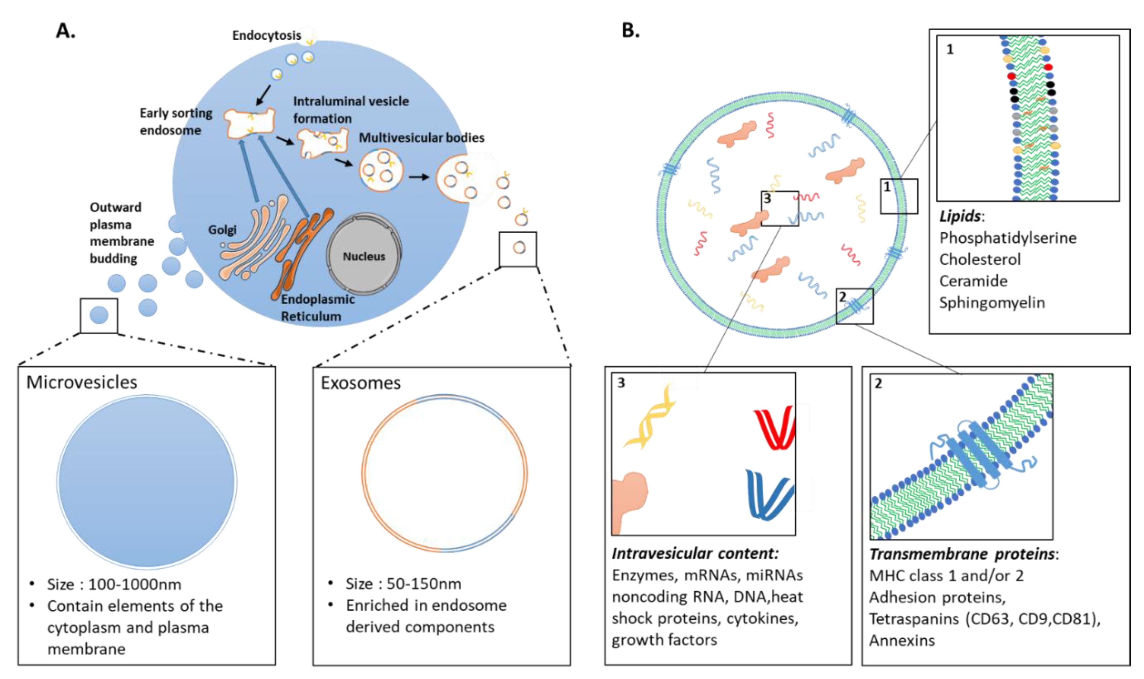
3.1. Extracellular Vesicles: Definition

Extracellular vesicles (EVs) form a heterogeneous group of double layered lipid membrane-enclosed vesicles, with distinct biophysical properties and functions both in physiology and under pathological conditions [136]. They have emerged as important mediators of intercellular communication due to their ability to shuttle a variety of nucleic acids (including mRNAs, miRNAs), proteins, and lipids between cells (Figure 1) [137,138,139]. EVs can transmit information to target cells through different mechanisms. First, the mere interaction of EVs with surface molecules on the cell membrane can trigger intracellular signaling cascades, without delivery of their content. EVs can also fuse with acceptor cells to release their cargo, either by direct fusion with the cell membrane, phagocytosis or through a variety of endocytic pathways, including clathrin- and caveolin-mediated endocytosis, macropinocytosis, micropinocytosis, and lipid raft-mediated internalization [140,141].
EVs are categorized into three major classes based on their biogenesis: (i) Exosomes are formed by the endocytic pathway through invagination of the endosomal membrane, which ultimately forms multivesicular bodies (MVBs; Figure 1). Upon the fusion of MVBs with the plasma membrane, exosomes are released into the extracellular environment. (ii) Microvesicles are shed directly by outward vesicle budding of the plasma membrane. Exosomes are typically 50–150 nm, whereas microvesicles are 100–1000 nm in size. (iii) Apoptotic bodies (size over 500 nm) are formed by blebbing of the plasma membrane of cells undergoing apoptosis. Recently updated guidelines of the International Society for Extracellular Vesicles (ISEV) recommend use of the term extracellular vesicle as “the generic term for particles naturally released from the cell that are delimited by a lipid bilayer and cannot replicate” [142]. Unless authors are able to assign an EV to a particular biogenesis pathway, with appropriate characterization of subcellular origin, the term EV should be used exclusively. Indeed, much overlap has been present in the literature with the term “exosome” used interchangeably with “extracellular vesicle” in many studies. For the sake of clarity and coherence, we chose here to refer to them as EVs.

3.2. Clinical Trials Using EVs

In recent years, a growing body of studies suggests that EVs might hold remarkable potential as therapeutics, either as active agents or delivery systems. EVs are primarily being developed as tools for anti-tumor/immunomodulatory therapies, drug delivery, and regenerative therapies [143]. In addition, EVs are extensively studied in the probing of pathophysiological states of the host as potential biomarkers in biological fluids for the diagnosis and monitoring of various diseases [144]. An increasing number of ongoing, planned or completed clinical trials have been undertaken in recent years. In order to evaluate the use of EVs in translational clinical trials, we searched for the keywords “exosomes”, “extracellular vesicles” or “microvesicles” on the ClinicalTrials.gov website. Results are presented for each EV subpopulation in relation with the disease context and source of EVs (Figure 2).
So far, most of the studies have investigated the use of EVs as reliable biomarkers in biological fluids, such as blood and urine, for diagnostic and prognostic. The immunomodulatory properties of EVs from dendritic and tumor cells are also being explored as cell-free anti-tumor agents. Promising results have been obtained in phase I and II clinical studies (reviewed in [145]). A phase II clinical trial (NCT01159288) evaluated the therapeutic potential of dendritic cell (DC)-derived EVs loaded with major histocompatibility complex (MHC) class I- and class II-restricted cancer antigens as maintenance immunotherapy after induction chemotherapy in patients bearing inoperable non-small cell lung cancer (NSCLC) [146]. This phase II trial showed that DC-derived EVs exerted NK cell effector functions in patients with advanced NSCLC, boosting the NK arm of antitumor immunity.
Recent attention has focused on the potential interest of EVs as therapeutic tools for acellular regenerative medicine. Thirty interventional clinical trials are based on the use of EVs for therapeutic purpose (Table S1). Only 16 of them are specifically evaluating the therapeutic efficacy and safety of stem-cell derived EVs in patients. Of note, none of these trials use EVs from either hPSCs or hPSC derivatives and the vast majority of them are based on MSCs. For example, a phase I clinical trial aims at studying the therapeutic potentials of condition medium from MSCs in wound healing on patients with chronic skin ulcer (NCT04134676). Similarly, a phase I/II trial uses MSC-derived EVs from normal donors to improve cutaneous wound healing of skin lesions in Epidermolysis Bullosa (EB) patients (NCT04173650). The safety and efficacy of MSC-derived EVs is also being investigated in a phase I clinical trial to promote functional recovery of large and refractory macular holes (NCT03437759). To date, none of these trials has published results yet.

3.3. EVs as Potential Therapeutic Tools: Selected Examples

Increasing evidence suggests that EVs could recapitulate the beneficial effect of their parental cells in a number of applications. To draw a parallel with our previous section, we will focus here on the recent developments obtained with EVs as a therapeutic tool for diseases affecting the eye, skin, heart and skeletal muscle. We also explore how to integrate cell-based and acellular therapies to take advantages of these two approaches and potentiate therapeutic results (Figure 3B).
Of note, studies described herein include, but are not limited to, PSCs and derivatives. Indeed, EVs from cells with well-described paracrine actions, for example, MSCs, have been shown to exert beneficial effects in various applications. Depending on the disease and affected tissue, the therapeutic potential of EVs should therefore be examined from a variety of cell sources.

3.3.1. Tissue Restoration in the Eye

Therapeutic effects of EVs have been evaluated for RP, AMD, and following injuries to the eye. Most of these studies focused on the potential of EVs to limit inflammation that would otherwise trigger the death of retinal cells [147].
As previously mentioned, different studies have started to reveal that the presence of cells might not be required to generate a beneficial effect. As an example, subretinal implantation of human neural progenitor cells preserved the vision of a model of RP through a paracrine effect [148]. The same effect is elicited by human fetal retinal progenitor cells in rats modeling RP and human patients [149,150]. As these transplantations were performed before PR degeneration, a replacement of dead cells was clearly not expected. Building on this, EVs derived from neural progenitor cells were injected subretinally in rats modeling RP before vision loss [151]. Following a single injection, the visual function and PR survival was temporarily improved (up to 28 days post-surgery). EVs were mostly internalized by Iba1+ microglial cells that had migrated from the inner retina to the subretinal space. EVs induced the downregulation of pro-inflammatory cytokines and inhibited microglia, whose suppressed activation is involved in PR survival in RP [152].
A choroidal neovascularization (CNV) is characteristic in wet AMD and can be induced in vivo with laser injuries in rodent (laser-induced CNV model). Human umbilical cord blood MSC (hUCMSC)-derived EVs injected once intravitreally were sufficient to reduce vessel leakage and the development of CNV via downregulation of VEGF-A [153]. Preservation of retinal functions and suppression of inflammation is equivalent when MSCs or their EVs are injected intravitreally [154]. In the same vein, retinal astroglial cell-derived EVs inhibited laser-induced CNV in mice when injected daily through the subtenon route for 7 days [155]. Interestingly, injections of EVs derived from RPE cells did not recapitulate these results [155]. Retinal neovascularization was also observed following oxygen-induced retinopathy in mice. The injection of EVs derived from hMSCs cultured under hypoxic conditions also reduced neovascularization [156].
Therefore, EVs are able to modulate retinal degeneration via the inflammatory response. This function is similar to transplanted cells, suggesting that the action of transplanted cells is likely through a paracrine effect (i.e., MSCs, retinal astroglial cells, human neural progenitors). An important point raised by these studies is that EVs or their parental cells need to be delivered at an early stage before complete degeneration. As EVs may not need complex surgeries or immunosuppression, it could be envisioned as a first line of treatment to support retinal survival and delay the requirement for a cell-based intervention. Future preclinical studies need however to determine a delivery route that allows repeated injections for long-term efficiency.
In the context of an advanced RPE cell degeneration as in late AMD, EVs may not be sufficient to recapitulate all RPE functions in order to preserve surrounding retina. Thus, endogenous RPE may be replaced through cell therapy (Figure 3A). A patch of RPE cells could be proposed as an ideal therapeutic substrate, using supporting scaffolds made of polymers or of biological composition. As EVs modulate inflammation during retinal degeneration, it should be determined whether a combined approach of RPE transplantation and EV therapy may improve visual outcomes. This is particularly true as RPE-derived EVs are not able to reduce the CNV when compared to EVs derived from paracrine-acting cells [155]. Such combined approaches may be of interest for multi-factorial diseases like AMD and may also preserve grafted cells from degeneration (Figure 3B).
Another potential EV therapy is related to PR transplantation studies for RP. Early studies showed the feasibility of transplanting post-mitotic PR precursors that achieved some degree of integration into the host mouse retina, ultimately resulting in partial visual function recovery [42,157,158,159]. The vast majority of transplanted PR precursors were later found to remain in the subretinal space where they engaged in a process of material transfer of functional proteins with host PRs [43,44,45,160,161]. The precise mechanism of this material exchange between PRs remains elusive but preclinical studies clearly demonstrated that this exchange occurs only with donor PR precursors and not with other cell types [7,160]. It could involve direct donor/host plasma membrane fusion or other methods of intercellular trafficking including EVs [162]. Therefore, it should be determined whether EVs secreted from PR precursors could recapitulate partial visual recovery obtained with PR precursors. If similar results were obtained, it would be an attractive strategy to preserve endogenous PRs without the need of cell grafting and all associated constraints (i.e., surgery, immunosuppression). When PRs have already degenerated, replacement strategies with PRs susceptible to integrate into the host retina will still be required. As discussed earlier, micro-structured scaffolds and/or addition of RPE cells could improve integration and structuration.
Taking advantage of EV ability to carry nucleic acids, they could be used as vehicles in gene therapy strategies for RP [9]. As an example, EV-associated AAV2 vector was delivered to the retina and outperformed conventional AAV2 [163]. Interestingly, these EV-AAV2 served as a robust gene delivery tool in the inner nuclear/outer plexiform and the outer nuclear layer, targeting retinal ganglion cells, bipolar cells, Müller cells as well as PRs [163]. The natural ability of EVs to deliver bioactive nucleic acids to multiple layers in the inner retina suggests that cell-free EV therapies may also benefit other traumatic or neurodegenerative ocular diseases [164].

3.3.2. Cutaneous Wound Healing

Major skin injuries, resulting from extensive burns, infection or trauma, require medical interventions to heal properly [54]. Therapeutic strategies aim at facilitating the 4 phases of cutaneous wound healing–homeostasis, inflammation, proliferation, remodeling-to accelerate wound repair and regeneration [165]. Molecular and cellular events in these phases are tightly coordinated and many cell types interact with each other in a highly coordinated sequence to restore the damaged tissue [166].
EVs hold the potential to promote all phases of wound healing and facilitate skin regeneration (reviewed in [166,167]). Transition from inflammatory to proliferative phase is a key step for successful wound healing. During the early stages of inflammation, the vast majority of macrophages differentiate towards a pro-inflammatory (M1) phenotype. As the wound matures, the ratio switches to an M2 phenotype that promotes tissue remodeling and wound healing [168]. EVs obtained from lipopolysaccharide-preconditioned MSCs could convert M1 macrophage polarization to an M2 phenotype, which alleviated inflammation, and enhanced diabetic cutaneous wound healing in rats by shuttling let-7b miRNA [169]. Similarly, MSC-derived EVs promoted cutaneous wound healing in mice by regulating macrophage polarization through miR-223 [170]. In line with these results, hUCMSCs significantly decreased the number of inflammatory cells and pro-inflammatory cytokines TNF-a, IL-1, IL-6 levels while increasing the production of the anti-inflammatory cytokine IL-10 in wounds of severe burn rats [171]. The same team later found that miR-181c expression in hUCMSC-derived EVs reduced burn-induced excessive inflammation by downregulating the TLR4 signaling pathway [172].
During the proliferative phase, re-epithelization, wound contraction and angiogenesis are essential processes for the restoration of normal tissue architecture. Early recruitment of resident keratinocytes and fibroblasts is particularly important as abnormalities in the intercellular epidermal-dermal crosstalk impairs the skin repair efficiency [173]. In this context, EVs from both fetal and adult stem cell sources can improve migration and proliferation of both fibroblasts and keratinocytes [174,175,176,177,178,179,180,181,182,183]. EVs from hUCMSCs and MSCs activate signaling pathways important in wound healing, including RAC-alpha serine/threonine-protein kinase (AKT) pathway [174,177,184] and Notch signaling [182]. Increased phosphorylation of extracellular signal-regulated kinase (ERK)-1/2 [175,180,184] and inhibition of phosphatase and tensin homolog (PTEN) [184] have also been reported. Additionally, an increased extracellular matrix (ECM) deposition by fibroblasts has been observed, facilitating wound contraction. Indeed, fibroblasts increased collagen I and III production following systemic administration of MSC-derived EVs at wound sites in a mice full-thickness wound model [176]. Similar results were obtained with EVs derived from hiPSC-MSCs [185] and hUCMSCs [186]. Human adipose MSCs-derived EVs also prevented the differentiation of fibroblasts into myofibroblasts, increased the ratio of transforming growth factor-β3 (TGF-β3) to TGF-β1 and upregulated the matrix metalloproteinases-3 (MMP3) expression of skin dermal fibroblasts through the activation of ERK/MAPK pathway [187]. As such, EVs could be used to promote extracellular matrix remodeling and reduce scar formation.
Transplantation of cellular skin substitutes have shown considerable potential to treat both acute and chronic wounds [58]. Complex multicellular 3D models are being developed with the goal of making engineered tissues similar to their natural counterpart (Figure 3A; reviewed in [59]). However, there is still an urgent need for improving the vascularization of these substitutes to prevent necrosis and provide better long-term function and integration in clinical practice. This is doubly important as patients with chronic skin wounds usually present defects in the angiogenesis process, which consequently leads to delayed wound healing. One possibility would be the use of pre-vascularized skin substitutes that combine dermal fibroblasts, endothelial cells, and epidermal keratinocytes [63,65,66,188]. An alternative strategy would be to supplement dermo-epidermal skin grafts with EVs conveying pro-angiogenic signals to activate tissue-resident endothelial progenitor cells (Figure 3B). Indeed, exogenous EVs were shown to promote local angiogenesis in murine models of wound healing [185,189,190]. For example, EVs derived from hUCMSC, hiPSC-MSC and human urine-derived stem cells (USC) enhanced in vitro endothelial cell proliferation, migration, and tube formation [178,185,191]. HUCMSC-derived EVs promoted angiogenesis in vivo to repair deep second-degree burn injury by delivering Wnt4 and activating Wnt/B-catenin signaling in endothelial cells [191]. EVs derived from human umbilical cord blood enriched in miR-21-3p promoted the proliferation and migration of fibroblasts as well as enhanced the angiogenic activities of endothelial cells in a full-thickness skin wound mice model, thus accelerating re-epithelialization and cutaneous wound healing [184]. Similarly, EVs derived from hiPSC-MSC applied to wound sites in a full-thickness skin defect rat model promoted not only the generation of newly formed vessels, but also accelerated their maturation [185]. In another study, human USC-derived EVs markedly enhanced the generation of newly formed blood vessels in diabetic mice, in part via the transfer of pro-angiogenic protein deleted in malignant brain tumors 1 (DMBT1) [178]. Alternatively, EVs derived from hESC facilitated pressure ulcer healing by reducing endothelial senescence and promoting local angiogenesis at wound site in aged mice [190].
At present, one of the main obstacles in the treatment of skin wounds is achieving healing over time, particularly in patients with underlying skin disorders. Biomaterial-based wound dressings could be loaded with EVs to achieve sustained release to the wound sites [192,193]. For instance, Tao et al. used the polymer chitosan to prolong delivery of EVs derived from miR-126-3p-overexpressing synovium MSCs to diabetic wounds [193]. They tested this system in a diabetic rat model and found that it increased formation of granulation tissue, which provides a scaffold for the assembly of neighboring cells at wound margins, along with angiogenesis [193].
Overall, all these proofs-of-concept experiments raised considerable interest of EVs for skin repair. Of interest, their delivery to skin wounds is relatively simple due to easy access and could be sustained over time by the use of biomaterial or repeated topical applications. However, additional preclinical studies are needed to evaluate the synergic effects of combined acellular and cellular strategies.

3.3.3. Heart

EVs have been investigated as promising therapeutic options for various cardiac diseases such as ischemic heart diseases and myocardial infarctions. One of the main objectives is to promote vascular repair mechanisms to reduce myocardial injury that would lead to cell death and therefore improve cardiac functions.
hESC-derived MSC conditioned medium (hESC-MSC-CoM), collected with clinically compatible processes, were shown to contain factors susceptible to modulate cardiovascular-related pathways [194]. Administration of hESC-MSC-CoM recapitulated the benefit of hESC-MSC injections in a context of post-myocardial infarction [195]. Indeed, in a porcine model of myocardial infarction, hESC-MSC-CoM intravenous treatment for 7 days enhanced capillary density, reduced the myocardial infarct size and preserved systolic and diastolic functions [195]. In addition, hESC-MSC-CoM reduced myocardial apoptosis and oxidative stress in another porcine model of ischemia and reperfusion injury [196]. This hESC-MSC-CoM contained large particles of 50–100 nm that were purified and characterized as EVs [197]. hESC-MSC-derived EVs similarly diminished the infarct size in an ex vivo mouse model of myocardial ischemia and reperfusion injury.
It was also proposed that ESC-derived EVs could stimulate endogenous myocardial regeneration [198]. Their delivery via an intramyorcardial route following mouse myocardial infarction stimulated endogenous repair (i.e., revascularization, cardiomyocyte proliferation/survival and reduced fibrosis). Interestingly, fibroblasts-derived EVs did not improve cardiac functions when compared to ESC-derived EVs in this model, highlighting differences between EV sources.
The functionality of cells from the cardiac lineage is superior to MSCs in the different heart disease models [199,200]. Therefore, EVs derived from these cells might achieve the most efficient heart recovery. Indeed, hPSC-CM-derived EVs recapitulated the therapeutic effects of their parental cells in the mouse model of chronic heart failure [201]. In this study, EVs or their parental cells were delivered once intramyocardially. Gene expression profiling identified 927 genes similarly upregulated in hearts treated with hPSC-CM-derived EVs and their parental cells as compared to control. The majority of enriched biological processes associated with these genes were predicted to improve heart regeneration and decrease fibrosis [201]. A recent study further highlighted the importance of determining which cellular source is the best candidate to produce therapeutic EVs. While both hPSC- and hPSC-CM-derived EVs protected CMs from hypoxia in vitro, only hPSC-CM-derived EVs completely improved the hypoxia-induced phenotype [202].
In order to maintain a sustained delivery, hPSC-CM-derived EVs were loaded into a collagen-based hydrogel patch [202]. Such system allows the release of EVs during 21 days in vitro. Patches loaded with EVs were implanted directly into the myocardium following an ischemic insult in a rat model of acute myocardial infarction. Rats recovered with this treatment, with improved heart contractile function and a reduction of the infarct size [202].
Overall, recent results indicate that EVs recapitulate the beneficial effects of their parental cells in the treatment of heart diseases (Figure 3B). Importantly, overall complexity associated with cell manufacturing, graft survival and patient immunosuppression are bypassed by this strategy. Future studies are nevertheless required to validate sustained EV release in large animal models as well as reproducibility across hPSC-CM-derived EV production protocols.

3.3.4. Skeletal Muscle

Severe muscle injuries and genetic defects like muscular dystrophies cause myofiber death. Spontaneous reparation to regenerate skeletal myofibers do occur but are insufficient [203]. A central goal of therapeutic approaches is to re-establish the muscle structural integrity and functionality by re-populating the satellite cell niche, promoting vascularization while inhibiting fibrosis formation, and stimulating the formation of contractile muscle fibers [100]. Several cellular candidates with myogenic or non-myogenic origins have been proposed for skeletal muscle regeneration, and their transplantation has been a widely investigated therapeutic strategy [100]. However, the massive donor cell death and cellular dispersion observed after delivery of cells via injection limit their therapeutic potential. Cell therapy products are still a long way from being able to reconstruct the muscle architecture, let alone to reconstruct it with nerve and sufficient vascularization. Overall, cell therapy could be considered for small muscles but is difficult to implement for diseases affecting all body muscles.
A mounting body of evidence suggests that EVs are actively produced by skeletal muscles cells and contribute to muscle repair and regeneration [203]. For example, EVs secreted during the differentiation of human skeletal myoblasts (HSkM) into myotubes contain specific biochemical cues that promote and regulate the myogenic differentiation of human adipose-derived stem cells (HASCs) [204]. Treatment of lacerated muscle sites with these differentiating HSkM-derived EVs led to an improved muscle regeneration with a large number of regenerative myofibers associated to minimal fibrosis compared to the control group [204]. In addition to the facilitation of myofiber repair, EVs also attenuate excessive ECM deposition for optimal muscle remodeling. In muscular dystrophies and severe muscle injuries, fibrogenic cells are overactivated and hyperproliferate, leading to the substitution of skeletal muscle with nonfunctional fibrotic tissue [205]. This excessive accumulation of extracellular matrix components not only alters muscle function but also reduces the amount of tissue available for therapy and repair. Establishing new anti-fibrotic therapeutic strategies is one of the major clinical options to improve muscle function in patients. In response to hypertrophic stimuli, satellite cells give rise to myogenic progenitor cells (MPCs) able to secrete EVs containing miR-206, which represses cell collagen expression through ribosomal binding protein 1 (Rrbp1) by neighboring fibroblasts, thus preventing excessive ECM deposition [206]. Similarly, fibroblasts derived from muscle biopsies of DMD patients secreted exosomes with increased levels of miR199a-5p, causing increased fibrosis in skeletal muscle and surrounding matrix [207]. These data indicate that EVs could be of interest as potential anti-fibrotic agents.
EVs are also evaluated as potential therapeutic agents to counteract muscle wasting and skeletal muscle dysfunction. Chronic kidney disease (CKD), which ultimately leads to end-stage renal failure, often leads to muscle wasting. Intramuscular injection of EV-encapsulated miR29, previously shown to have anti-fibrotic activity, could attenuate UUO-induced body weight loss and muscle atrophy [208]. Similar results were obtained after injection of EV-miR26a in CKD mice [209]. DMD is a heritable myodegenerative disease characterized by the absence of functional dystrophin leading to progressive muscle weakness and degeneration. Recent data suggest that a treatment with EVs from cardiosphere-derived cells (CDCs) originally targeted at DMD cardiomyopathy could potentially benefit both cardiac and skeletal muscle [210]. CDC-derived EVs injected into the soleus of mdx mouse model of DMD enhanced muscle regeneration, decreased inflammation and fibrosis, allowing complete restoration of contractile forces. More surprisingly, detectable levels of full-length dystrophin were evident in the diaphragm and soleus up to three weeks after systemic CDC-derived EV delivery [210]. Dystrophin protein and transcript were undetectable in CDC-derived EVs. Moreover, analysis of exon-intron junctions for dystrophins transcripts after CDC-derived EV treatment showed no exon skipping or alternative splicing. However, RNA-seq of CDC-EVs revealed a 144-fold increase in miR-148a. Intramyocardial injection of miR-148a restored expression of dystrophin in mdx hearts 3 weeks after administration, implicating this miRNA as a potential mediator of enhanced full-length dystrophin protein synthesis [210]. Targeting EV-derived miRNAs appears as a promising strategy to improve muscle function [211].
Finally, EVs could be used as efficient delivery tools of functional cargoes in vivo to restore expression of missing proteins in patients. For example, Gao et al. demonstrated that systemic administration of exosomes loaded with CP05-conjugated dystrophin splice increased dystrophin protein expression in dystrophin-deficient mdx mice with functional improvements [212].
It has become clear that EVs enable intercellular signaling that facilitate myofiber regeneration, limit excessive ECM deposits and improve muscle functions. More strikingly, their systemic delivery improves muscle function in diseases like DMD or CDK. Future studies are now required to characterize their biogenesis, compositions and biological activities on recipient cells in both physiological and pathophysiological conditions to determine whether they might be envisioned for therapy [213].

3.4. Challenges

As discussed, some of the benefits observed with cell therapy are likely due to paracrine effects that can be recapitulated by EVs derived from these cells, rather than long-term engraftment and survival of transplanted cells [214]. Overall, EVs and grafted cells can elicit different outcomes according to the target organ (Figure 3B). In some instances, therapeutic effects could be similar and therefore, EV therapy may have the highest benefice/risk ratio. In other instances, both therapies are complementary and a combination of both may achieve the best therapeutic effects.
Despite their promising roles, several challenges associated with EVs as acellular therapy products still need to be overcome.

3.4.1. Manufacturing EVs: Considerations

As products of cells, the identity and functions of EVs are directly correlated to their cell source. Consequently, it is essential to identify the most appropriate cell source for EV production [9]. As illustrated in Section 3.3, studies have identified a growing list of cell sources, some being more relevant than others, that could be suitable in acellular approaches for regenerative medicine. Our capacity to decipher in depth the various mechanisms by which EVs mediate biological and regenerative functions will be determinant to identify the best candidates in the future. It is also crucial to optimize the upstream processing conditions to increase the notably low EV yields [215]. Finding the optimal conditions for EV production by a specific cell type remains a challenge. Indeed, it is always a compromise between optimal conditions for growth and phenotype and those for EV production and isolation [216]. This is particularly true for the protocols that rely on the induction of a cell stress to obtain specific EVs (for example, hypoxia [156]).
Another challenge is the development of robust procedures to isolate and purify EVs with high yield and purity while preserving their structure and activity [215]. As with GMP-compliant cell manufacturing, large quantities of EVs should be produced with defined medium conditions, devoid as much as possible of xenogeneic substances and serum-derived vesicles, which otherwise have a high risk of contaminating the isolated EV sample [9]. Of note, while EVs secreted by cells cultured in serum-free conditions did not exhibit significant biophysical or size differences compared with cells cultured with serum, the expression levels of certain vesicular proteins (e.g., small GTPases, G-protein complexes, mRNA processing proteins and splicing factors) were found differentially expressed in EVs [217]. The use of commercial exosome-depleted fetal bovine serum (FBS) is another alternative to serum-free conditions. However, they are obtained by various exosome-reduction means, with each inducing their own effect upon the serum constituents and characteristics [218]. Alternatively, cells could be cultured during the EV release period with medium that has been pre-depleted of EVs [142]. However, FBS-derived EV elimination protocols have profound impacts on the cells themselves as they were shown to decrease cell growth and survival-promoting effects of FBS [219,220].
EVs are isolated from a variety of different sources, including body fluids with highly variable compositions (e.g., plasma, milk, urine, saliva) and cell culture media [221]. Importantly, the non-EV contaminants found in EV preparations differ substantially depending on their source. Specific EV isolation procedures should, therefore, be carefully determined accordingly to the starting material [221]. So far, there is no consensus on a “gold standard” technology to isolate EVs, for either therapeutic application or basic research [222,223]. Differential centrifugation/ultracentrifugation, polymer-based precipitation and density-gradient centrifugation are the most commonly used techniques but have very limited scalability and are associated with low EV recovery, disruption of EV integrity and risk of undesirable co-isolation of contaminants [9,136,223,224,225].
To purify EVs in a GMP-grade scalable manner, studies have employed ultrafiltration or tangential flow filtration (TFF) to concentrate EVs based on their size [226,227,228,229]. For example, GMP-EV-cardiac progenitor cell manufacturing was implemented for up to 8 L of conditioned medium, allowing high final product yield (≥58%) with concomitant consistent reduction of contaminants (total protein removal 97–98%) [228]. Subsequent size exclusion chromatography (SEC) allows the separation of EVs from other media components without altering their integrity [227,229,230,231]. However, size-based isolation techniques do not purify a specific EV subpopulation but rather yield complex mixtures because of the overlap in size of EVs (described in Section 3.1; [232]). Additional purification steps may also still be necessary to remove contaminants with overlapping sizes, such as bovine serum-EVs. Recently, alternative isolation techniques based on EV-surface markers have been described and seem to be promising but still in infancy [233,234]. Overall, existing techniques to isolate EVs do not have a one-size-fits-all model. The optimal isolation method(s) and acceptable level of impurity need to be carefully considered to prevent loss of function due to damage to EV-intrinsic effectors or loss of associated factors that act with EVs to exert function [142,143,221].
As with cells and other cell-derived products, maintenance of EV biological activity during storage is critical for their therapeutic use [235]. Stability should rely on the monitoring of both changes in physicochemical parameters (size, particle concentration, and morphology) and EV bioactivity. A number of studies have evaluated the impact of different storage temperatures (4, −20, and −80 °C) and repetitive free-thaw cycles on the composition and functionality of EVs isolated from various body fluids (reviewed in [235,236]). Current evidences suggest that storage at −80 °C is best-suited. Nonetheless, our current understanding of storage-mediated effects is still limited and standard criterion of EV preservation should be established in the future.

3.4.2. EV-Based Therapeutics: Regulatory Aspects

Current legislation does not provide specific regulatory guidelines for EV-based therapies. In the European Union (EU) and United States of America, they enter in the framework of the biological medicinal products regulation under the definition of “biological medicine” (i.e., a medicine that contains one or more active substances made by or derived from a biological cell) [222]. The pharmaceutical classification of any biological product is determined by its active substance(s). In that regard, EV-based therapeutics are particularly challenging as it is not clear in many cases whether the biological effect depends on the vesicle internal content (EV-associated cargo), the vesicle membranes, or a combination of both [143]. As such, they share characteristics from both cell and gene therapies, which makes them difficult to classify in an existing pharmaceutical category.
Three subcategories of EV products are possible, each with specific regulatory guidance: 1/native EVs from unmodified cells, 2/EVs from genetically manipulated cells which do not contain transgene products, and 3/EVs from genetically modified cells containing transgene products [222]. In addition to the existing guidance on the manufacture of biological medicinal products, safety and quality standards for tissue- or cell-based products (DIRECTIVE 2004/23/EC and DIRECTIVE 2006/17/EC in the EU) may serve as roadmaps for EV-based therapeutics derived from human tissues and cells [222].
Stem cell therapy and tissue engineering are intimately intertwined in the development of regenerative medicine products. In recent years, research has focused on the use of biocompatible scaffolds functionalized with EVs, with or without cells, to improve regenerative capacity of the grafted products. Regulatory strategies for advanced therapy medicinal products (ATMPs)-encompassing gene therapy, somatic cell therapy, and tissue-engineered products–could be applied to such complex EV-based products (DIRECTIVE 2001/83/EC). EVs produced by genetically modified cells with biologically active transgene products could also fall within this subcategory [143]. Of note, EVs are considered as an excipient instead of an active substance when used as drug delivery systems loaded with molecules.

4. Conclusions

As outlined in this review, a number of key steps to develop regenerative therapies has been achieved. Preclinical evidences for many indications were the basis for first-in-man clinical trials. Main concerns related to the use of hPSCs were ruled out in clinical trials (i.e., risks of teratoma formation and cell dispersion). So far, simplified cell therapy systems have been used with some promising therapeutic results. However, preclinical evidences highlight the need for more complex cell therapy products containing various cells and/or biomaterials, structured as the native tissue. In parallel, the roles played by EVs as intercellular effectors of paracrine signaling has led to a strong interest in their use as cell-free therapeutic products to stimulate endogenous factors that would limit cell death or improve tissue regeneration. Strikingly, as these two strategies could act at different levels depending on the pathological context, their complementarity opens new perspectives to maximize therapeutic outcomes. EVs could be substituted to cell therapy in a number of indications including heart diseases, simplifying treatment complexity. However, much preclinical work is still required to characterize optimal “acellular” and cellular therapies and their potential synergistic effects. In addition, definition of appropriate conditions of GMP production need to be elaborated and customized for each application. All these future improvements may help to achieve the ultimate goal of regenerative medicine: the replacement of a degenerated tissue.

5. Patents

WH, CM and KB are inventors of a patent (FR3078712) related to medical devices for the preparation of retinal tissues for regenerative medicine. LM, CM and KB are inventors of a pending patent related to the automated differentiation of hPSC into RPE cells (EP3754014). LM and CB are inventors of a pending patent related to the automated differentiation of hPSC into keratinocytes (EP20305217.0). SD and CB are inventors of two pending patents related to the differentiation of hPSCs into keratinocytes (EP20305218.0) and fibroblasts (EP 20305214.7) and to the formation of a skin substitute composed of hPSCs derived cells (EP 20305213.9). AD and CB are inventors of a pending patent related to the differentiation of hPSCs into endothelial cells (EP 20305215.4).

Supplementary Materials

The following are available online at https://www.mdpi.com/2073-4409/10/2/240/s1, Table S1: List of planned or initiated clinical trials using EVs as therapeutics.

Author Contributions

All authors contributed to the manuscript writing and approved the final version. All authors have read and agreed to the published version of the manuscript.

Funding

This work was supported by a grant from the ANR (RebuildingRETINA: ANR-19-CE18-0004) to KB. EH and SM are recipient of PhD fellowships from the French Ministry of Higher Education and Research. I-Stem is part of the Biotherapies Institute for Rare Diseases supported by the Association Française contre les Myopathies-Téléthon.

Informed Consent Statement

Not applicable.

Data Availability Statement

Not applicable.

Acknowledgments

Some elements of figures were created using Servier Medical Art templates (https://smart.servier.com), which are licensed under a Creative Commons Attribution 3.0 Unported License.

Conflicts of Interest

CB collaborates with the company URGO to develop cell therapy products. The other authors declare no conflict of interest.

References

  1. Kobold, S.; Guhr, A.; Mah, N.; Bultjer, N.; Seltmann, S.; Seiler Wulczyn, A.E.M.; Stacey, G.; Jie, H.; Liu, W.; Loser, P.; et al. A Manually Curated Database on Clinical Studies Involving Cell Products Derived from Human Pluripotent Stem Cells. Stem Cell Rep. 2020, 15, 546–555. [Google Scholar] [CrossRef]
  2. Schwartz, S.D.; Hubschman, J.P.; Heilwell, G.; Franco-Cardenas, V.; Pan, C.K.; Ostrick, R.M.; Mickunas, E.; Gay, R.; Klimanskaya, I.; Lanza, R. Embryonic stem cell trials for macular degeneration: A preliminary report. Lancet 2012, 379, 713–720. [Google Scholar] [CrossRef]
  3. Ben M’Barek, K.; Monville, C. Cell Therapy for Retinal Dystrophies: From Cell Suspension Formulation to Complex Retinal Tissue Bioengineering. Stem Cells Int. 2019, 2019, 4568979. [Google Scholar] [CrossRef]
  4. Ghareeb, A.E.; Lako, M.; Steel, D.H. Coculture techniques for modeling retinal development and disease, and enabling regenerative medicine. Stem Cells Transl. Med. 2020, 9, 1531–1548. [Google Scholar] [CrossRef]
  5. Sun, B.K.; Siprashvili, Z.; Khavari, P.A. Advances in skin grafting and treatment of cutaneous wounds. Science 2014, 346, 941–945. [Google Scholar] [CrossRef] [PubMed]
  6. Sun, J.; Mandai, M.; Kamao, H.; Hashiguchi, T.; Shikamura, M.; Kawamata, S.; Sugita, S.; Takahashi, M. Protective Effects of Human iPS-Derived Retinal Pigmented Epithelial Cells in Comparison with Human Mesenchymal Stromal Cells and Human Neural Stem Cells on the Degenerating Retina in rd1 mice. Stem Cells 2015, 33, 1543–1553. [Google Scholar] [CrossRef] [PubMed]
  7. Morizur, L.; Herardot, E.; Monville, C.; Ben M’Barek, K. Human pluripotent stem cells: A toolbox to understand and treat retinal degeneration. Mol. Cell Neurosci. 2020, 107, 103523. [Google Scholar] [CrossRef] [PubMed]
  8. Hargett, L.A.; Bauer, N.N. On the origin of microparticles: From “platelet dust” to mediators of intercellular communication. Pulm. Circ. 2013, 3, 329–340. [Google Scholar] [CrossRef] [PubMed] [Green Version]
  9. Wiklander, O.P.B.; Brennan, M.A.; Lotvall, J.; Breakefield, X.O.; El Andaloussi, S. Advances in therapeutic applications of extracellular vesicles. Sci. Transl. Med. 2019, 11. [Google Scholar] [CrossRef] [PubMed]
  10. S, E.L.A.; Mager, I.; Breakefield, X.O.; Wood, M.J. Extracellular vesicles: Biology and emerging therapeutic opportunities. Nat. Rev. Drug Discov. 2013, 12, 347–357. [Google Scholar] [CrossRef]
  11. Bachoud-Levi, A.C.; Perrier, A.L. Regenerative medicine in Huntington’s disease: Current status on fetal grafts and prospects for the use of pluripotent stem cell. Rev. Neurol. (Paris) 2014, 170, 749–762. [Google Scholar] [CrossRef] [PubMed]
  12. Bachoud-Levi, A.C.; Massart, R.; Rosser, A. Cell therapy in Huntington’s disease: Taking stock of past studies to move the field forward. Stem Cells 2020. [Google Scholar] [CrossRef]
  13. Nakagawa, M.; Koyanagi, M.; Tanabe, K.; Takahashi, K.; Ichisaka, T.; Aoi, T.; Okita, K.; Mochiduki, Y.; Takizawa, N.; Yamanaka, S. Generation of induced pluripotent stem cells without Myc from mouse and human fibroblasts. Nat. Biotechnol. 2008, 26, 101–106. [Google Scholar] [CrossRef] [PubMed]
  14. Takahashi, K.; Tanabe, K.; Ohnuki, M.; Narita, M.; Ichisaka, T.; Tomoda, K.; Yamanaka, S. Induction of pluripotent stem cells from adult human fibroblasts by defined factors. Cell 2007, 131, 861–872. [Google Scholar] [CrossRef] [Green Version]
  15. Thomson, J.A.; Itskovitz-Eldor, J.; Shapiro, S.S.; Waknitz, M.A.; Swiergiel, J.J.; Marshall, V.S.; Jones, J.M. Embryonic stem cell lines derived from human blastocysts. Science 1998, 282, 1145–1147. [Google Scholar] [CrossRef] [Green Version]
  16. Flaxman, S.R.; Bourne, R.R.A.; Resnikoff, S.; Ackland, P.; Braithwaite, T.; Cicinelli, M.V.; Das, A.; Jonas, J.B.; Keeffe, J.; Kempen, J.H.; et al. Global causes of blindness and distance vision impairment 1990-2020: A systematic review and meta-analysis. Lancet Glob. Health 2017, 5, e1221–e1234. [Google Scholar] [CrossRef] [Green Version]
  17. Gagliardi, G.; Ben M’Barek, K.; Goureau, O. Photoreceptor cell replacement in macular degeneration and retinitis pigmentosa: A pluripotent stem cell-based approach. Prog. Retin. Eye Res. 2019, 71, 1–25. [Google Scholar] [CrossRef]
  18. Ben M’Barek, K.; Habeler, W.; Plancheron, A.; Jarraya, M.; Regent, F.; Terray, A.; Yang, Y.; Chatrousse, L.; Domingues, S.; Masson, Y.; et al. Human ESC-derived retinal epithelial cell sheets potentiate rescue of photoreceptor cell loss in rats with retinal degeneration. Sci. Transl. Med. 2017, 9. [Google Scholar] [CrossRef] [Green Version]
  19. Klimanskaya, I.; Hipp, J.; Rezai, K.A.; West, M.; Atala, A.; Lanza, R. Derivation and comparative assessment of retinal pigment epithelium from human embryonic stem cells using transcriptomics. Cloning Stem Cells 2004, 6, 217–245. [Google Scholar] [CrossRef]
  20. Buchholz, D.E.; Hikita, S.T.; Rowland, T.J.; Friedrich, A.M.; Hinman, C.R.; Johnson, L.V.; Clegg, D.O. Derivation of functional retinal pigmented epithelium from induced pluripotent stem cells. Stem Cells 2009, 27, 2427–2434. [Google Scholar] [CrossRef]
  21. Buchholz, D.E.; Pennington, B.O.; Croze, R.H.; Hinman, C.R.; Coffey, P.J.; Clegg, D.O. Rapid and efficient directed differentiation of human pluripotent stem cells into retinal pigmented epithelium. Stem Cells Transl. Med. 2013, 2, 384–393. [Google Scholar] [CrossRef] [PubMed]
  22. Leach, L.L.; Buchholz, D.E.; Nadar, V.P.; Lowenstein, S.E.; Clegg, D.O. Canonical/beta-catenin Wnt pathway activation improves retinal pigmented epithelium derivation from human embryonic stem cells. Investig. Ophthalmol. Vis. Sci. 2015, 56, 1002–1013. [Google Scholar] [CrossRef] [PubMed]
  23. Idelson, M.; Alper, R.; Obolensky, A.; Ben-Shushan, E.; Hemo, I.; Yachimovich-Cohen, N.; Khaner, H.; Smith, Y.; Wiser, O.; Gropp, M.; et al. Directed differentiation of human embryonic stem cells into functional retinal pigment epithelium cells. Cell Stem Cell 2009, 5, 396–408. [Google Scholar] [CrossRef] [Green Version]
  24. Maruotti, J.; Sripathi, S.R.; Bharti, K.; Fuller, J.; Wahlin, K.J.; Ranganathan, V.; Sluch, V.M.; Berlinicke, C.A.; Davis, J.; Kim, C.; et al. Small-molecule-directed, efficient generation of retinal pigment epithelium from human pluripotent stem cells. Proc. Natl. Acad. Sci. USA 2015, 112, 10950–10955. [Google Scholar] [CrossRef] [PubMed] [Green Version]
  25. Crombie, D.E.; Daniszewski, M.; Liang, H.H.; Kulkarni, T.; Li, F.; Lidgerwood, G.E.; Conquest, A.; Hernandez, D.; Hung, S.S.; Gill, K.P.; et al. Development of a Modular Automated System for Maintenance and Differentiation of Adherent Human Pluripotent Stem Cells. SLAS Discov. 2017, 22, 1016–1025. [Google Scholar] [CrossRef] [Green Version]
  26. Regent, F.; Morizur, L.; Lesueur, L.; Habeler, W.; Plancheron, A.; Ben M’Barek, K.; Monville, C. Automation of human pluripotent stem cell differentiation toward retinal pigment epithelial cells for large-scale productions. Sci. Rep. 2019, 9, 10646. [Google Scholar] [CrossRef] [Green Version]
  27. Matsumoto, E.; Koide, N.; Hanzawa, H.; Kiyama, M.; Ohta, M.; Kuwabara, J.; Takeda, S.; Takahashi, M. Fabricating retinal pigment epithelial cell sheets derived from human induced pluripotent stem cells in an automated closed culture system for regenerative medicine. PLoS ONE 2019, 14, e0212369. [Google Scholar] [CrossRef] [Green Version]
  28. Carr, A.J.; Vugler, A.A.; Hikita, S.T.; Lawrence, J.M.; Gias, C.; Chen, L.L.; Buchholz, D.E.; Ahmado, A.; Semo, M.; Smart, M.J.; et al. Protective effects of human iPS-derived retinal pigment epithelium cell transplantation in the retinal dystrophic rat. PLoS ONE 2009, 4, e8152. [Google Scholar] [CrossRef]
  29. Lu, B.; Malcuit, C.; Wang, S.; Girman, S.; Francis, P.; Lemieux, L.; Lanza, R.; Lund, R. Long-term safety and function of RPE from human embryonic stem cells in preclinical models of macular degeneration. Stem Cells 2009, 27, 2126–2135. [Google Scholar] [CrossRef]
  30. Lund, R.D.; Wang, S.; Klimanskaya, I.; Holmes, T.; Ramos-Kelsey, R.; Lu, B.; Girman, S.; Bischoff, N.; Sauve, Y.; Lanza, R. Human embryonic stem cell-derived cells rescue visual function in dystrophic RCS rats. Cloning Stem Cells 2006, 8, 189–199. [Google Scholar] [CrossRef] [Green Version]
  31. Diniz, B.; Thomas, P.; Thomas, B.; Ribeiro, R.; Hu, Y.; Brant, R.; Ahuja, A.; Zhu, D.; Liu, L.; Koss, M.; et al. Subretinal implantation of retinal pigment epithelial cells derived from human embryonic stem cells: Improved survival when implanted as a monolayer. Investig. Ophthalmol. Vis. Sci. 2013, 54, 5087–5096. [Google Scholar] [CrossRef] [Green Version]
  32. Sharma, R.; Khristov, V.; Rising, A.; Jha, B.S.; Dejene, R.; Hotaling, N.; Li, Y.; Stoddard, J.; Stankewicz, C.; Wan, Q.; et al. Clinical-grade stem cell-derived retinal pigment epithelium patch rescues retinal degeneration in rodents and pigs. Sci. Transl. Med. 2019, 11. [Google Scholar] [CrossRef] [PubMed]
  33. Stanzel, B.V.; Liu, Z.; Somboonthanakij, S.; Wongsawad, W.; Brinken, R.; Eter, N.; Corneo, B.; Holz, F.G.; Temple, S.; Stern, J.H.; et al. Human RPE stem cells grown into polarized RPE monolayers on a polyester matrix are maintained after grafting into rabbit subretinal space. Stem Cell Rep. 2014, 2, 64–77. [Google Scholar] [CrossRef] [PubMed] [Green Version]
  34. da Cruz, L.; Fynes, K.; Georgiadis, O.; Kerby, J.; Luo, Y.H.; Ahmado, A.; Vernon, A.; Daniels, J.T.; Nommiste, B.; Hasan, S.M.; et al. Phase 1 clinical study of an embryonic stem cell-derived retinal pigment epithelium patch in age-related macular degeneration. Nat. Biotechnol. 2018, 36, 328–337. [Google Scholar] [CrossRef] [PubMed] [Green Version]
  35. Fernandes, R.A.B.; Stefanini, F.R.; Falabella, P.; Koss, M.J.; Wells, T.; Diniz, B.; Ribeiro, R.; Schor, P.; Maia, M.; Penha, F.M.; et al. Development of a new tissue injector for subretinal transplantation of human embryonic stem cell derived retinal pigmented epithelium. Int. J. Retina Vitr. 2017, 3, 41. [Google Scholar] [CrossRef] [Green Version]
  36. Ben M’Barek, K.; Bertin, S.; Brazhnikova, E.; Jaillard, C.; Habeler, W.; Plancheron, A.; Fovet, C.M.; Demilly, J.; Jarraya, M.; Bejanariu, A.; et al. Clinical-grade production and safe delivery of human ESC derived RPE sheets in primates and rodents. Biomaterials 2020, 230, 119603. [Google Scholar] [CrossRef]
  37. Nakano, T.; Ando, S.; Takata, N.; Kawada, M.; Muguruma, K.; Sekiguchi, K.; Saito, K.; Yonemura, S.; Eiraku, M.; Sasai, Y. Self-formation of optic cups and storable stratified neural retina from human ESCs. Cell Stem Cell 2012, 10, 771–785. [Google Scholar] [CrossRef] [Green Version]
  38. Zhong, X.; Gutierrez, C.; Xue, T.; Hampton, C.; Vergara, M.N.; Cao, L.H.; Peters, A.; Park, T.S.; Zambidis, E.T.; Meyer, J.S.; et al. Generation of three-dimensional retinal tissue with functional photoreceptors from human iPSCs. Nat. Commun. 2014, 5, 4047. [Google Scholar] [CrossRef] [Green Version]
  39. Capowski, E.E.; Samimi, K.; Mayerl, S.J.; Phillips, M.J.; Pinilla, I.; Howden, S.E.; Saha, J.; Jansen, A.D.; Edwards, K.L.; Jager, L.D.; et al. Reproducibility and staging of 3D human retinal organoids across multiple pluripotent stem cell lines. Development 2019, 146. [Google Scholar] [CrossRef] [Green Version]
  40. Reichman, S.; Slembrouck, A.; Gagliardi, G.; Chaffiol, A.; Terray, A.; Nanteau, C.; Potey, A.; Belle, M.; Rabesandratana, O.; Duebel, J.; et al. Generation of Storable Retinal Organoids and Retinal Pigmented Epithelium from Adherent Human iPS Cells in Xeno-Free and Feeder-Free Conditions. Stem Cells 2017, 35, 1176–1188. [Google Scholar] [CrossRef] [Green Version]
  41. Welby, E.; Lakowski, J.; Di Foggia, V.; Budinger, D.; Gonzalez-Cordero, A.; Lun, A.T.L.; Epstein, M.; Patel, A.; Cuevas, E.; Kruczek, K.; et al. Isolation and Comparative Transcriptome Analysis of Human Fetal and iPSC-Derived Cone Photoreceptor Cells. Stem Cell Rep. 2017, 9, 1898–1915. [Google Scholar] [CrossRef] [PubMed] [Green Version]
  42. MacLaren, R.E.; Pearson, R.A.; MacNeil, A.; Douglas, R.H.; Salt, T.E.; Akimoto, M.; Swaroop, A.; Sowden, J.C.; Ali, R.R. Retinal repair by transplantation of photoreceptor precursors. Nature 2006, 444, 203–207. [Google Scholar] [CrossRef]
  43. Singh, M.S.; Balmer, J.; Barnard, A.R.; Aslam, S.A.; Moralli, D.; Green, C.M.; Barnea-Cramer, A.; Duncan, I.; MacLaren, R.E. Transplanted photoreceptor precursors transfer proteins to host photoreceptors by a mechanism of cytoplasmic fusion. Nat. Commun. 2016, 7, 13537. [Google Scholar] [CrossRef] [PubMed] [Green Version]
  44. Santos-Ferreira, T.; Llonch, S.; Borsch, O.; Postel, K.; Haas, J.; Ader, M. Retinal transplantation of photoreceptors results in donor-host cytoplasmic exchange. Nat. Commun. 2016, 7, 13028. [Google Scholar] [CrossRef] [PubMed] [Green Version]
  45. Decembrini, S.; Martin, C.; Sennlaub, F.; Chemtob, S.; Biel, M.; Samardzija, M.; Moulin, A.; Behar-Cohen, F.; Arsenijevic, Y. Cone Genesis Tracing by the Chrnb4-EGFP Mouse Line: Evidences of Cellular Material Fusion after Cone Precursor Transplantation. Mol. Ther. 2017, 25, 634–653. [Google Scholar] [CrossRef] [Green Version]
  46. Tu, H.Y.; Watanabe, T.; Shirai, H.; Yamasaki, S.; Kinoshita, M.; Matsushita, K.; Hashiguchi, T.; Onoe, H.; Matsuyama, T.; Kuwahara, A.; et al. Medium- to long-term survival and functional examination of human iPSC-derived retinas in rat and primate models of retinal degeneration. EBioMedicine 2019, 39, 562–574. [Google Scholar] [CrossRef] [PubMed] [Green Version]
  47. Iraha, S.; Tu, H.Y.; Yamasaki, S.; Kagawa, T.; Goto, M.; Takahashi, R.; Watanabe, T.; Sugita, S.; Yonemura, S.; Sunagawa, G.A.; et al. Establishment of Immunodeficient Retinal Degeneration Model Mice and Functional Maturation of Human ESC-Derived Retinal Sheets after Transplantation. Stem Cell Rep. 2018, 10, 1059–1074. [Google Scholar] [CrossRef] [PubMed] [Green Version]
  48. McLelland, B.T.; Lin, B.; Mathur, A.; Aramant, R.B.; Thomas, B.B.; Nistor, G.; Keirstead, H.S.; Seiler, M.J. Transplanted hESC-Derived Retina Organoid Sheets Differentiate, Integrate, and Improve Visual Function in Retinal Degenerate Rats. Investig. Ophthalmol. Vis. Sci. 2018, 59, 2586–2603. [Google Scholar] [CrossRef] [Green Version]
  49. Jung, Y.H.; Phillips, M.J.; Lee, J.; Xie, R.; Ludwig, A.L.; Chen, G.; Zheng, Q.; Kim, T.J.; Zhang, H.; Barney, P.; et al. 3D Microstructured Scaffolds to Support Photoreceptor Polarization and Maturation. Adv. Mater. 2018, 30, e1803550. [Google Scholar] [CrossRef]
  50. Worthington, K.S.; Wiley, L.A.; Kaalberg, E.E.; Collins, M.M.; Mullins, R.F.; Stone, E.M.; Tucker, B.A. Two-photon polymerization for production of human iPSC-derived retinal cell grafts. Acta Biomater. 2017, 55, 385–395. [Google Scholar] [CrossRef]
  51. Thompson, J.R.; Worthington, K.S.; Green, B.J.; Mullin, N.K.; Jiao, C.; Kaalberg, E.E.; Wiley, L.A.; Han, I.C.; Russell, S.R.; Sohn, E.H.; et al. Two-photon polymerized poly(caprolactone) retinal cell delivery scaffolds and their systemic and retinal biocompatibility. Acta Biomater. 2019, 94, 204–218. [Google Scholar] [CrossRef] [PubMed]
  52. Singh, D.; Wang, S.B.; Xia, T.; Tainsh, L.; Ghiassi-Nejad, M.; Xu, T.; Peng, S.; Adelman, R.A.; Rizzolo, L.J. A biodegradable scaffold enhances differentiation of embryonic stem cells into a thick sheet of retinal cells. Biomaterials 2018, 154, 158–168. [Google Scholar] [CrossRef] [PubMed]
  53. Garita-Hernandez, M.; Lampic, M.; Chaffiol, A.; Guibbal, L.; Routet, F.; Santos-Ferreira, T.; Gasparini, S.; Borsch, O.; Gagliardi, G.; Reichman, S.; et al. Restoration of visual function by transplantation of optogenetically engineered photoreceptors. Nat. Commun. 2019, 10, 4524. [Google Scholar] [CrossRef] [Green Version]
  54. Chu, G.Y.; Chen, Y.F.; Chen, H.Y.; Chan, M.H.; Gau, C.S.; Weng, S.M. Stem cell therapy on skin: Mechanisms, recent advances and drug reviewing issues. J. Food Drug Anal. 2018, 26, 14–20. [Google Scholar] [CrossRef] [PubMed]
  55. Gurtner, G.C.; Werner, S.; Barrandon, Y.; Longaker, M.T. Wound repair and regeneration. Nature 2008, 453, 314–321. [Google Scholar] [CrossRef]
  56. Przekora, A. A Concise Review on Tissue Engineered Artificial Skin Grafts for Chronic Wound Treatment: Can We Reconstruct Functional Skin Tissue In Vitro? Cells 2020, 9, 1622. [Google Scholar] [CrossRef]
  57. Dai, C.; Shih, S.; Khachemoune, A. Skin substitutes for acute and chronic wound healing: An updated review. J. Dermatolog. Treat. 2020, 31, 639–648. [Google Scholar] [CrossRef]
  58. Nourian Dehkordi, A.; Mirahmadi Babaheydari, F.; Chehelgerdi, M.; Raeisi Dehkordi, S. Skin tissue engineering: Wound healing based on stem-cell-based therapeutic strategies. Stem Cell Res. Ther. 2019, 10, 111. [Google Scholar] [CrossRef] [Green Version]
  59. Vig, K.; Chaudhari, A.; Tripathi, S.; Dixit, S.; Sahu, R.; Pillai, S.; Dennis, V.A.; Singh, S.R. Advances in Skin Regeneration Using Tissue Engineering. Int. J. Mol. Sci. 2017, 18, 789. [Google Scholar] [CrossRef]
  60. Guenou, H.; Nissan, X.; Larcher, F.; Feteira, J.; Lemaitre, G.; Saidani, M.; Del Rio, M.; Barrault, C.C.; Bernard, F.X.; Peschanski, M.; et al. Human embryonic stem-cell derivatives for full reconstruction of the pluristratified epidermis: A preclinical study. Lancet 2009, 374, 1745–1753. [Google Scholar] [CrossRef]
  61. Umegaki-Arao, N.; Pasmooij, A.M.; Itoh, M.; Cerise, J.E.; Guo, Z.; Levy, B.; Gostynski, A.; Rothman, L.R.; Jonkman, M.F.; Christiano, A.M. Induced pluripotent stem cells from human revertant keratinocytes for the treatment of epidermolysis bullosa. Sci. Transl. Med. 2014, 6, 264ra164. [Google Scholar] [CrossRef] [PubMed]
  62. Sebastiano, V.; Zhen, H.H.; Haddad, B.; Bashkirova, E.; Melo, S.P.; Wang, P.; Leung, T.L.; Siprashvili, Z.; Tichy, A.; Li, J.; et al. Human COL7A1-corrected induced pluripotent stem cells for the treatment of recessive dystrophic epidermolysis bullosa. Sci. Transl. Med. 2014, 6, 264ra163. [Google Scholar] [CrossRef] [PubMed] [Green Version]
  63. Miyazaki, H.; Tsunoi, Y.; Akagi, T.; Sato, S.; Akashi, M.; Saitoh, D. A novel strategy to engineer pre-vascularized 3-dimensional skin substitutes to achieve efficient, functional engraftment. Sci. Rep. 2019, 9, 7797. [Google Scholar] [CrossRef] [PubMed]
  64. Itoh, M.; Umegaki-Arao, N.; Guo, Z.; Liu, L.; Higgins, C.A.; Christiano, A.M. Generation of 3D skin equivalents fully reconstituted from human induced pluripotent stem cells (iPSCs). PLoS ONE 2013, 8, e77673. [Google Scholar] [CrossRef] [Green Version]
  65. Abaci, H.E.; Guo, Z.; Coffman, A.; Gillette, B.; Lee, W.H.; Sia, S.K.; Christiano, A.M. Human Skin Constructs with Spatially Controlled Vasculature Using Primary and iPSC-Derived Endothelial Cells. Adv. Healthc. Mater. 2016, 5, 1800–1807. [Google Scholar] [CrossRef] [Green Version]
  66. Kim, B.S.; Kwon, Y.W.; Kong, J.S.; Park, G.T.; Gao, G.; Han, W.; Kim, M.B.; Lee, H.; Kim, J.H.; Cho, D.W. 3D cell printing of in vitro stabilized skin model and in vivo pre-vascularized skin patch using tissue-specific extracellular matrix bioink: A step towards advanced skin tissue engineering. Biomaterials 2018, 168, 38–53. [Google Scholar] [CrossRef]
  67. Hosaka, C.; Kunisada, M.; Koyanagi-Aoi, M.; Masaki, T.; Takemori, C.; Taniguchi-Ikeda, M.; Aoi, T.; Nishigori, C. Induced pluripotent stem cell-derived melanocyte precursor cells undergoing differentiation into melanocytes. Pigment Cell Melanoma Res. 2019, 32, 623–633. [Google Scholar] [CrossRef]
  68. Ohta, S.; Imaizumi, Y.; Okada, Y.; Akamatsu, W.; Kuwahara, R.; Ohyama, M.; Amagai, M.; Matsuzaki, Y.; Yamanaka, S.; Okano, H.; et al. Generation of human melanocytes from induced pluripotent stem cells. PLoS ONE 2011, 6, e16182. [Google Scholar] [CrossRef]
  69. Nissan, X.; Larribere, L.; Saidani, M.; Hurbain, I.; Delevoye, C.; Feteira, J.; Lemaitre, G.; Peschanski, M.; Baldeschi, C. Functional melanocytes derived from human pluripotent stem cells engraft into pluristratified epidermis. Proc. Natl. Acad. Sci. USA 2011, 108, 14861–14866. [Google Scholar] [CrossRef] [Green Version]
  70. Lee, J.; Rabbani, C.C.; Gao, H.; Steinhart, M.R.; Woodruff, B.M.; Pflum, Z.E.; Kim, A.; Heller, S.; Liu, Y.; Shipchandler, T.Z.; et al. Hair-bearing human skin generated entirely from pluripotent stem cells. Nature 2020, 582, 399–404. [Google Scholar] [CrossRef]
  71. Yang, R.; Zheng, Y.; Burrows, M.; Liu, S.; Wei, Z.; Nace, A.; Guo, W.; Kumar, S.; Cotsarelis, G.; Xu, X. Generation of folliculogenic human epithelial stem cells from induced pluripotent stem cells. Nat. Commun. 2014, 5, 3071. [Google Scholar] [CrossRef] [PubMed] [Green Version]
  72. GBD 2017 Causes of Death Collaborators. Global, regional, and national age-sex-specific mortality for 282 causes of death in 195 countries and territories, 1980-2017: A systematic analysis for the Global Burden of Disease Study 2017. Lancet 2018, 392, 1736–1788. [Google Scholar] [CrossRef] [Green Version]
  73. Nakamura, K.; Murry, C.E. Function Follows Form- A Review of Cardiac Cell Therapy. Circ. J. 2019, 83, 2399–2412. [Google Scholar] [CrossRef] [PubMed] [Green Version]
  74. Muller, P.; Lemcke, H.; David, R. Stem Cell Therapy in Heart Diseases—Cell Types, Mechanisms and Improvement Strategies. Cell Physiol. Biochem. 2018, 48, 2607–2655. [Google Scholar] [CrossRef]
  75. Kadota, S.; Tanaka, Y.; Shiba, Y. Heart regeneration using pluripotent stem cells. J. Cardiol. 2020, 76, 459–463. [Google Scholar] [CrossRef]
  76. Yang, L.; Soonpaa, M.H.; Adler, E.D.; Roepke, T.K.; Kattman, S.J.; Kennedy, M.; Henckaerts, E.; Bonham, K.; Abbott, G.W.; Linden, R.M.; et al. Human cardiovascular progenitor cells develop from a KDR+ embryonic-stem-cell-derived population. Nature 2008, 453, 524–528. [Google Scholar] [CrossRef]
  77. Laflamme, M.A.; Chen, K.Y.; Naumova, A.V.; Muskheli, V.; Fugate, J.A.; Dupras, S.K.; Reinecke, H.; Xu, C.; Hassanipour, M.; Police, S.; et al. Cardiomyocytes derived from human embryonic stem cells in pro-survival factors enhance function of infarcted rat hearts. Nat. Biotechnol. 2007, 25, 1015–1024. [Google Scholar] [CrossRef]
  78. Karakikes, I.; Ameen, M.; Termglinchan, V.; Wu, J.C. Human induced pluripotent stem cell-derived cardiomyocytes: Insights into molecular, cellular, and functional phenotypes. Circ. Res. 2015, 117, 80–88. [Google Scholar] [CrossRef] [Green Version]
  79. Machiraju, P.; Greenway, S.C. Current methods for the maturation of induced pluripotent stem cell-derived cardiomyocytes. World J. Stem Cells 2019, 11, 33–43. [Google Scholar] [CrossRef]
  80. Burridge, P.W.; Matsa, E.; Shukla, P.; Lin, Z.C.; Churko, J.M.; Ebert, A.D.; Lan, F.; Diecke, S.; Huber, B.; Mordwinkin, N.M.; et al. Chemically defined generation of human cardiomyocytes. Nat. Methods 2014, 11, 855–860. [Google Scholar] [CrossRef] [Green Version]
  81. Kempf, H.; Kropp, C.; Olmer, R.; Martin, U.; Zweigerdt, R. Cardiac differentiation of human pluripotent stem cells in scalable suspension culture. Nat. Protoc. 2015, 10, 1345–1361. [Google Scholar] [CrossRef] [PubMed]
  82. Verma, V.; Purnamawati, K.; Manasi; Shim, W. Steering signal transduction pathway towards cardiac lineage from human pluripotent stem cells: A review. Cell Signal 2013, 25, 1096–1107. [Google Scholar] [CrossRef] [PubMed]
  83. Ellis, P.; Fagan, B.M.; Magness, S.T.; Hutton, S.; Taranova, O.; Hayashi, S.; McMahon, A.; Rao, M.; Pevny, L. SOX2, a persistent marker for multipotential neural stem cells derived from embryonic stem cells, the embryo or the adult. Dev. Neurosci. 2004, 26, 148–165. [Google Scholar] [CrossRef] [PubMed]
  84. Lian, X.; Hsiao, C.; Wilson, G.; Zhu, K.; Hazeltine, L.B.; Azarin, S.M.; Raval, K.K.; Zhang, J.; Kamp, T.J.; Palecek, S.P. Robust cardiomyocyte differentiation from human pluripotent stem cells via temporal modulation of canonical Wnt signaling. Proc. Natl. Acad. Sci. USA 2012, 109, E1848–E1857. [Google Scholar] [CrossRef] [Green Version]
  85. Kadota, S.; Minami, I.; Morone, N.; Heuser, J.E.; Agladze, K.; Nakatsuji, N. Development of a reentrant arrhythmia model in human pluripotent stem cell-derived cardiac cell sheets. Eur. Heart J. 2013, 34, 1147–1156. [Google Scholar] [CrossRef] [Green Version]
  86. Menasche, P.; Vanneaux, V.; Hagege, A.; Bel, A.; Cholley, B.; Parouchev, A.; Cacciapuoti, I.; Al-Daccak, R.; Benhamouda, N.; Blons, H.; et al. Transplantation of Human Embryonic Stem Cell-Derived Cardiovascular Progenitors for Severe Ischemic Left Ventricular Dysfunction. J. Am. Coll. Cardiol. 2018, 71, 429–438. [Google Scholar] [CrossRef]
  87. Guan, X.; Xu, W.; Zhang, H.; Wang, Q.; Yu, J.; Zhang, R.; Chen, Y.; Xia, Y.; Wang, J.; Wang, D. Transplantation of human induced pluripotent stem cell-derived cardiomyocytes improves myocardial function and reverses ventricular remodeling in infarcted rat hearts. Stem Cell Res. Ther. 2020, 11, 73. [Google Scholar] [CrossRef] [Green Version]
  88. Shiba, Y.; Gomibuchi, T.; Seto, T.; Wada, Y.; Ichimura, H.; Tanaka, Y.; Ogasawara, T.; Okada, K.; Shiba, N.; Sakamoto, K.; et al. Allogeneic transplantation of iPS cell-derived cardiomyocytes regenerates primate hearts. Nature 2016, 538, 388–391. [Google Scholar] [CrossRef]
  89. Liu, Y.W.; Chen, B.; Yang, X.; Fugate, J.A.; Kalucki, F.A.; Futakuchi-Tsuchida, A.; Couture, L.; Vogel, K.W.; Astley, C.A.; Baldessari, A.; et al. Erratum: Human embryonic stem cell-derived cardiomyocytes restore function in infarcted hearts of non-human primates. Nat. Biotechnol. 2018, 36, 899. [Google Scholar] [CrossRef]
  90. Chong, J.J.; Yang, X.; Don, C.W.; Minami, E.; Liu, Y.W.; Weyers, J.J.; Mahoney, W.M.; Van Biber, B.; Cook, S.M.; Palpant, N.J.; et al. Human embryonic-stem-cell-derived cardiomyocytes regenerate non-human primate hearts. Nature 2014, 510, 273–277. [Google Scholar] [CrossRef]
  91. Ishida, M.; Miyagawa, S.; Saito, A.; Fukushima, S.; Harada, A.; Ito, E.; Ohashi, F.; Watabe, T.; Hatazawa, J.; Matsuura, K.; et al. Transplantation of Human-induced Pluripotent Stem Cell-derived Cardiomyocytes Is Superior to Somatic Stem Cell Therapy for Restoring Cardiac Function and Oxygen Consumption in a Porcine Model of Myocardial Infarction. Transplantation 2019, 103, 291–298. [Google Scholar] [CrossRef] [PubMed]
  92. Romagnuolo, R.; Masoudpour, H.; Porta-Sanchez, A.; Qiang, B.; Barry, J.; Laskary, A.; Qi, X.; Masse, S.; Magtibay, K.; Kawajiri, H.; et al. Human Embryonic Stem Cell-Derived Cardiomyocytes Regenerate the Infarcted Pig Heart but Induce Ventricular Tachyarrhythmias. Stem Cell Rep. 2019, 12, 967–981. [Google Scholar] [CrossRef] [PubMed] [Green Version]
  93. Kehat, I.; Khimovich, L.; Caspi, O.; Gepstein, A.; Shofti, R.; Arbel, G.; Huber, I.; Satin, J.; Itskovitz-Eldor, J.; Gepstein, L. Electromechanical integration of cardiomyocytes derived from human embryonic stem cells. Nat. Biotechnol. 2004, 22, 1282–1289. [Google Scholar] [CrossRef]
  94. Zhang, J.Z.; Termglinchan, V.; Shao, N.Y.; Itzhaki, I.; Liu, C.; Ma, N.; Tian, L.; Wang, V.Y.; Chang, A.C.Y.; Guo, H.; et al. A Human iPSC Double-Reporter System Enables Purification of Cardiac Lineage Subpopulations with Distinct Function and Drug Response Profiles. Cell Stem Cell 2019, 24, 802–811. [Google Scholar] [CrossRef]
  95. Gerbin, K.A.; Yang, X.; Murry, C.E.; Coulombe, K.L. Enhanced Electrical Integration of Engineered Human Myocardium via Intramyocardial versus Epicardial Delivery in Infarcted Rat Hearts. PLoS ONE 2015, 10, e0131446. [Google Scholar] [CrossRef] [Green Version]
  96. Bargehr, J.; Ong, L.P.; Colzani, M.; Davaapil, H.; Hofsteen, P.; Bhandari, S.; Gambardella, L.; Le Novere, N.; Iyer, D.; Sampaziotis, F.; et al. Epicardial cells derived from human embryonic stem cells augment cardiomyocyte-driven heart regeneration. Nat. Biotechnol. 2019, 37, 895–906. [Google Scholar] [CrossRef] [PubMed]
  97. Park, S.J.; Kim, R.Y.; Park, B.W.; Lee, S.; Choi, S.W.; Park, J.H.; Choi, J.J.; Kim, S.W.; Jang, J.; Cho, D.W.; et al. Dual stem cell therapy synergistically improves cardiac function and vascular regeneration following myocardial infarction. Nat. Commun. 2019, 10, 3123. [Google Scholar] [CrossRef] [Green Version]
  98. Judson, R.N.; Rossi, F.M.V. Towards stem cell therapies for skeletal muscle repair. NPJ Regen. Med. 2020, 5, 10. [Google Scholar] [CrossRef]
  99. Chal, J.; Pourquie, O. Making muscle: Skeletal myogenesis in vivo and in vitro. Development 2017, 144, 2104–2122. [Google Scholar] [CrossRef] [Green Version]
  100. Qazi, T.H.; Duda, G.N.; Ort, M.J.; Perka, C.; Geissler, S.; Winkler, T. Cell therapy to improve regeneration of skeletal muscle injuries. J. Cachexia Sarcopenia Muscle 2019, 10, 501–516. [Google Scholar] [CrossRef] [Green Version]
  101. Sacco, A.; Doyonnas, R.; Kraft, P.; Vitorovic, S.; Blau, H.M. Self-renewal and expansion of single transplanted muscle stem cells. Nature 2008, 456, 502–506. [Google Scholar] [CrossRef] [PubMed] [Green Version]
  102. Cerletti, M.; Jurga, S.; Witczak, C.A.; Hirshman, M.F.; Shadrach, J.L.; Goodyear, L.J.; Wagers, A.J. Highly efficient, functional engraftment of skeletal muscle stem cells in dystrophic muscles. Cell 2008, 134, 37–47. [Google Scholar] [CrossRef] [PubMed] [Green Version]
  103. Montarras, D.; Morgan, J.; Collins, C.; Relaix, F.; Zaffran, S.; Cumano, A.; Partridge, T.; Buckingham, M. Direct isolation of satellite cells for skeletal muscle regeneration. Science 2005, 309, 2064–2067. [Google Scholar] [CrossRef] [PubMed]
  104. Arpke, R.W.; Darabi, R.; Mader, T.L.; Zhang, Y.; Toyama, A.; Lonetree, C.L.; Nash, N.; Lowe, D.A.; Perlingeiro, R.C.; Kyba, M. A new immuno-, dystrophin-deficient model, the NSG-mdx(4Cv) mouse, provides evidence for functional improvement following allogeneic satellite cell transplantation. Stem Cells 2013, 31, 1611–1620. [Google Scholar] [CrossRef] [PubMed] [Green Version]
  105. Chal, J.; Oginuma, M.; Al Tanoury, Z.; Gobert, B.; Sumara, O.; Hick, A.; Bousson, F.; Zidouni, Y.; Mursch, C.; Moncuquet, P.; et al. Differentiation of pluripotent stem cells to muscle fiber to model Duchenne muscular dystrophy. Nat. Biotechnol. 2015, 33, 962–969. [Google Scholar] [CrossRef] [Green Version]
  106. Chal, J.; Al Tanoury, Z.; Hestin, M.; Gobert, B.; Aivio, S.; Hick, A.; Cherrier, T.; Nesmith, A.P.; Parker, K.K.; Pourquie, O. Generation of human muscle fibers and satellite-like cells from human pluripotent stem cells in vitro. Nat. Protoc. 2016, 11, 1833–1850. [Google Scholar] [CrossRef] [Green Version]
  107. Hicks, M.R.; Hiserodt, J.; Paras, K.; Fujiwara, W.; Eskin, A.; Jan, M.; Xi, H.; Young, C.S.; Evseenko, D.; Nelson, S.F.; et al. ERBB3 and NGFR mark a distinct skeletal muscle progenitor cell in human development and hPSCs. Nat. Cell. Biol. 2018, 20, 46–57. [Google Scholar] [CrossRef] [Green Version]
  108. Wu, J.; Matthias, N.; Lo, J.; Ortiz-Vitali, J.L.; Shieh, A.W.; Wang, S.H.; Darabi, R. A Myogenic Double-Reporter Human Pluripotent Stem Cell Line Allows Prospective Isolation of Skeletal Muscle Progenitors. Cell Rep. 2018, 25, 1966–1981.e4. [Google Scholar] [CrossRef] [Green Version]
  109. van der Wal, E.; Herrero-Hernandez, P.; Wan, R.; Broeders, M.; In ‘t Groen, S.L.M.; van Gestel, T.J.M.; van, I.W.F.J.; Cheung, T.H.; van der Ploeg, A.T.; Schaaf, G.J.; et al. Large-Scale Expansion of Human iPSC-Derived Skeletal Muscle Cells for Disease Modeling and Cell-Based Therapeutic Strategies. Stem Cell Rep. 2018, 10, 1975–1990. [Google Scholar] [CrossRef]
  110. Quarta, M.; Cromie, M.; Chacon, R.; Blonigan, J.; Garcia, V.; Akimenko, I.; Hamer, M.; Paine, P.; Stok, M.; Shrager, J.B.; et al. Bioengineered constructs combined with exercise enhance stem cell-mediated treatment of volumetric muscle loss. Nat. Commun. 2017, 8, 15613. [Google Scholar] [CrossRef]
  111. Wolf, M.T.; Dearth, C.L.; Sonnenberg, S.B.; Loboa, E.G.; Badylak, S.F. Naturally derived and synthetic scaffolds for skeletal muscle reconstruction. Adv. Drug Deliv. Rev. 2015, 84, 208–221. [Google Scholar] [CrossRef] [PubMed] [Green Version]
  112. Alper, J. Geron gets green light for human trial of ES cell-derived product. Nat. Biotechnol. 2009, 27, 213–214. [Google Scholar] [CrossRef]
  113. Scott, C.T.; Magnus, D. Wrongful termination: Lessons from the Geron clinical trial. Stem Cells Transl. Med. 2014, 3, 1398–1401. [Google Scholar] [CrossRef] [PubMed]
  114. Desgres, M.; Menasche, P. Clinical Translation of Pluripotent Stem Cell Therapies: Challenges and Considerations. Cell Stem Cell 2019, 25, 594–606. [Google Scholar] [CrossRef]
  115. Qiu, T.G. Transplantation of human embryonic stem cell-derived retinal pigment epithelial cells (MA09-hRPE) in macular degeneration. NPJ Regen. Med. 2019, 4, 19. [Google Scholar] [CrossRef] [PubMed] [Green Version]
  116. Kashani, A.H.; Lebkowski, J.S.; Rahhal, F.M.; Avery, R.L.; Salehi-Had, H.; Dang, W.; Lin, C.M.; Mitra, D.; Zhu, D.; Thomas, B.B.; et al. A bioengineered retinal pigment epithelial monolayer for advanced, dry age-related macular degeneration. Sci. Transl. Med. 2018, 10. [Google Scholar] [CrossRef] [Green Version]
  117. Mandai, M.; Watanabe, A.; Kurimoto, Y.; Hirami, Y.; Morinaga, C.; Daimon, T.; Fujihara, M.; Akimaru, H.; Sakai, N.; Shibata, Y.; et al. Autologous Induced Stem-Cell-Derived Retinal Cells for Macular Degeneration. N. Engl. J. Med. 2017, 376, 1038–1046. [Google Scholar] [CrossRef]
  118. Sneddon, J.B.; Tang, Q.; Stock, P.; Bluestone, J.A.; Roy, S.; Desai, T.; Hebrok, M. Stem Cell Therapies for Treating Diabetes: Progress and Remaining Challenges. Cell Stem Cell 2018, 22, 810–823. [Google Scholar] [CrossRef] [Green Version]
  119. Bravery, C.A. Do human leukocyte antigen-typed cellular therapeutics based on induced pluripotent stem cells make commercial sense? Stem Cells Dev. 2015, 24, 1–10. [Google Scholar] [CrossRef]
  120. Malat, G.; Culkin, C. The ABCs of Immunosuppression: A Primer for Primary Care Physicians. Med. Clin. N. Am. 2016, 100, 505–518. [Google Scholar] [CrossRef]
  121. Schwartz, S.D.; Regillo, C.D.; Lam, B.L.; Eliott, D.; Rosenfeld, P.J.; Gregori, N.Z.; Hubschman, J.P.; Davis, J.L.; Heilwell, G.; Spirn, M.; et al. Human embryonic stem cell-derived retinal pigment epithelium in patients with age-related macular degeneration and Stargardt’s macular dystrophy: Follow-up of two open-label phase 1/2 studies. Lancet 2015, 385, 509–516. [Google Scholar] [CrossRef]
  122. Taylor, C.J.; Bolton, E.M.; Pocock, S.; Sharples, L.D.; Pedersen, R.A.; Bradley, J.A. Banking on human embryonic stem cells: Estimating the number of donor cell lines needed for HLA matching. Lancet 2005, 366, 2019–2025. [Google Scholar] [CrossRef]
  123. Wilmut, I.; Leslie, S.; Martin, N.G.; Peschanski, M.; Rao, M.; Trounson, A.; Turner, D.; Turner, M.L.; Yamanaka, S.; Taylor, C.J. Development of a global network of induced pluripotent stem cell haplobanks. Regen. Med. 2015, 10, 235–238. [Google Scholar] [CrossRef] [PubMed] [Green Version]
  124. Taylor, C.J.; Peacock, S.; Chaudhry, A.N.; Bradley, J.A.; Bolton, E.M. Generating an iPSC bank for HLA-matched tissue transplantation based on known donor and recipient HLA types. Cell Stem Cell 2012, 11, 147–152. [Google Scholar] [CrossRef] [Green Version]
  125. Sugita, S.; Mandai, M.; Hirami, Y.; Takagi, S.; Maeda, T.; Fujihara, M.; Matsuzaki, M.; Yamamoto, M.; Iseki, K.; Hayashi, N.; et al. HLA-Matched Allogeneic iPS Cells-Derived RPE Transplantation for Macular Degeneration. J. Clin. Med. 2020, 9, 2217. [Google Scholar] [CrossRef]
  126. Gornalusse, G.G.; Hirata, R.K.; Funk, S.E.; Riolobos, L.; Lopes, V.S.; Manske, G.; Prunkard, D.; Colunga, A.G.; Hanafi, L.A.; Clegg, D.O.; et al. HLA-E-expressing pluripotent stem cells escape allogeneic responses and lysis by NK cells. Nat. Biotechnol. 2017, 35, 765–772. [Google Scholar] [CrossRef] [Green Version]
  127. Xu, H.; Wang, B.; Ono, M.; Kagita, A.; Fujii, K.; Sasakawa, N.; Ueda, T.; Gee, P.; Nishikawa, M.; Nomura, M.; et al. Targeted Disruption of HLA Genes via CRISPR-Cas9 Generates iPSCs with Enhanced Immune Compatibility. Cell Stem Cell 2019, 24, 566–578.e7. [Google Scholar] [CrossRef] [Green Version]
  128. Han, X.; Wang, M.; Duan, S.; Franco, P.J.; Kenty, J.H.; Hedrick, P.; Xia, Y.; Allen, A.; Ferreira, L.M.R.; Strominger, J.L.; et al. Generation of hypoimmunogenic human pluripotent stem cells. Proc. Natl. Acad. Sci. USA 2019, 116, 10441–10446. [Google Scholar] [CrossRef] [Green Version]
  129. Deuse, T.; Hu, X.; Gravina, A.; Wang, D.; Tediashvili, G.; De, C.; Thayer, W.O.; Wahl, A.; Garcia, J.V.; Reichenspurner, H.; et al. Hypoimmunogenic derivatives of induced pluripotent stem cells evade immune rejection in fully immunocompetent allogeneic recipients. Nat. Biotechnol. 2019, 37, 252–258. [Google Scholar] [CrossRef]
  130. Shi, Z.D.; Tchao, J.; Wu, L.; Carman, A.J. Precision installation of a highly efficient suicide gene safety switch in human induced pluripotent stem cells. Stem Cells Transl. Med. 2020, 9, 1378–1388. [Google Scholar] [CrossRef]
  131. Jing, H.; He, X.; Zheng, J. Exosomes and regenerative medicine: State of the art and perspectives. Transl. Res. 2018, 196, 1–16. [Google Scholar] [CrossRef] [PubMed]
  132. Brini, A.T.; Amodeo, G.; Ferreira, L.M.; Milani, A.; Niada, S.; Moschetti, G.; Franchi, S.; Borsani, E.; Rodella, L.F.; Panerai, A.E.; et al. Therapeutic effect of human adipose-derived stem cells and their secretome in experimental diabetic pain. Sci. Rep. 2017, 7, 9904. [Google Scholar] [CrossRef] [PubMed] [Green Version]
  133. Maumus, M.; Rozier, P.; Boulestreau, J.; Jorgensen, C.; Noel, D. Mesenchymal Stem Cell-Derived Extracellular Vesicles: Opportunities and Challenges for Clinical Translation. Front. Bioeng. Biotechnol. 2020, 8, 997. [Google Scholar] [CrossRef]
  134. Cai, Y.; Liu, W.; Lian, L.; Xu, Y.; Bai, X.; Xu, S.; Zhang, J. Stroke treatment: Is exosome therapy superior to stem cell therapy? Biochimie 2020, 179, 190–204. [Google Scholar] [CrossRef] [PubMed]
  135. Kennedy, T.L.; Russell, A.J.; Riley, P. Experimental limitations of extracellular vesicle-based therapies for the treatment of myocardial infarction. Trends Cardiovasc. Med. 2020. [Google Scholar] [CrossRef]
  136. Fuster-Matanzo, A.; Gessler, F.; Leonardi, T.; Iraci, N.; Pluchino, S. Acellular approaches for regenerative medicine: On the verge of clinical trials with extracellular membrane vesicles? Stem Cell Res. Ther. 2015, 6, 227. [Google Scholar] [CrossRef] [Green Version]
  137. Thery, C.; Zitvogel, L.; Amigorena, S. Exosomes: Composition, biogenesis and function. Nat. Rev. Immunol. 2002, 2, 569–579. [Google Scholar] [CrossRef]
  138. Valadi, H.; Ekstrom, K.; Bossios, A.; Sjostrand, M.; Lee, J.J.; Lotvall, J.O. Exosome-mediated transfer of mRNAs and microRNAs is a novel mechanism of genetic exchange between cells. Nat. Cell Biol. 2007, 9, 654–659. [Google Scholar] [CrossRef] [Green Version]
  139. Skog, J.; Wurdinger, T.; van Rijn, S.; Meijer, D.H.; Gainche, L.; Sena-Esteves, M.; Curry, W.T., Jr.; Carter, B.S.; Krichevsky, A.M.; Breakefield, X.O. Glioblastoma microvesicles transport RNA and proteins that promote tumour growth and provide diagnostic biomarkers. Nat. Cell Biol. 2008, 10, 1470–1476. [Google Scholar] [CrossRef]
  140. Mulcahy, L.A.; Pink, R.C.; Carter, D.R. Routes and mechanisms of extracellular vesicle uptake. J. Extracell. Vesicles 2014, 3. [Google Scholar] [CrossRef] [Green Version]
  141. Mathieu, M.; Martin-Jaular, L.; Lavieu, G.; Thery, C. Specificities of secretion and uptake of exosomes and other extracellular vesicles for cell-to-cell communication. Nat. Cell Biol. 2019, 21, 9–17. [Google Scholar] [CrossRef] [PubMed]
  142. Thery, C.; Witwer, K.W.; Aikawa, E.; Alcaraz, M.J.; Anderson, J.D.; Andriantsitohaina, R.; Antoniou, A.; Arab, T.; Archer, F.; Atkin-Smith, G.K.; et al. Minimal information for studies of extracellular vesicles 2018 (MISEV2018): A position statement of the International Society for Extracellular Vesicles and update of the MISEV2014 guidelines. J. Extracell. Vesicles 2018, 7, 1535750. [Google Scholar] [CrossRef] [PubMed] [Green Version]
  143. Reiner, A.T.; Witwer, K.W.; van Balkom, B.W.M.; de Beer, J.; Brodie, C.; Corteling, R.L.; Gabrielsson, S.; Gimona, M.; Ibrahim, A.G.; de Kleijn, D.; et al. Concise Review: Developing Best-Practice Models for the Therapeutic Use of Extracellular Vesicles. Stem Cells Transl. Med. 2017, 6, 1730–1739. [Google Scholar] [CrossRef] [PubMed] [Green Version]
  144. Gandham, S.; Su, X.; Wood, J.; Nocera, A.L.; Alli, S.C.; Milane, L.; Zimmerman, A.; Amiji, M.; Ivanov, A.R. Technologies and Standardization in Research on Extracellular Vesicles. Trends Biotechnol. 2020, 38, 1066–1098. [Google Scholar] [CrossRef] [PubMed]
  145. Markov, O.; Oshchepkova, A.; Mironova, N. Immunotherapy Based on Dendritic Cell-Targeted/-Derived Extracellular Vesicles-A Novel Strategy for Enhancement of the Anti-tumor Immune Response. Front. Pharmacol. 2019, 10, 1152. [Google Scholar] [CrossRef] [Green Version]
  146. Besse, B.; Charrier, M.; Lapierre, V.; Dansin, E.; Lantz, O.; Planchard, D.; Le Chevalier, T.; Livartoski, A.; Barlesi, F.; Laplanche, A.; et al. Dendritic cell-derived exosomes as maintenance immunotherapy after first line chemotherapy in NSCLC. Oncoimmunology 2016, 5, e1071008. [Google Scholar] [CrossRef] [Green Version]
  147. Mead, B.; Tomarev, S. Extracellular vesicle therapy for retinal diseases. Prog. Retin. Eye Res. 2020, 100849. [Google Scholar] [CrossRef]
  148. Wang, S.; Girman, S.; Lu, B.; Bischoff, N.; Holmes, T.; Shearer, R.; Wright, L.S.; Svendsen, C.N.; Gamm, D.M.; Lund, R.D. Long-term vision rescue by human neural progenitors in a rat model of photoreceptor degeneration. Investig. Ophthalmol. Vis. Sci. 2008, 49, 3201–3206. [Google Scholar] [CrossRef]
  149. Liu, Y.; Chen, S.J.; Li, S.Y.; Qu, L.H.; Meng, X.H.; Wang, Y.; Xu, H.W.; Liang, Z.Q.; Yin, Z.Q. Long-term safety of human retinal progenitor cell transplantation in retinitis pigmentosa patients. Stem Cell Res. Ther. 2017, 8, 209. [Google Scholar] [CrossRef] [Green Version]
  150. Luo, J.; Baranov, P.; Patel, S.; Ouyang, H.; Quach, J.; Wu, F.; Qiu, A.; Luo, H.; Hicks, C.; Zeng, J.; et al. Human retinal progenitor cell transplantation preserves vision. J. Biol. Chem. 2014, 289, 6362–6371. [Google Scholar] [CrossRef] [Green Version]
  151. Bian, B.; Zhao, C.; He, X.; Gong, Y.; Ren, C.; Ge, L.; Zeng, Y.; Li, Q.; Chen, M.; Weng, C.; et al. Exosomes derived from neural progenitor cells preserve photoreceptors during retinal degeneration by inactivating microglia. J. Extracell. Vesicles 2020, 9, 1748931. [Google Scholar] [CrossRef] [PubMed] [Green Version]
  152. Peng, B.; Xiao, J.; Wang, K.; So, K.F.; Tipoe, G.L.; Lin, B. Suppression of microglial activation is neuroprotective in a mouse model of human retinitis pigmentosa. J. Neurosci. Off. J. Soc. Neurosci. 2014, 34, 8139–8150. [Google Scholar] [CrossRef] [PubMed] [Green Version]
  153. He, G.H.; Zhang, W.; Ma, Y.X.; Yang, J.; Chen, L.; Song, J.; Chen, S. Mesenchymal stem cells-derived exosomes ameliorate blue light stimulation in retinal pigment epithelium cells and retinal laser injury by VEGF-dependent mechanism. Int. J. Ophthalmol. 2018, 11, 559–566. [Google Scholar] [CrossRef] [PubMed]
  154. Yu, B.; Shao, H.; Su, C.; Jiang, Y.; Chen, X.; Bai, L.; Zhang, Y.; Li, Q.; Zhang, X.; Li, X. Exosomes derived from MSCs ameliorate retinal laser injury partially by inhibition of MCP-1. Sci. Rep. 2016, 6, 34562. [Google Scholar] [CrossRef] [Green Version]
  155. Hajrasouliha, A.R.; Jiang, G.; Lu, Q.; Lu, H.; Kaplan, H.J.; Zhang, H.G.; Shao, H. Exosomes from retinal astrocytes contain antiangiogenic components that inhibit laser-induced choroidal neovascularization. J. Biol. Chem. 2013, 288, 28058–28067. [Google Scholar] [CrossRef] [Green Version]
  156. Moisseiev, E.; Anderson, J.D.; Oltjen, S.; Goswami, M.; Zawadzki, R.J.; Nolta, J.A.; Park, S.S. Protective Effect of Intravitreal Administration of Exosomes Derived from Mesenchymal Stem Cells on Retinal Ischemia. Curr. Eye Res. 2017, 42, 1358–1367. [Google Scholar] [CrossRef]
  157. Bartsch, U.; Oriyakhel, W.; Kenna, P.F.; Linke, S.; Richard, G.; Petrowitz, B.; Humphries, P.; Farrar, G.J.; Ader, M. Retinal cells integrate into the outer nuclear layer and differentiate into mature photoreceptors after subretinal transplantation into adult mice. Exp. Eye Res. 2008, 86, 691–700. [Google Scholar] [CrossRef]
  158. Pearson, R.A.; Barber, A.C.; Rizzi, M.; Hippert, C.; Xue, T.; West, E.L.; Duran, Y.; Smith, A.J.; Chuang, J.Z.; Azam, S.A.; et al. Restoration of vision after transplantation of photoreceptors. Nature 2012, 485, 99–103. [Google Scholar] [CrossRef] [Green Version]
  159. Barber, A.C.; Hippert, C.; Duran, Y.; West, E.L.; Bainbridge, J.W.; Warre-Cornish, K.; Luhmann, U.F.; Lakowski, J.; Sowden, J.C.; Ali, R.R.; et al. Repair of the degenerate retina by photoreceptor transplantation. Proc. Natl. Acad. Sci. USA 2013, 110, 354–359. [Google Scholar] [CrossRef] [Green Version]
  160. Pearson, R.A.; Gonzalez-Cordero, A.; West, E.L.; Ribeiro, J.R.; Aghaizu, N.; Goh, D.; Sampson, R.D.; Georgiadis, A.; Waldron, P.V.; Duran, Y.; et al. Donor and host photoreceptors engage in material transfer following transplantation of post-mitotic photoreceptor precursors. Nat. Commun. 2016, 7, 13029. [Google Scholar] [CrossRef] [Green Version]
  161. Ortin-Martinez, A.; Tsai, E.L.; Nickerson, P.E.; Bergeret, M.; Lu, Y.; Smiley, S.; Comanita, L.; Wallace, V.A. A Reinterpretation of Cell Transplantation: GFP Transfer From Donor to Host Photoreceptors. Stem Cells 2017, 35, 932–939. [Google Scholar] [CrossRef] [PubMed] [Green Version]
  162. Singh, M.S.; Park, S.S.; Albini, T.A.; Canto-Soler, M.V.; Klassen, H.; MacLaren, R.E.; Takahashi, M.; Nagiel, A.; Schwartz, S.D.; Bharti, K. Retinal stem cell transplantation: Balancing safety and potential. Prog. Retin. Eye Res. 2020, 75, 100779. [Google Scholar] [CrossRef] [PubMed]
  163. Wassmer, S.J.; Carvalho, L.S.; Gyorgy, B.; Vandenberghe, L.H.; Maguire, C.A. Exosome-associated AAV2 vector mediates robust gene delivery into the murine retina upon intravitreal injection. Sci. Rep. 2017, 7, 45329. [Google Scholar] [CrossRef] [PubMed]
  164. van der Merwe, Y.; Steketee, M.B. Extracellular Vesicles: Biomarkers, Therapeutics, and Vehicles in the Visual System. Curr. Ophthalmol. Rep. 2017, 5, 276–282. [Google Scholar] [CrossRef] [Green Version]
  165. Gonzalez, A.C.; Costa, T.F.; Andrade, Z.A.; Medrado, A.R. Wound healing—A literature review. An. Bras. Dermatol. 2016, 91, 614–620. [Google Scholar] [CrossRef] [Green Version]
  166. Ha, D.H.; Kim, H.K.; Lee, J.; Kwon, H.H.; Park, G.H.; Yang, S.H.; Jung, J.Y.; Choi, H.; Lee, J.H.; Sung, S.; et al. Mesenchymal Stem/Stromal Cell-Derived Exosomes for Immunomodulatory Therapeutics and Skin Regeneration. Cells 2020, 9, 1157. [Google Scholar] [CrossRef]
  167. Ferreira, A.D.F.; Gomes, D.A. Stem Cell Extracellular Vesicles in Skin Repair. Bioengineering 2018, 6, 4. [Google Scholar] [CrossRef] [Green Version]
  168. Hesketh, M.; Sahin, K.B.; West, Z.E.; Murray, R.Z. Macrophage Phenotypes Regulate Scar Formation and Chronic Wound Healing. Int. J. Mol. Sci. 2017, 18, 1545. [Google Scholar] [CrossRef] [Green Version]
  169. Ti, D.; Hao, H.; Tong, C.; Liu, J.; Dong, L.; Zheng, J.; Zhao, Y.; Liu, H.; Fu, X.; Han, W. LPS-preconditioned mesenchymal stromal cells modify macrophage polarization for resolution of chronic inflammation via exosome-shuttled let-7b. J. Transl. Med. 2015, 13, 308. [Google Scholar] [CrossRef] [Green Version]
  170. He, X.; Dong, Z.; Cao, Y.; Wang, H.; Liu, S.; Liao, L.; Jin, Y.; Yuan, L.; Li, B. MSC-Derived Exosome Promotes M2 Polarization and Enhances Cutaneous Wound Healing. Stem Cells Int. 2019, 2019, 7132708. [Google Scholar] [CrossRef] [Green Version]
  171. Liu, L.; Yu, Y.; Hou, Y.; Chai, J.; Duan, H.; Chu, W.; Zhang, H.; Hu, Q.; Du, J. Human umbilical cord mesenchymal stem cells transplantation promotes cutaneous wound healing of severe burned rats. PLoS ONE 2014, 9, e88348. [Google Scholar] [CrossRef] [PubMed] [Green Version]
  172. Li, X.; Liu, L.; Yang, J.; Yu, Y.; Chai, J.; Wang, L.; Ma, L.; Yin, H. Exosome Derived From Human Umbilical Cord Mesenchymal Stem Cell Mediates MiR-181c Attenuating Burn-induced Excessive Inflammation. EBioMedicine 2016, 8, 72–82. [Google Scholar] [CrossRef] [PubMed] [Green Version]
  173. Werner, S.; Krieg, T.; Smola, H. Keratinocyte-fibroblast interactions in wound healing. J. Investig. Dermatol. 2007, 127, 998–1008. [Google Scholar] [CrossRef] [PubMed] [Green Version]
  174. Zhang, B.; Wang, M.; Gong, A.; Zhang, X.; Wu, X.; Zhu, Y.; Shi, H.; Wu, L.; Zhu, W.; Qian, H.; et al. HucMSC-Exosome Mediated-Wnt4 Signaling Is Required for Cutaneous Wound Healing. Stem Cells 2015, 33, 2158–2168. [Google Scholar] [CrossRef] [PubMed]
  175. Shabbir, A.; Cox, A.; Rodriguez-Menocal, L.; Salgado, M.; Van Badiavas, E. Mesenchymal Stem Cell Exosomes Induce Proliferation and Migration of Normal and Chronic Wound Fibroblasts, and Enhance Angiogenesis In Vitro. Stem Cells Dev. 2015, 24, 1635–1647. [Google Scholar] [CrossRef]
  176. Hu, L.; Wang, J.; Zhou, X.; Xiong, Z.; Zhao, J.; Yu, R.; Huang, F.; Zhang, H.; Chen, L. Exosomes derived from human adipose mensenchymal stem cells accelerates cutaneous wound healing via optimizing the characteristics of fibroblasts. Sci. Rep. 2016, 6, 32993. [Google Scholar] [CrossRef]
  177. Ferreira, A.D.F.; Cunha, P.D.S.; Carregal, V.M.; da Silva, P.C.; de Miranda, M.C.; Kunrath-Lima, M.; de Melo, M.I.A.; Faraco, C.C.F.; Barbosa, J.L.; Frezard, F.; et al. Extracellular Vesicles from Adipose-Derived Mesenchymal Stem/Stromal Cells Accelerate Migration and Activate AKT Pathway in Human Keratinocytes and Fibroblasts Independently of miR-205 Activity. Stem Cells Int. 2017, 2017, 9841035. [Google Scholar] [CrossRef]
  178. Chen, C.Y.; Rao, S.S.; Ren, L.; Hu, X.K.; Tan, Y.J.; Hu, Y.; Luo, J.; Liu, Y.W.; Yin, H.; Huang, J.; et al. Exosomal DMBT1 from human urine-derived stem cells facilitates diabetic wound repair by promoting angiogenesis. Theranostics 2018, 8, 1607–1623. [Google Scholar] [CrossRef]
  179. Cooper, D.R.; Wang, C.; Patel, R.; Trujillo, A.; Patel, N.A.; Prather, J.; Gould, L.J.; Wu, M.H. Human Adipose-Derived Stem Cell Conditioned Media and Exosomes Containing MALAT1 Promote Human Dermal Fibroblast Migration and Ischemic Wound Healing. Adv. Wound Care (New Rochelle) 2018, 7, 299–308. [Google Scholar] [CrossRef] [Green Version]
  180. Kim, S.; Lee, S.K.; Kim, H.; Kim, T.M. Exosomes Secreted from Induced Pluripotent Stem Cell-Derived Mesenchymal Stem Cells Accelerate Skin Cell Proliferation. Int. J. Mol. Sci. 2018, 19, 3119. [Google Scholar] [CrossRef] [Green Version]
  181. Kobayashi, H.; Ebisawa, K.; Kambe, M.; Kasai, T.; Suga, H.; Nakamura, K.; Narita, Y.; Ogata, A.; Kamei, Y. Editors’ Choice Effects of exosomes derived from the induced pluripotent stem cells on skin wound healing. Nagoya J. Med. Sci. 2018, 80, 141–153. [Google Scholar] [CrossRef] [PubMed]
  182. Wang, X.; Jiao, Y.; Pan, Y.; Zhang, L.; Gong, H.; Qi, Y.; Wang, M.; Gong, H.; Shao, M.; Wang, X.; et al. Fetal Dermal Mesenchymal Stem Cell-Derived Exosomes Accelerate Cutaneous Wound Healing by Activating Notch Signaling. Stem Cells Int. 2019, 2019, 2402916. [Google Scholar] [CrossRef] [PubMed]
  183. Yan, Y.; Wu, R.; Bo, Y.; Zhang, M.; Chen, Y.; Wang, X.; Huang, M.; Liu, B.; Zhang, L. Induced pluripotent stem cells-derived microvesicles accelerate deep second-degree burn wound healing in mice through miR-16-5p-mediated promotion of keratinocytes migration. Theranostics 2020, 10, 9970–9983. [Google Scholar] [CrossRef] [PubMed]
  184. Hu, Y.; Rao, S.S.; Wang, Z.X.; Cao, J.; Tan, Y.J.; Luo, J.; Li, H.M.; Zhang, W.S.; Chen, C.Y.; Xie, H. Exosomes from human umbilical cord blood accelerate cutaneous wound healing through miR-21-3p-mediated promotion of angiogenesis and fibroblast function. Theranostics 2018, 8, 169–184. [Google Scholar] [CrossRef] [PubMed]
  185. Zhang, J.; Guan, J.; Niu, X.; Hu, G.; Guo, S.; Li, Q.; Xie, Z.; Zhang, C.; Wang, Y. Exosomes released from human induced pluripotent stem cells-derived MSCs facilitate cutaneous wound healing by promoting collagen synthesis and angiogenesis. J. Transl. Med. 2015, 13, 49. [Google Scholar] [CrossRef] [Green Version]
  186. Kim, Y.J.; Yoo, S.M.; Park, H.H.; Lim, H.J.; Kim, Y.L.; Lee, S.; Seo, K.W.; Kang, K.S. Exosomes derived from human umbilical cord blood mesenchymal stem cells stimulates rejuvenation of human skin. Biochem. Biophys. Res. Commun. 2017, 493, 1102–1108. [Google Scholar] [CrossRef]
  187. Wang, L.; Hu, L.; Zhou, X.; Xiong, Z.; Zhang, C.; Shehada, H.M.A.; Hu, B.; Song, J.; Chen, L. Exosomes secreted by human adipose mesenchymal stem cells promote scarless cutaneous repair by regulating extracellular matrix remodelling. Sci. Rep. 2017, 7, 13321. [Google Scholar] [CrossRef]
  188. Marino, D.; Luginbuhl, J.; Scola, S.; Meuli, M.; Reichmann, E. Bioengineering dermo-epidermal skin grafts with blood and lymphatic capillaries. Sci. Transl. Med. 2014, 6, 221ra214. [Google Scholar] [CrossRef] [Green Version]
  189. Zhang, J.; Chen, C.; Hu, B.; Niu, X.; Liu, X.; Zhang, G.; Zhang, C.; Li, Q.; Wang, Y. Exosomes Derived from Human Endothelial Progenitor Cells Accelerate Cutaneous Wound Healing by Promoting Angiogenesis Through Erk1/2 Signaling. Int. J. Biol. Sci. 2016, 12, 1472–1487. [Google Scholar] [CrossRef]
  190. Chen, B.; Sun, Y.; Zhang, J.; Zhu, Q.; Yang, Y.; Niu, X.; Deng, Z.; Li, Q.; Wang, Y. Human embryonic stem cell-derived exosomes promote pressure ulcer healing in aged mice by rejuvenating senescent endothelial cells. Stem Cell Res. Ther. 2019, 10, 142. [Google Scholar] [CrossRef] [Green Version]
  191. Zhang, B.; Wu, X.; Zhang, X.; Sun, Y.; Yan, Y.; Shi, H.; Zhu, Y.; Wu, L.; Pan, Z.; Zhu, W.; et al. Human umbilical cord mesenchymal stem cell exosomes enhance angiogenesis through the Wnt4/beta-catenin pathway. Stem Cells Transl. Med. 2015, 4, 513–522. [Google Scholar] [CrossRef] [PubMed]
  192. Shi, Q.; Qian, Z.; Liu, D.; Sun, J.; Wang, X.; Liu, H.; Xu, J.; Guo, X. GMSC-Derived Exosomes Combined with a Chitosan/Silk Hydrogel Sponge Accelerates Wound Healing in a Diabetic Rat Skin Defect Model. Front. Physiol. 2017, 8, 904. [Google Scholar] [CrossRef] [PubMed]
  193. Tao, S.C.; Guo, S.C.; Li, M.; Ke, Q.F.; Guo, Y.P.; Zhang, C.Q. Chitosan Wound Dressings Incorporating Exosomes Derived from MicroRNA-126-Overexpressing Synovium Mesenchymal Stem Cells Provide Sustained Release of Exosomes and Heal Full-Thickness Skin Defects in a Diabetic Rat Model. Stem Cells Transl. Med. 2017, 6, 736–747. [Google Scholar] [CrossRef] [PubMed]
  194. Sze, S.K.; de Kleijn, D.P.; Lai, R.C.; Khia Way Tan, E.; Zhao, H.; Yeo, K.S.; Low, T.Y.; Lian, Q.; Lee, C.N.; Mitchell, W.; et al. Elucidating the secretion proteome of human embryonic stem cell-derived mesenchymal stem cells. Mol. Cell Proteom. MCP 2007, 6, 1680–1689. [Google Scholar] [CrossRef] [Green Version]
  195. Timmers, L.; Lim, S.K.; Hoefer, I.E.; Arslan, F.; Lai, R.C.; van Oorschot, A.A.; Goumans, M.J.; Strijder, C.; Sze, S.K.; Choo, A.; et al. Human mesenchymal stem cell-conditioned medium improves cardiac function following myocardial infarction. Stem Cell Res. 2011, 6, 206–214. [Google Scholar] [CrossRef] [PubMed] [Green Version]
  196. Timmers, L.; Lim, S.K.; Arslan, F.; Armstrong, J.S.; Hoefer, I.E.; Doevendans, P.A.; Piek, J.J.; El Oakley, R.M.; Choo, A.; Lee, C.N.; et al. Reduction of myocardial infarct size by human mesenchymal stem cell conditioned medium. Stem Cell Res. 2007, 1, 129–137. [Google Scholar] [CrossRef] [Green Version]
  197. Lai, R.C.; Arslan, F.; Lee, M.M.; Sze, N.S.; Choo, A.; Chen, T.S.; Salto-Tellez, M.; Timmers, L.; Lee, C.N.; El Oakley, R.M.; et al. Exosome secreted by MSC reduces myocardial ischemia/reperfusion injury. Stem Cell Res. 2010, 4, 214–222. [Google Scholar] [CrossRef] [Green Version]
  198. Khan, M.; Nickoloff, E.; Abramova, T.; Johnson, J.; Verma, S.K.; Krishnamurthy, P.; Mackie, A.R.; Vaughan, E.; Garikipati, V.N.; Benedict, C.; et al. Embryonic stem cell-derived exosomes promote endogenous repair mechanisms and enhance cardiac function following myocardial infarction. Circ. Res. 2015, 117, 52–64. [Google Scholar] [CrossRef] [PubMed] [Green Version]
  199. Oskouei, B.N.; Lamirault, G.; Joseph, C.; Treuer, A.V.; Landa, S.; Da Silva, J.; Hatzistergos, K.; Dauer, M.; Balkan, W.; McNiece, I.; et al. Increased potency of cardiac stem cells compared with bone marrow mesenchymal stem cells in cardiac repair. Stem Cells Transl. Med. 2012, 1, 116–124. [Google Scholar] [CrossRef] [PubMed]
  200. Li, T.S.; Cheng, K.; Malliaras, K.; Smith, R.R.; Zhang, Y.; Sun, B.; Matsushita, N.; Blusztajn, A.; Terrovitis, J.; Kusuoka, H.; et al. Direct comparison of different stem cell types and subpopulations reveals superior paracrine potency and myocardial repair efficacy with cardiosphere-derived cells. J. Am. Coll. Cardiol. 2012, 59, 942–953. [Google Scholar] [CrossRef] [Green Version]
  201. Kervadec, A.; Bellamy, V.; El Harane, N.; Arakelian, L.; Vanneaux, V.; Cacciapuoti, I.; Nemetalla, H.; Perier, M.C.; Toeg, H.D.; Richart, A.; et al. Cardiovascular progenitor-derived extracellular vesicles recapitulate the beneficial effects of their parent cells in the treatment of chronic heart failure. J. Heart Lung Transpl. 2016, 35, 795–807. [Google Scholar] [CrossRef] [PubMed]
  202. Liu, B.; Lee, B.W.; Nakanishi, K.; Villasante, A.; Williamson, R.; Metz, J.; Kim, J.; Kanai, M.; Bi, L.; Brown, K.; et al. Cardiac recovery via extended cell-free delivery of extracellular vesicles secreted by cardiomyocytes derived from induced pluripotent stem cells. Nat. Biomed. Eng. 2018, 2, 293–303. [Google Scholar] [CrossRef] [PubMed]
  203. Bittel, D.C.; Jaiswal, J.K. Contribution of Extracellular Vesicles in Rebuilding Injured Muscles. Front. Physiol. 2019, 10, 828. [Google Scholar] [CrossRef] [PubMed] [Green Version]
  204. Choi, J.S.; Yoon, H.I.; Lee, K.S.; Choi, Y.C.; Yang, S.H.; Kim, I.S.; Cho, Y.W. Exosomes from differentiating human skeletal muscle cells trigger myogenesis of stem cells and provide biochemical cues for skeletal muscle regeneration. J. Control. Release 2016, 222, 107–115. [Google Scholar] [CrossRef] [PubMed]
  205. Kharraz, Y.; Guerra, J.; Pessina, P.; Serrano, A.L.; Munoz-Canoves, P. Understanding the process of fibrosis in Duchenne muscular dystrophy. Biomed. Res. Int. 2014, 2014, 965631. [Google Scholar] [CrossRef]
  206. Fry, C.S.; Kirby, T.J.; Kosmac, K.; McCarthy, J.J.; Peterson, C.A. Myogenic Progenitor Cells Control Extracellular Matrix Production by Fibroblasts during Skeletal Muscle Hypertrophy. Cell Stem Cell 2017, 20, 56–69. [Google Scholar] [CrossRef] [Green Version]
  207. Zanotti, S.; Gibertini, S.; Blasevich, F.; Bragato, C.; Ruggieri, A.; Saredi, S.; Fabbri, M.; Bernasconi, P.; Maggi, L.; Mantegazza, R.; et al. Exosomes and exosomal miRNAs from muscle-derived fibroblasts promote skeletal muscle fibrosis. Matrix Biol. 2018, 74, 77–100. [Google Scholar] [CrossRef]
  208. Wang, H.; Wang, B.; Zhang, A.; Hassounah, F.; Seow, Y.; Wood, M.; Ma, F.; Klein, J.D.; Price, S.R.; Wang, X.H. Exosome-Mediated miR-29 Transfer Reduces Muscle Atrophy and Kidney Fibrosis in Mice. Mol. Ther. 2019, 27, 571–583. [Google Scholar] [CrossRef] [Green Version]
  209. Wang, B.; Zhang, A.; Wang, H.; Klein, J.D.; Tan, L.; Wang, Z.M.; Du, J.; Naqvi, N.; Liu, B.C.; Wang, X.H. miR-26a Limits Muscle Wasting and Cardiac Fibrosis through Exosome-Mediated microRNA Transfer in Chronic Kidney Disease. Theranostics 2019, 9, 1864–1877. [Google Scholar] [CrossRef]
  210. Aminzadeh, M.A.; Rogers, R.G.; Fournier, M.; Tobin, R.E.; Guan, X.; Childers, M.K.; Andres, A.M.; Taylor, D.J.; Ibrahim, A.; Ding, X.; et al. Exosome-Mediated Benefits of Cell Therapy in Mouse and Human Models of Duchenne Muscular Dystrophy. Stem Cell Rep. 2018, 10, 942–955. [Google Scholar] [CrossRef] [Green Version]
  211. Murphy, C.; Withrow, J.; Hunter, M.; Liu, Y.; Tang, Y.L.; Fulzele, S.; Hamrick, M.W. Emerging role of extracellular vesicles in musculoskeletal diseases. Mol. Asp. Med. 2018, 60, 123–128. [Google Scholar] [CrossRef] [PubMed]
  212. Gao, X.; Ran, N.; Dong, X.; Zuo, B.; Yang, R.; Zhou, Q.; Moulton, H.M.; Seow, Y.; Yin, H. Anchor peptide captures, targets, and loads exosomes of diverse origins for diagnostics and therapy. Sci. Transl. Med. 2018, 10. [Google Scholar] [CrossRef] [Green Version]
  213. Rome, S.; Forterre, A.; Mizgier, M.L.; Bouzakri, K. Skeletal Muscle-Released Extracellular Vesicles: State of the Art. Front. Physiol. 2019, 10, 929. [Google Scholar] [CrossRef]
  214. Cho, H.; Blatchley, M.R.; Duh, E.J.; Gerecht, S. Acellular and cellular approaches to improve diabetic wound healing. Adv. Drug Deliv. Rev. 2019, 146, 267–288. [Google Scholar] [CrossRef] [PubMed]
  215. Paganini, C.; Capasso Palmiero, U.; Pocsfalvi, G.; Touzet, N.; Bongiovanni, A.; Arosio, P. Scalable Production and Isolation of Extracellular Vesicles: Available Sources and Lessons from Current Industrial Bioprocesses. Biotechnol. J. 2019, 14, e1800528. [Google Scholar] [CrossRef] [PubMed]
  216. Ludwig, N.; Whiteside, T.L.; Reichert, T.E. Challenges in Exosome Isolation and Analysis in Health and Disease. Int. J. Mol. Sci. 2019, 20, 4684. [Google Scholar] [CrossRef] [Green Version]
  217. Li, J.; Lee, Y.; Johansson, H.J.; Mager, I.; Vader, P.; Nordin, J.Z.; Wiklander, O.P.; Lehtio, J.; Wood, M.J.; Andaloussi, S.E. Serum-free culture alters the quantity and protein composition of neuroblastoma-derived extracellular vesicles. J. Extracell. Vesicles 2015, 4, 26883. [Google Scholar] [CrossRef] [PubMed]
  218. Whitford, W.; Guterstam, P. Exosome manufacturing status. Future Med. Chem. 2019, 11, 1225–1236. [Google Scholar] [CrossRef] [Green Version]
  219. Shelke, G.V.; Lasser, C.; Gho, Y.S.; Lotvall, J. Importance of exosome depletion protocols to eliminate functional and RNA-containing extracellular vesicles from fetal bovine serum. J. Extracell. Vesicles 2014, 3. [Google Scholar] [CrossRef] [Green Version]
  220. Eitan, E.; Zhang, S.; Witwer, K.W.; Mattson, M.P. Extracellular vesicle-depleted fetal bovine and human sera have reduced capacity to support cell growth. J. Extracell. Vesicles 2015, 4, 26373. [Google Scholar] [CrossRef]
  221. Mateescu, B.; Kowal, E.J.; van Balkom, B.W.; Bartel, S.; Bhattacharyya, S.N.; Buzas, E.I.; Buck, A.H.; de Candia, P.; Chow, F.W.; Das, S.; et al. Obstacles and opportunities in the functional analysis of extracellular vesicle RNA—An ISEV position paper. J. Extracell. Vesicles 2017, 6, 1286095. [Google Scholar] [CrossRef] [PubMed] [Green Version]
  222. Lener, T.; Gimona, M.; Aigner, L.; Borger, V.; Buzas, E.; Camussi, G.; Chaput, N.; Chatterjee, D.; Court, F.A.; Del Portillo, H.A.; et al. Applying extracellular vesicles based therapeutics in clinical trials—An ISEV position paper. J. Extracell. Vesicles 2015, 4, 30087. [Google Scholar] [CrossRef] [PubMed]
  223. Li, P.; Kaslan, M.; Lee, S.H.; Yao, J.; Gao, Z. Progress in Exosome Isolation Techniques. Theranostics 2017, 7, 789–804. [Google Scholar] [CrossRef] [PubMed]
  224. Gardiner, C.; Di Vizio, D.; Sahoo, S.; Thery, C.; Witwer, K.W.; Wauben, M.; Hill, A.F. Techniques used for the isolation and characterization of extracellular vesicles: Results of a worldwide survey. J. Extracell. Vesicles 2016, 5, 32945. [Google Scholar] [CrossRef] [PubMed]
  225. Brennan, K.; Martin, K.; FitzGerald, S.P.; O’Sullivan, J.; Wu, Y.; Blanco, A.; Richardson, C.; Mc Gee, M.M. A comparison of methods for the isolation and separation of extracellular vesicles from protein and lipid particles in human serum. Sci. Rep. 2020, 10, 1039. [Google Scholar] [CrossRef] [Green Version]
  226. Lamparski, H.G.; Metha-Damani, A.; Yao, J.Y.; Patel, S.; Hsu, D.H.; Ruegg, C.; Le Pecq, J.B. Production and characterization of clinical grade exosomes derived from dendritic cells. J. Immunol. Methods 2002, 270, 211–226. [Google Scholar] [CrossRef]
  227. Lobb, R.J.; Becker, M.; Wen, S.W.; Wong, C.S.; Wiegmans, A.P.; Leimgruber, A.; Moller, A. Optimized exosome isolation protocol for cell culture supernatant and human plasma. J. Extracell. Vesicles 2015, 4, 27031. [Google Scholar] [CrossRef]
  228. Andriolo, G.; Provasi, E.; Lo Cicero, V.; Brambilla, A.; Soncin, S.; Torre, T.; Milano, G.; Biemmi, V.; Vassalli, G.; Turchetto, L.; et al. Exosomes From Human Cardiac Progenitor Cells for Therapeutic Applications: Development of a GMP-Grade Manufacturing Method. Front. Physiol. 2018, 9, 1169. [Google Scholar] [CrossRef] [Green Version]
  229. Watson, D.C.; Yung, B.C.; Bergamaschi, C.; Chowdhury, B.; Bear, J.; Stellas, D.; Morales-Kastresana, A.; Jones, J.C.; Felber, B.K.; Chen, X.; et al. Scalable, cGMP-compatible purification of extracellular vesicles carrying bioactive human heterodimeric IL-15/lactadherin complexes. J. Extracell. Vesicles 2018, 7, 1442088. [Google Scholar] [CrossRef]
  230. Nordin, J.Z.; Lee, Y.; Vader, P.; Mager, I.; Johansson, H.J.; Heusermann, W.; Wiklander, O.P.; Hallbrink, M.; Seow, Y.; Bultema, J.J.; et al. Ultrafiltration with size-exclusion liquid chromatography for high yield isolation of extracellular vesicles preserving intact biophysical and functional properties. Nanomedicine 2015, 11, 879–883. [Google Scholar] [CrossRef] [Green Version]
  231. Mol, E.A.; Goumans, M.J.; Doevendans, P.A.; Sluijter, J.P.G.; Vader, P. Higher functionality of extracellular vesicles isolated using size-exclusion chromatography compared to ultracentrifugation. Nanomedicine 2017, 13, 2061–2065. [Google Scholar] [CrossRef] [PubMed]
  232. Yanez-Mo, M.; Siljander, P.R.; Andreu, Z.; Zavec, A.B.; Borras, F.E.; Buzas, E.I.; Buzas, K.; Casal, E.; Cappello, F.; Carvalho, J.; et al. Biological properties of extracellular vesicles and their physiological functions. J. Extracell. Vesicles 2015, 4, 27066. [Google Scholar] [CrossRef] [PubMed] [Green Version]
  233. Wiklander, O.P.B.; Bostancioglu, R.B.; Welsh, J.A.; Zickler, A.M.; Murke, F.; Corso, G.; Felldin, U.; Hagey, D.W.; Evertsson, B.; Liang, X.M.; et al. Systematic Methodological Evaluation of a Multiplex Bead-Based Flow Cytometry Assay for Detection of Extracellular Vesicle Surface Signatures. Front. Immunol. 2018, 9, 1326. [Google Scholar] [CrossRef] [Green Version]
  234. Gorgens, A.; Bremer, M.; Ferrer-Tur, R.; Murke, F.; Tertel, T.; Horn, P.A.; Thalmann, S.; Welsh, J.A.; Probst, C.; Guerin, C.; et al. Optimisation of imaging flow cytometry for the analysis of single extracellular vesicles by using fluorescence-tagged vesicles as biological reference material. J. Extracell. Vesicles 2019, 8, 1587567. [Google Scholar] [CrossRef] [Green Version]
  235. Jeyaram, A.; Jay, S.M. Preservation and Storage Stability of Extracellular Vesicles for Therapeutic Applications. AAPS J. 2017, 20, 1. [Google Scholar] [CrossRef] [PubMed]
  236. Kusuma, G.D.; Barabadi, M.; Tan, J.L.; Morton, D.A.V.; Frith, J.E.; Lim, R. To Protect and to Preserve: Novel Preservation Strategies for Extracellular Vesicles. Front. Pharmacol. 2018, 9, 1199. [Google Scholar] [CrossRef] [PubMed] [Green Version]
Figure 1. Biogenesis and general composition of EVs. (A) Scheme describing the biogenesis of EVs: Microvesicles are produced via the outward budding of the plasma membrane whereas exosomes arise from the fusion of multivesicular bodies with the plasma membrane. Early sorting endosomes receive materials from endoplasmic reticulum, golgi and the endocytic pathway. Multivesicular bodies are generated through the formation of intraluminal vesicles in the late sorting endosome. (B) Illustration of nucleic acids, proteins and lipids that can be present in EVs (this list is not exhaustive). Exact nature of EV cargo depends on the cell type, culture conditions (i.e., stress) and pathological state.
Cells 10 00240 g001
Figure 2. Planned or initiated clinical trials based on EVs. Diagrams representing the proportion of clinical trials targeting a group of diseases (A) or using a specific EV source (as biomarker or therapeutic); (B). These diagrams were obtained following a database search on the Clinicaltrial.gov website with indicated search terms (exosomes, extracellular vesicles, microvesicles). The majority of clinical trials use EVs as biomarkers from the blood or urine.
Cells 10 00240 g002
Figure 3. Disease-specific regenerative medicine strategy: toward tailored approaches. (A) Source material, mode of delivery and advantages of EV and hPSC-based therapies. (B) Therapeutic effects of EVs and cell therapies in preclinical animal models for selected organs (eye, skin, heart, skeletal muscle) and expected cumulative effects.
Cells 10 00240 g003
Publisher’s Note: MDPI stays neutral with regard to jurisdictional claims in published maps and institutional affiliations.

