
First Occurrence of Titanian Hydroxylclinohumite in Marble-Hosting Gem Spinel Deposits, Luc Yen, Vietnam
 ,
,  , and
, and
Abstract
:1. Introduction
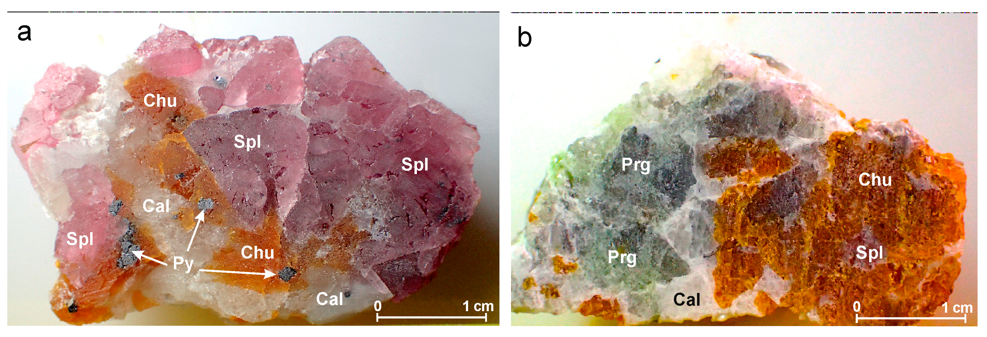
2. Geological Setting and Petrography
3. Materials and Methods
3.1. Microprobe Analysis
3.2. Single-Crystal X-ray Diffraction
3.3. Raman Spectrometry
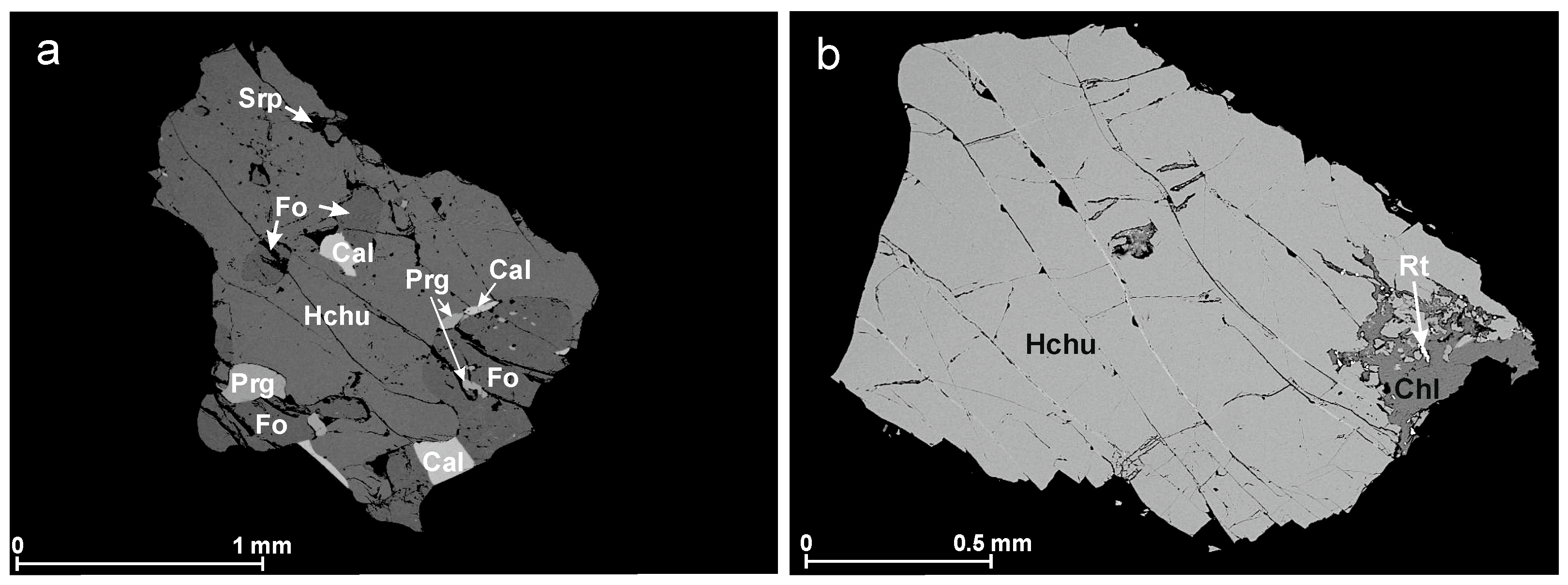
4. Results
4.1. Major Element Mineral Chemistry
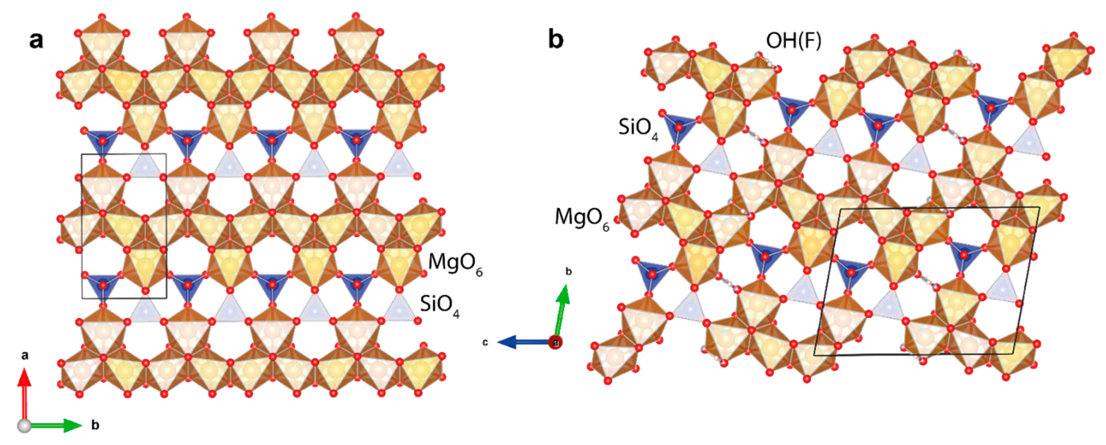
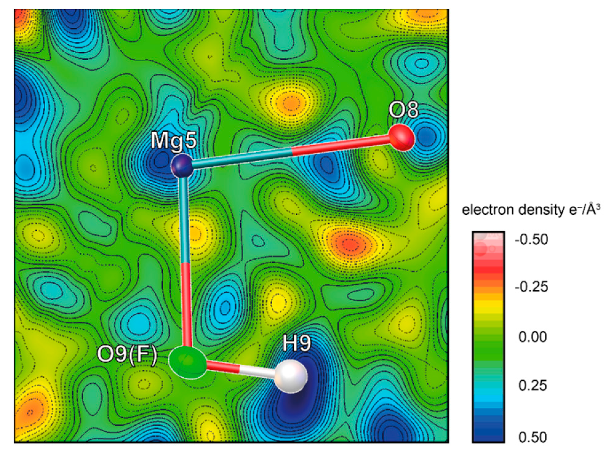
4.2. Structure Description and Refinement
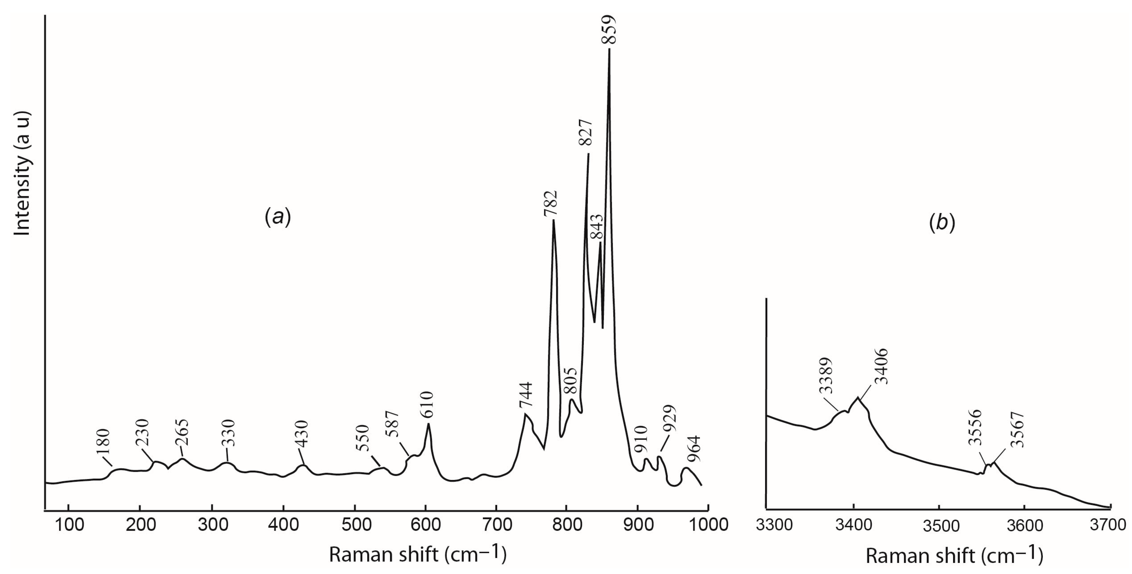
4.3. Raman Spectroscopy
5. Discussion
6. Conclusions
Supplementary Materials
Author Contributions
Funding
Data Availability Statement
Acknowledgments
Conflicts of Interest
References
- McGetchin, T.R.; Silver, L.T.; Chodos, A.A. Titanoclinohumite: A possible mineralogical site for water in the upper mantle. J. Geophys. Res. 1970, 75, 244–259. [Google Scholar] [CrossRef] [Green Version]
- Aoki, K.; Fujino, K.; Akaogi, M. Titanochondrodite and titanoclinohumite derived from the upper mantle in the Buell Park kimberlite, Arizona, USA. Contrib. Miner. Petrol. 1976, 56, 243–254. [Google Scholar] [CrossRef]
- Papike, J.; Cameron, M. Crystal chemistry of silicate minerals of geophysical interest. Rev. Geophys. Sp. Phys. 1976, 14, 37–80. [Google Scholar] [CrossRef]
- Kanzaki, M. Stability of hydrous magnesium silicates in the mantle transition zone. Phys. Earth Plan. Inter. 1991, 66, 307–312. [Google Scholar] [CrossRef]
- Wunder, B.; Medenbach, O.; Daniels, P.; Schreyer, W. First synthesis of the hydroxyl end-member of humite, Mg7Si3O12(OH)2. Amer. Miner. 1995, 80, 638–640. [Google Scholar]
- Zheng, Y.F. Metamorphic chemical geodynamics in continental subduction zones. Chem. Geol. 2012, 328, 5–48. [Google Scholar] [CrossRef]
- Arai, S.; Ishimaru, S.; Mizukami, T. Methane and propane micro-inclusions in olivine in titanoclinohumite-bearing dunites from the Sanbagawa high-P metamorphic belt, Japan: Hydrocarbon activity in a subduction zone and Ti mobility. Earth Planet. Sci. Lett. 2012, 353, 1–11. [Google Scholar] [CrossRef]
- Hughes, L.; Pawley, A. Fluorine partitioning between humite-group minerals and aqueous fluids: Implications for volatile storage in the upper mantle. Contrib. Miner. Petrol. 2019, 174, 78. [Google Scholar] [CrossRef] [Green Version]
- Gekimyants, V.M.; Sokolova, E.V.; Spiridonov, E.M.; Ferraris, G.; Chukanov, N.V.; Prencipe, M.; Avdonin, V.N.; Polenov, Y.A. Hydroxylclinohumite Mg9(SiO4)4(OH,F)2-a new mineral of the humite group. Zapiski RMO. 1999, 128, 64–70. (In Russian) [Google Scholar]
- Borneman-Starynkevich, I.D.; Myasnikov, V.S. On isomorphic substitution in Clinohumite. Compt. Rend. Acad. Sci. URSS 1950, 71, 137–140. (In Russian) [Google Scholar]
- Beeker, P. Klinohumite vom Laperwitzbach, Dorfertal, Osttirol. Karinthin 1976, 75, 257–260. (In German) [Google Scholar]
- Mitchell, R.H. Manganoan magnesian ilmenite and titanian clinohumite from the Jacupiranga carbonatite, Sao Paulo, Brazil. Amer. Miner. 1978, 63, 544–547. [Google Scholar]
- Möckel, J.R. Structural petrology of the garnet peridotite of Alpe Arami (Ticino), Switzerland. Leid. Geol. Med. 1969, 42, 61–130. [Google Scholar]
- Trommsdorff, V.; Evans, B.W. Titanian hydroxyl-clinohumite formation and breakdown in antigorite rocks (Malenco-Italy). Contrib. Miner. Petrol. 1980, 72, 229–242. [Google Scholar] [CrossRef]
- Ehlers, K.; Hoinkes, G. Titanian Chondrodite and Clinohumite in Marbles from the Ötztal Crystalline Basement. Miner. Petrol. 1987, 36, 13–25. [Google Scholar] [CrossRef]
- Jones, N.W.; Ribbe, P.H.; Gibbs, G.V. Crystal chemistry of the humite minerals. Amer. Miner. 1969, 54, 391–411. [Google Scholar]
- Fujino, K.; Takeuchi, Y. Crystal chemistry of titanian chondrodite and titanian clinohumite of high pressure origin. Amer. Miner. 1978, 63, 535–543. [Google Scholar]
- Ribbe, P.H. Titanium, fluorine and hydroxyl in humite minerals. Amer. Miner. 1979, 64, 1027–1035. [Google Scholar]
- Huong, L.T.-T.; Haeger, T.; Phan, T.-L. Study of impurity in blue spinel from the Luc Yen mining area, Yen Bai province, Vietnam. Vietnam J. Earth Sci. 2018, 40, 47–55. [Google Scholar] [CrossRef] [Green Version]
- Chauvire, B.; Rondeau, B.; Fritsch, E.; Ressigeac, P.; Devidal, J.-L. Blue spinel from the Luc Yen district of Vietnam. Gems Gemol. 2015, 51, 2–17. [Google Scholar] [CrossRef]
- Huong, L.T.-T.; Häger, T.; Hofmeister, W.; Hauzenberger, C.; Schwarz, D.; Long, P.V.; Wehrmeister, W.; Khoi, N.N.; Nhung, N.T. Gemstones from Vietnam: An update. Gems Gemol. 2012, 48, 158–176. [Google Scholar] [CrossRef] [Green Version]
- Sokolov, P.; Kuksa, K.; Marakhovskaya, O.; Gussias, G.A. In search of cobalt blue spinel in Vietnam. Color Summer 2019, 43, 43–49. [Google Scholar]
- Peretti, A.; Günther, D. Spinel from Namya. Contrib. Gem. 2003, 2, 15–18. [Google Scholar]
- Malsy, A.-K.; Klemm, L. Distinction of gem spinels from the Himalayan mountain belt. Chim. Int. J. Chem. 2010, 64, 741–746. [Google Scholar] [CrossRef]
- Sutthirat, C.; Lapngamchana, S.; Pisutha-Arnond, V.; Khoi, N.N. Petrography and some mineral chemistry of gem-bearing marble from Luc Yen deposit, northern Vietnam. In Proceedings of the International Symposia on Geoscience Resources and Environments of Asian Terranes (GREAT 2008), Bangkok, Thailand, 24–26 November 2008; pp. 283–288. [Google Scholar]
- Häger, T.; Hauzenberger, C.; Lehmann, C.; Zimmer, D.; Nguyen Ngoc Khoi, D.A.T.; Le Thi-Thu Huong, H.W. Causes of colour of natural untreated spinels from Vietnam in comparison to flame fusion and flux grown synthetics. In Proceedings of the 33rd International Gemmological Conference, Hanoi, Vietnam, 10 October 2013; pp. 89–91. [Google Scholar]
- Garnier, V.; Ohnenstetter, D.; Giuliani, G.; Maluski, H.; Deloule, E.; Phan Trong, T.; Pham Van, L.; Hoàng Quang, V. Age and significance of ruby-bearing marble from the Red River Shear Zone, northern Vietnam. Can. Miner. 2005, 43, 1315–1329. [Google Scholar] [CrossRef]
- Garnier, V.; Giuliani, G.; Ohnenstetter, D.; Fallick, A.E.; Dubessy, J.; Banks, D.; Vinh, H.Q.; Lhomme, T.; Maluski, H.; Pêcher, A. Marble-hosted ruby deposits from central and southeast Asia: Towards a new genetic model. Ore Geol. Rev. 2008, 34, 169–191. [Google Scholar] [CrossRef]
- Giuliani, G.; Fallick, A.E.; Boyce, A.J.; Pardieu, V.; Pham, V.L. Pink and red spinels in marble: Trace elements, oxygen isotopes, and sources. Can. Miner. 2017, 55, 743–761. [Google Scholar] [CrossRef]
- Fallick, A.E.; Giuliani, G.; Rigaudier, T.; Boyce, A.J.; Pham, V.L.; Pardieu, V. Remarkably uniform oxygen isotope systematics for co-existing pairs of gem-spinel and calcite in marble, with special reference to Vietnamese deposits. Comp. Rend. Geosci. 2019, 351, 27–36. [Google Scholar] [CrossRef]
- Krivovichev, V.G.; Kuksa, K.A.; Sokolov, P.B.; Marakhovskay, O.Y.; Klimacheva, M.E. Marble-hosted noble spinel deposits from Luc Yen district (Vietnam): Mineral systems and some aspects of genesis. Zapiski RMO. 2022, 147, 37–49. (In Russian) [Google Scholar]
- Hofmeister, A.; Mao, H. Evaluation of shear moduli and other properties of silicates with the spinel structure from IR spectroscopy. Amer. Miner. 2001, 86, 622–639. [Google Scholar] [CrossRef]
- Kuksa, K.; Sokolov, P.; Marakhovskaya, O.; Gussias, G.; Brownscombe, W. Mineralogy, geochemistry and genesis of the Luc Yen noble spinel deposit, Vietnam. Mineralogy 2019, 5, 56–69. (In Russian) [Google Scholar] [CrossRef] [Green Version]
- Long, P.V.; Giuliani, G. Update on gemstone mining in Luc Yen, Vietnam. Gems Gemol. 2013, 49, 31–46. [Google Scholar] [CrossRef]
- Hauzenberger, C.A.; Häger, T.; Hofmeister, W.; Quang, V.X.; Rohan Fernando, G.W.A. Origin and formation of gem quality corundum from Vietnam. In Proceedings of the International Workshop, “Geo- and Material-Science on Gem-Minerals of Vietnam”, Hanoi, Vietnam, 1–8 October 2003; pp. 1–8. [Google Scholar]
- Hurai, V.; Wierzbicka-Wieczorek, M.; Pentrák, M.; Huraiová, M.; Thomas, R.; Swierczewska, A.; Luptáková, J. X-ray Diffraction and Vibrational Spectroscopic Characteristics of Hydroxylclinohumite from Ruby-Bearing Marbles (Luc Yen District, Vietnam). Intern. J. Miner. 2014, 2014, 648530. [Google Scholar] [CrossRef] [Green Version]
- Ottolini, L.; C’amara, F.; Bigi, S. An investigation of matrix effects in the analysis of fluorine in humite-group minerals by EMPA, SIMS, and SREF. Amer. Miner. 2000, 85, 89–102. [Google Scholar] [CrossRef]
- Long, P.V.; Giuliani, G.; Fallick, A.E.; Boyce, A.J.; Pardieu, V. Trace elements and oxygen isotopes of gem spinels in marble from the Luc Yen-An Phu areas, Yen Bai province, North Vietnam. Vietnam J. Earth Sci. 2018, 40, 165–177. [Google Scholar] [CrossRef] [Green Version]
- Krivovichev, V.G.; Kuksa, K.A.; Sokolov, P.B.; Marakhovskaya, O.Y.; Zolotarev, A.A.; Bocharov, V.N.; Semenova, T.F.; Klimacheva, M.E.; Gussiås, G.A. Preiswerkite: A First Occurrence in Marble Hosting Gem Spinel Deposits, Luc Yen, Vietnam. Minerals 2022, 12, 1024. [Google Scholar] [CrossRef]
- Agilent Technologies CrysAlis CCD and CrysAlis RED; Oxford Diffraction Ltd.: Yarnton, UK, 2014.
- Sheldrick, G.M. Crystal structure refinement with SHELXL. Acta Crystallogr. Sect. C Str. Chem. 2015, 71, 3–8. [Google Scholar] [CrossRef] [Green Version]
- Izumi, F.; Momma, K. Three-Dimensional Visualization in Powder Diffraction. Solid State Phen. 2007, 130, 15–20. [Google Scholar] [CrossRef]
- Robinson, K.; Gibbs, G.V.; Ribbe, P.H. The crystal structures of the humite minerals. IV. Clinohumite and titanoclinohumite. Amer. Miner. 1973, 58, 43–49. [Google Scholar]
- White, T.J.; Hyde, B.G. Electron microscope study of the humite minerals: I Mg-rich specimens. Phys. Chem. Minerals 1982, 8, 55–63. [Google Scholar] [CrossRef]
- Gaspar, J.C. Titanian clinohumite in the carbonatites of the Jacupiranga Complex, Brazil: Mineral chemistry and comparison with titanian clinohumite from other environments. Amer. Miner. 1992, 77, 168–178. [Google Scholar]
- Bragg, W.L.; West, J. The structure of certain silicates. Proc. R. Soc. London. Ser. A 1927, 114, 450–473. [Google Scholar] [CrossRef]
- Langer, K.; Platonov, A.N.; Matsyuk, S.; Wildner, M. The crystal chemistry of the humite minerals: Fe2+-Ti4+ charge transfer and structural allocation of Ti4+ in chondrodite and clinohumite. Eur. J. Miner. 2002, 14, 1027–1032. [Google Scholar] [CrossRef]
- Mikhailova, J.A.; Ivanyuk, G.Y.; Kalashnikov, A.O.; Pakhomovsky, Y.A.; Bazai, A.V.; Panikorovskii, T.L.; Yakovenchuk, V.N.; Konoplev, N.G.; Goryainov, P.M. Three-D mineralogical mapping of the kovdor phoscorite-carbonatite complex, NW Russia: I. Forsterite. Minerals 2018, 8, 260. [Google Scholar] [CrossRef] [Green Version]
- Friedrich, A.; Lager, G.A.; Kunz, M.; Chakoumakos, B.C.; Smyth, J.R.; Schultz, A.J. Temperature-dependent single-crystal neutron diffraction study of natural chondrodite and clinohumites. Amer. Miner. 2001, 86, 981–989. [Google Scholar] [CrossRef]
- Berry, A.J.; James, M. Refinement of hydrogen positions in synthetic hydroxyl-clinohumite by powder neutron diffraction. Amer. Miner. 2001, 86, 181–184. [Google Scholar] [CrossRef]
- Ferraris, G.; Prencipe, M.; Sokolova, E.; Gekimyants, V.M.; Spiridonov, E.M. Hydroxylclinohumite, a new member of the humite group: Twinning, crystal structure and crystal chemistry of the clinohumite subgroup. Zeitschrift für Krist.-Cryst. Mater. 2000, 215, 169–173. [Google Scholar] [CrossRef]
- Libowitzky, E. Correlation of O-H Stretching Frequencies and O-H…O Hydrogen Bond Lengths in Minerals. Hydrog. Bond Res. 1999, 1059, 103–115. [Google Scholar] [CrossRef]
- Liu, D.; Pang, Y.; Ye, Y.; Jin, Z.; Smyth, J.R.; Yang, Y.; Zhang, Z.; Wang, Z. In-situ high-temperature vibrational spectra for synthetic and natural clinohumite: Implications for dense hydrous magnesium silicates in subduction zones. Amer. Miner. 2019, 104, 53–63. [Google Scholar] [CrossRef]
- Frost, R.; Palmer, S.; Bouzaid, J.; Reddy, J. A Raman spectroscopic study of humite minerals. J. Raman Spectr. 2007, 38, 68–77. [Google Scholar] [CrossRef] [Green Version]
- Ye, Y.; Smyth, J.R.; Jacobsen, S.D.; Goujon, C. Crystal chemistry, thermal expansion, and Raman spectra of hydroxyl-clinohumite: Implications for water in Earth’s interior. Contrib. Miner. Petrol. 2013, 165, 563–574. [Google Scholar] [CrossRef]
- Pekov, I.V.; Gerasimova, E.I.; Chukanov, N.V.; Kabalov, Y.K.; Zubkova, N.V.; Zadov, A.E.; Yapaskurt, V.O.; Gekimyants, V.M.; Pushcharovskii, D.Y. Hydroxylchondrodite Mg5(SiO4)2(OH)2: A new mineral of the humite group and its crystal structure. Doklady Earth Sciences 2011, 436, 230–236. [Google Scholar] [CrossRef]
- Duffy, C.J.; Greenwood, H.J. Phase equilibria in the system MgO-MgF2-SiO2-H2O. Amer. Miner. 1979, 64, 1156–1174. [Google Scholar]
- Rice, J.M. Phase equilibria involving humite minerals in impure dolomitic limestones: Part I. Calculated stability of clinohumite. Contrib. Miner. Petrol. 1980, 71, 219–235. [Google Scholar] [CrossRef]
- Bucher, K.; Rodney, G. Petrogenesis of Metamorphic Rocks, 8th ed.; Springer: Berlin/Heidelberg, Germany, 2011; p. 428. [Google Scholar] [CrossRef] [Green Version]
- Spear, F.S. Metamorphic Phase Equilibria and Pressure-Temperature-Time Paths; Mineralogical Society of America: Washington, IL, USA, 1995; p. 799. [Google Scholar]
- Korzhinskiy, D.S. Physico-Chemical Basis of the Analysis of the Paragenesis of Minerals; Consultants Bureau, New York and Chapman & Hall: London, UK, 1959; p. 142. [Google Scholar]
- Korzhinskiy, D.S. Teoreticheskiye Osnovy Analiza Paragenezisov Mineralov (Theoretical Basis of Analysis of Mineral Para-Geneses); Nauka: Moscow, Russia, 1973; p. 288. [Google Scholar]
- Giuliani, G.; Dubessy, J.; Ohnenstetter, D.; Banks, D.; Branquet, Y.; Feneyrol, J.; Fallick, A.E.; Martelat, J.-E. The role of evaporites in the formation of gems during metamorphism of carbonate platforms: A review. Miner. Deposita 2018, 53, 1–20. [Google Scholar] [CrossRef]
- Yamamoto, K.; Akimoto, S. The system MgO-SiO2-H2O at high pressures and temperatures-stability field for hydroxylchondrodite, hydroxyl-clinohumite and 10 A-phase. Amer. J. Sci. 1977, 277, 288–312. [Google Scholar] [CrossRef]
- Hauzenberger, C.A.; Häger, T.; Baumgartner, L.P.; Hofmeister, W. High-grade metamorphism and stable-isotope geochemistry of N-Vietnamese gem-bearing rocks. In Material Characterization by Solid State Spectroscopy: Gems and Mineral of Vietnam; Hofmeister, W., Dao, N.Q., Quang, V.X., Eds.; International Workshop: Hanoi, Vietnam, 2001; pp. 124–138. [Google Scholar]


| V-11-2d | V-11-2a | V-12-03 | σ | ||
|---|---|---|---|---|---|
| SiO2 wt% | 38.58 | 38.64 | 38.91 | 38.71 | 0.17 |
| TiO2 | 3.33 | 3.90 | 3.93 | 3.72 | 0.34 |
| MgO | 54.79 | 54.57 | 54.63 | 54.66 | 0.11 |
| * FeO | 0.35 | 0.42 | 0.00 | 0.26 | 0.22 |
| F | 1.85 | 1.50 | 1.37 | 1.57 | 0.25 |
| ** H2O | 1.47 | 1.60 | 1.61 | 1.56 | 0.08 |
| Total | 100.41 | 100.63 | 100.45 | 100.50 | 0.12 |
| -O=F2 | 0.76 | 0.64 | 0.58 | 0.66 | 0.09 |
| Total | 99.65 | 99.99 | 99.87 | 99.84 | 0.17 |
| *** Si apfu | 4.07 | 4.07 | 4.10 | 4.08 | 0.02 |
| Ti | 0.27 | 0.30 | 0.31 | 0.29 | 0.02 |
| Mg | 8.63 | 8.59 | 8.59 | 8.60 | 0.02 |
| Fe | 0.03 | 0.04 | 0.00 | 0.02 | 0.02 |
| OH | 1.04 | 1.13 | 1.13 | 1.10 | 0.05 |
| F | 0.62 | 0.50 | 0.46 | 0.53 | 0.08 |
| O | 0.34 | 0.37 | 0.41 | 0.37 | 0.03 |
| Mg# | 0.97 | 0.96 | 0.96 | 0.96 | 0.01 |
| Identification Code | V-11-2d | V-11-2a | V-12-03 |
|---|---|---|---|
| Temperature/K | 293(2) | ||
| Crystal system | monoclinic | ||
| Space group | P21/b11 | ||
| a/Å | 4.7381(2) | 4.74060(10) | 4.7386(2) |
| b/Å | 10.2423(4) | 10.2431(3) | 10.2416(3) |
| c/Å | 13.6531(6) | 13.6551(4) | 13.6597(5) |
| α/° | 100.917(4) | 100.935(3) | 100.905(3) |
| Volume/Å3 | 650.58(5) | 651.03(3) | 650.95(4) |
| Z | 2 | ||
| ρcalcg/cm3 | 3.199 | 3.197 | 3.200 |
| μ/mm−1 | 1.118 | 1.122 | 1.135 |
| F(000) | 623.0 | 623.0 | 624.0 |
| Crystal size/mm3 | 0.15 × 0.13 × 0.1 | 0.22 × 0.15 × 0.14 | 0.17 × 0.12 × 0.1 |
| Radiation | Mo Kα (λ = 0.71073) | ||
| 2Θ range for data collection/° | 8.1 to 67.142 | 8.098 to 66.374 | 8.1 to 66.498 |
| Index ranges | −7 ≤ h ≤ 6, −15 ≤ k ≤ 14, −19 ≤ l ≤ 18 | −5 ≤ h ≤ 7, −14 ≤ k ≤ 12, −18 ≤ l ≤ 20 | −7 ≤ h ≤ 7, −14 ≤ k ≤ 15, −15 ≤ l ≤ 19 |
| Reflections collected | 7228 | 7179 | 7001 |
| Independent reflections | 2188 [Rint = 0.0308, Rsigma = 0.0301] | 2158 [Rint = 0.0230, Rsigma = 0.0236] | 2145 [Rint = 0.0247, Rsigma = 0.0257] |
| Data/restraints/parameters | 2188/3/147 | 2158/3/147 | 2145/3/147 |
| Goodness-of-fit on F2 | 1.096 | 1.060 | 1.101 |
| Final R indexes [I >= 2σ (I)] | R1 = 0.0266, wR2 = 0.0689 | R1 = 0.0230, wR2 = 0.0570 | R1 = 0.0249, wR2 = 0.0721 |
| Final R indexes [all data] | R1 = 0.0313, wR2 = 0.0719 | R1 = 0.0269, wR2 = 0.0594 | R1 = 0.0296, wR2 = 0.0752 |
| Largest diff. peak/hole/e Å−3 | 0.51/−0.49 | 0.54/−0.45 | 0.48/−0.45 |
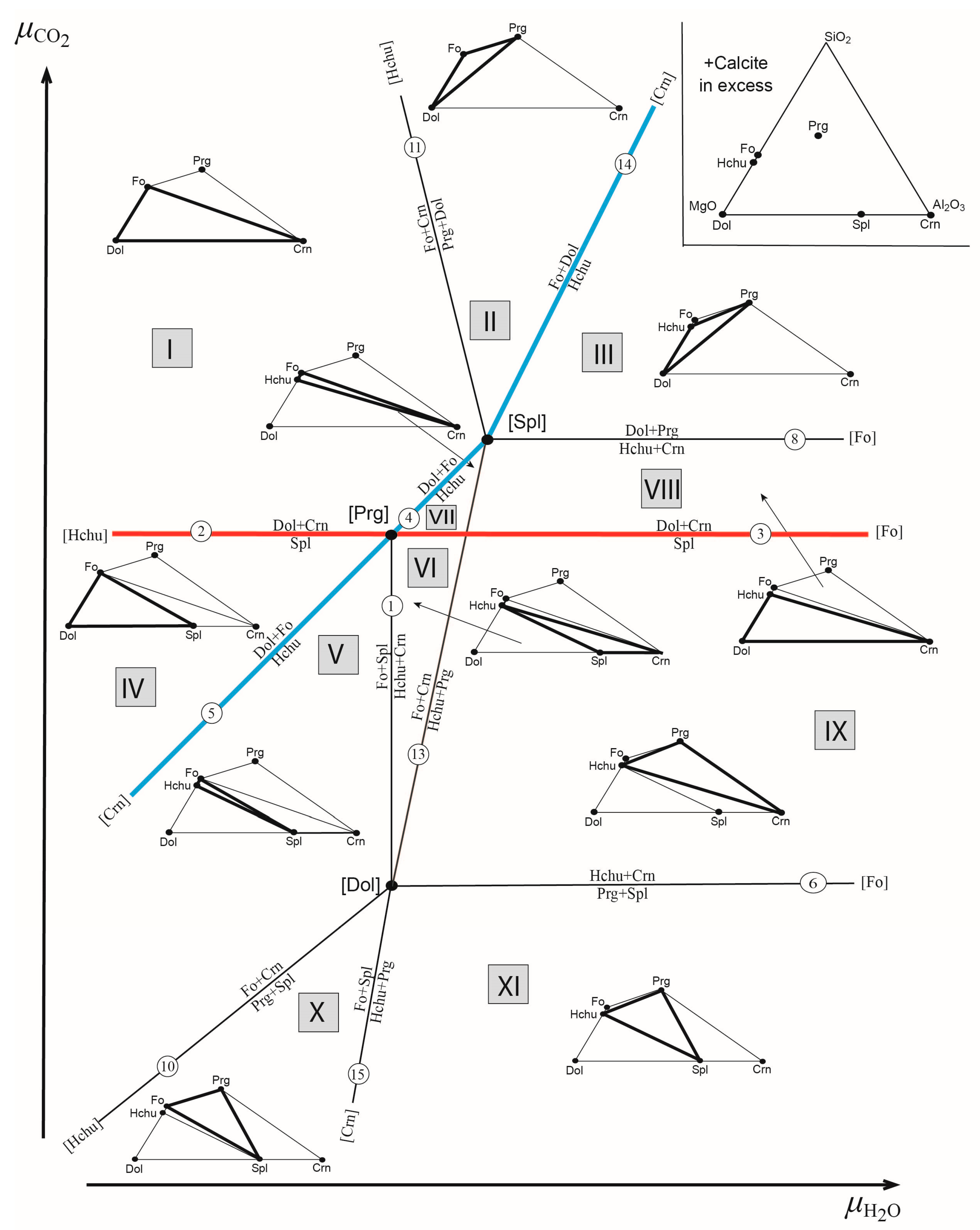
| Mineral | Symbol | Formula | Mg | Al | Si |
|---|---|---|---|---|---|
| Hydroxylclinohumite | Hchu | Mg9(SiO4)4(OH)2 | 9 | 0 | 4 |
| Pargasite | Prg | NaCa2(Mg4Al)Σ5(Al2Si6O22)(OH)2 | 4 | 3 | 6 |
| Forsterite | Fo | Mg2SiO4 | 2 | 0 | 1 |
| Dolomite | Dol | CaMg(CO3)2 | 1 | 0 | 0 |
| Spinel | Spl | MgAl2O4 | 1 | 2 | 0 |
| Corundum | Crn | Al2O3 | 0 | 2 | 0 |
| N | Symbols * | Chemical Reactions |
|---|---|---|
| 1 | [PrgDol] | 4Fo + Spl + H2O = Hchu + Crn |
| 2 | [PrgHchu] | Dol + Crn = Spl + (Cal) + CO2 |
| 3 | [PrgFo] | Dol + Crn = Spl + (Cal) + CO2 |
| 4 | [PrgSpl] | Dol + 4Fo + H2O = Hchu + (Cal) + CO2 |
| 5 | [PrgCrn] | Dol + 4Fo + H2O = Hchu + (Cal) + CO2 |
| 6 | [FoDol] | 3Hchu + 22Crn + 4(Cal) + 2Na+ = 2Prg + 19Spl + 4CO2 + 2H+ |
| 7 | [FoHchU] | Dol + Crn = Spl + (Cal) + CO2 |
| 8 | [FoSpl] | 19Dol + 2Prg + 2H+ = 3Hchu + 3Crn + 23(Cal) + 15CO2 + 2Na+ |
| 9 | [FoCrn] | 22Dol + 2Prg + 2H+ = 3Hchu + 3Spl + 26(Cal) + 18CO2 + 2Na+ |
| 10 | [HchuDol] | 2Prg + 16Spl + 4CO2 + 2H+= 12Fo + 19Crn + 4(Cal) + 3H2O + 2Na+ |
| 11 | [HchuSpl] | 2Prg + 16Dol + 2H+ = 12Fo + 3Crn + 20(Cal) + 3H2O + 12CO2 + 2Na+ |
| 12 | [HchuCrn] | 2Prg + 19Dol + 2H+= 12Fo + 3Spl + 23(Cal) + 2H2O + 15CO2 + 2Na+ |
| 13 | [SplDol] | 76Fo + 3Crn + 4(Cal) + 19H2O + 2Na+ = 16Hchu + 2Prg + 2H+ + 4CO2 |
| 14 | [SplCrn] | Hchu + (Cal) + CO2 = 4Fo + Dol + 2H2O |
| 15 | [DolCrn] | 88Fo + 3Spl + 4(Cal) + 22H2O + 2Na+ = 19Hchu + 2Prg + 4CO2 + 2H+ |
Disclaimer/Publisher’s Note: The statements, opinions and data contained in all publications are solely those of the individual author(s) and contributor(s) and not of MDPI and/or the editor(s). MDPI and/or the editor(s) disclaim responsibility for any injury to people or property resulting from any ideas, methods, instructions or products referred to in the content. |
© 2023 by the authors. Licensee MDPI, Basel, Switzerland. This article is an open access article distributed under the terms and conditions of the Creative Commons Attribution (CC BY) license (https://creativecommons.org/licenses/by/4.0/).
Share and Cite
Krivovichev, V.G.; Kuksa, K.A.; Sokolov, P.B.; Panikorovskii, T.L.; Bocharov, V.N.; Gussiås, G.A. First Occurrence of Titanian Hydroxylclinohumite in Marble-Hosting Gem Spinel Deposits, Luc Yen, Vietnam. Minerals 2023, 13, 901. https://doi.org/10.3390/min13070901
Krivovichev VG, Kuksa KA, Sokolov PB, Panikorovskii TL, Bocharov VN, Gussiås GA. First Occurrence of Titanian Hydroxylclinohumite in Marble-Hosting Gem Spinel Deposits, Luc Yen, Vietnam. Minerals. 2023; 13(7):901. https://doi.org/10.3390/min13070901
Chicago/Turabian StyleKrivovichev, Vladimir G., Katherine A. Kuksa, Pavel B. Sokolov, Taras L. Panikorovskii, Vladimir N. Bocharov, and Geir Atle Gussiås. 2023. "First Occurrence of Titanian Hydroxylclinohumite in Marble-Hosting Gem Spinel Deposits, Luc Yen, Vietnam" Minerals 13, no. 7: 901. https://doi.org/10.3390/min13070901