
Case Report: MRI, CEUS, and CT Imaging Features of Metanephric Adenoma with Histopathological Correlation and Literature Review


Abstract
:1. Introduction
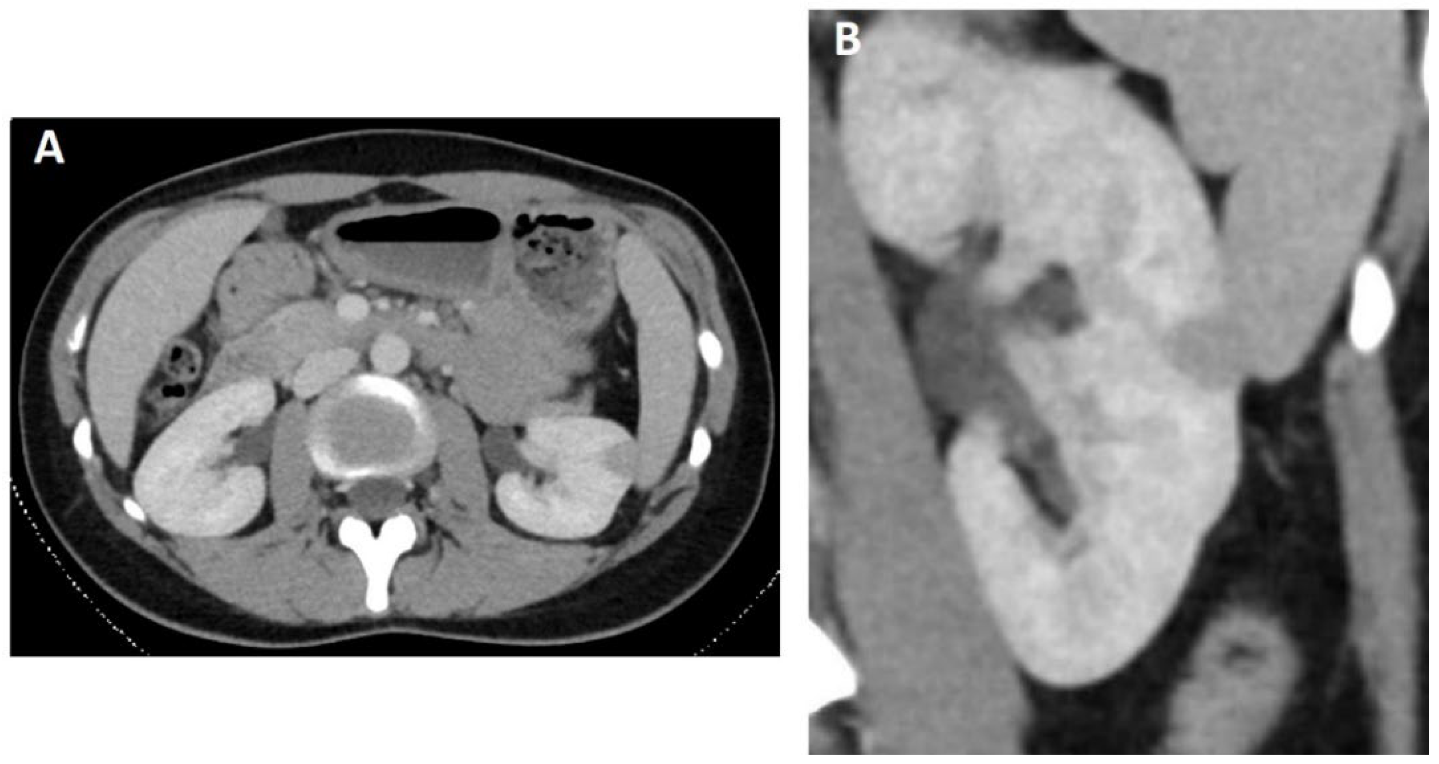
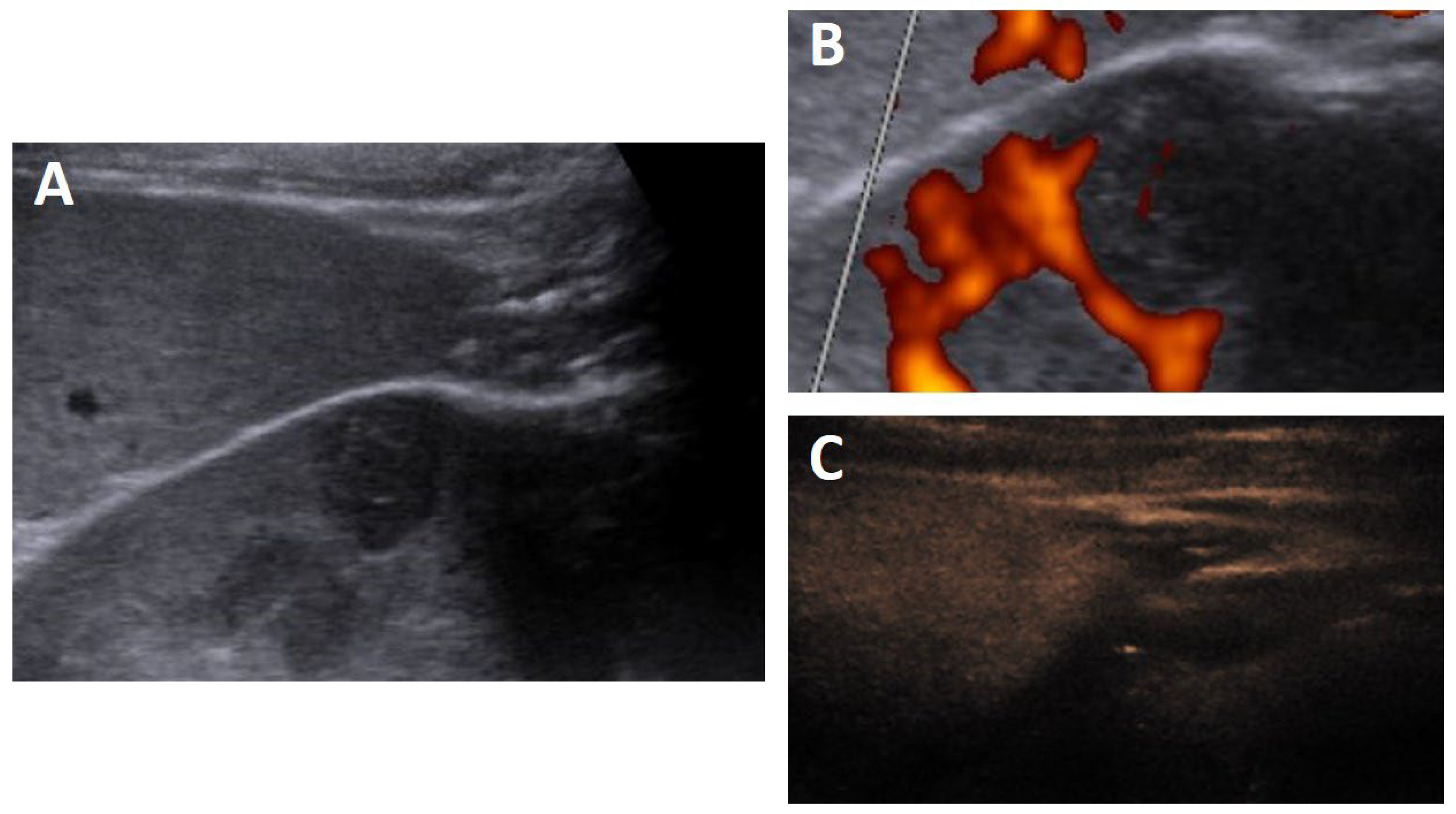
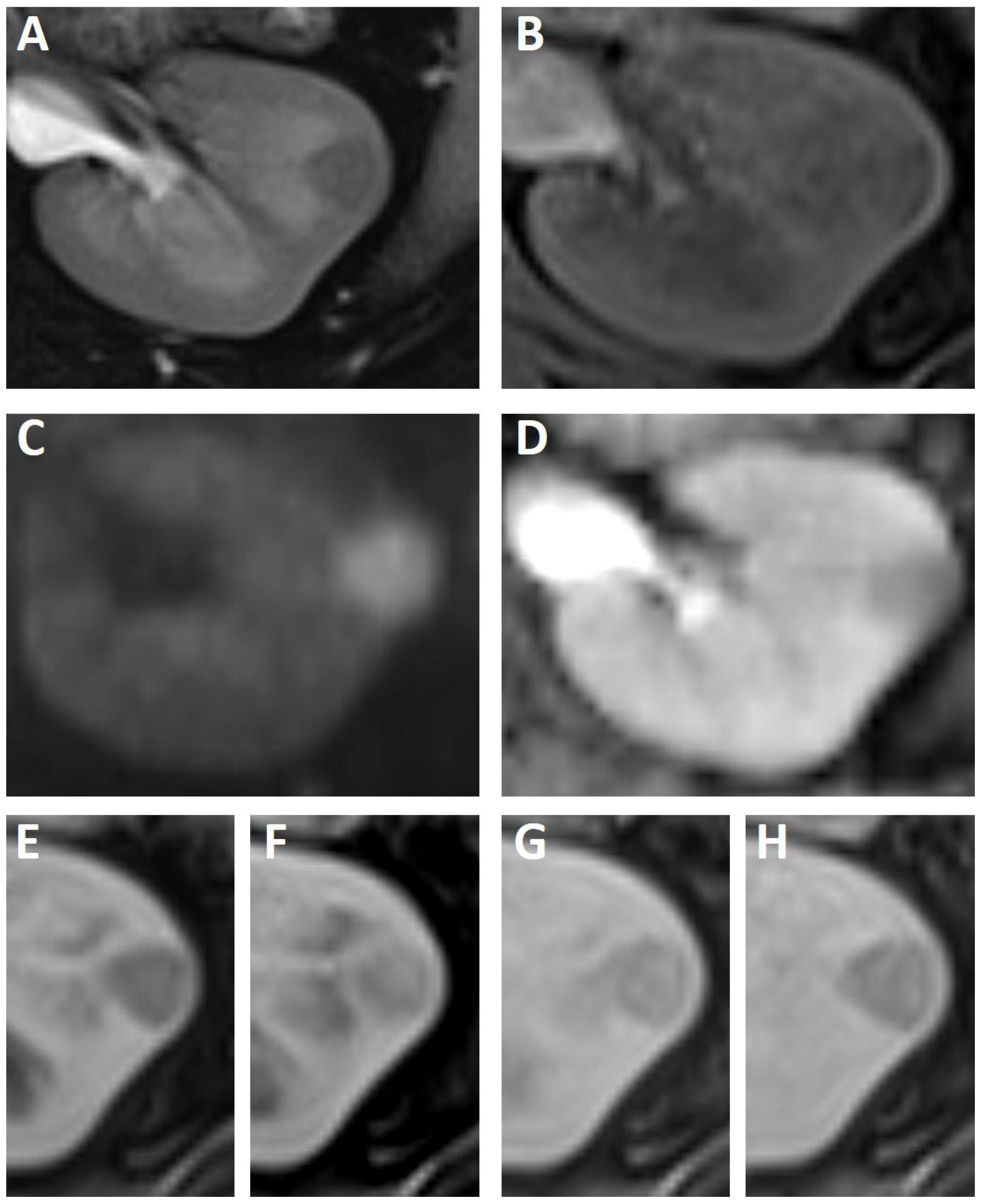
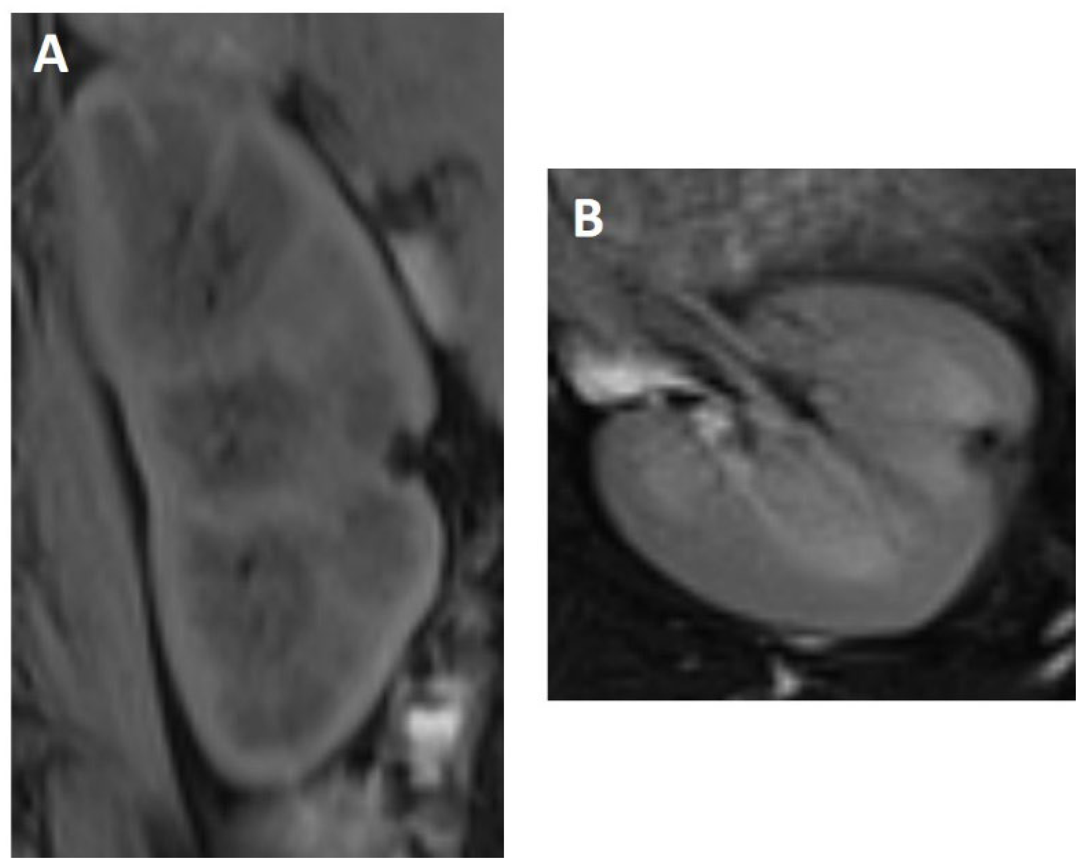
2. Case Study
3. Discussion
4. Conclusions
Author Contributions
Funding
Informed Consent Statement
Data Availability Statement
Acknowledgments
Conflicts of Interest
References
- Brisigotti, M.; Cozzutto, C.; Fabbretti, G.; Sergi, C.; Callea, F. Metanephric adenoma. Histol. Histopathol. 1992, 7, 689–692. [Google Scholar] [PubMed]
- Spaner, S.J.; Yu, Y.; Cook, A.J.; Boag, G. Pediatric metanephric adenoma: Case report and review of the literature. Int. Urol. Nephrol. 2014, 46, 677–680. [Google Scholar] [CrossRef] [PubMed]
- Mantoan Padilha, M.; Billis, A.; Allende, D.; Zhou, M.; Magi-Galluzzi, C. Metanephric adenoma and solid variant of papillary renal cell carcinoma: Common and distinctive features. Histopathology 2013, 62, 941–953. [Google Scholar] [CrossRef] [PubMed]
- Amin, M.B.; Amin, M.B.; Tamboli, P.; Javidan, J.; Stricker, H.; de-Peralta Venturina, M.; Deshpande, A.; Menon, M. Prognostic impact of histologic subtyping of adult renal epithelial neoplasms: An experience of 405 cases. Am. J. Surg. Pathol. 2002, 26, 281–291. [Google Scholar] [CrossRef] [PubMed]
- Snyder, M.E.; Bach, A.; Kattan, M.W.; Raj, G.V.; Reuter, V.E.; Russo, P. Incidence of benign lesions for clinically localized renal masses smaller than 7 cm in radiological diameter: Influence of sex. J. Urol. 2006, 176, 2391–2395, discussion 2395–2396. [Google Scholar] [CrossRef] [PubMed]
- Pins, M.R.; Jones, E.C.; Martul, E.V.; Kamat, B.R.; Umlas, J.; Renshaw, A.A. Metanephric adenoma-like tumors of the kidney: Report of 3 malignancies with emphasis on discriminating features. Arch. Pathol. Lab. Med. 1999, 123, 415–420. [Google Scholar] [CrossRef]
- Nakagawa, T.; Kanai, Y.; Fujimoto, H.; Kitamura, H.; Furukawa, H.; Maeda, S.; Oyama, T.; Takesaki, T.; Hasegawa, T. Malignant mixed epithelial and stromal tumours of the kidney: A report of the first two cases with a fatal clinical outcome. Histopathology 2004, 44, 302–304. [Google Scholar] [CrossRef]
- Schmelz, H.U.; Stoschek, M.; Schwerer, M.; Danz, B.; Hauck, E.W.; Weidner, W.; Sparwasser, C. Metanephric adenoma of the kidney: Case report and review of the literature. Int. Urol. Nephrol. 2005, 37, 213–217. [Google Scholar] [CrossRef]
- Fan, H.; Shao, Q.Q.; Li, H.Z.; Xiao, Y.; Zhang, Y.S. The Clinical Characteristics of Metanephric Adenoma: A Case Report and Literature Review. Medicine 2016, 95, e3486. [Google Scholar] [CrossRef]
- Wang, P.; Tian, Y.; Xiao, Y.; Zhang, Y.; Sun, F.A.; Tang, K. A metanephric adenoma of the kidney associated with polycythemia: A case report. Oncol. Lett. 2016, 11, 352–354. [Google Scholar] [CrossRef] [Green Version]
- Yoshioka, K.; Miyakawa, A.; Ohno, Y.; Namiki, K.; Horiguchi, Y.; Murai, M.; Mukai, M.; Tachibana, M. Production of erythropoietin and multiple cytokines by metanephric adenoma results in erythrocytosis. Pathol. Int. 2007, 57, 529–536. [Google Scholar] [CrossRef]
- Paner, G.P.; Turk, T.M.; Clark, J.I.; Lindgren, V.; Picken, M.M. Passive seeding in metanephric adenoma: A review of pseudometastatic lesions in perinephric lymph nodes. Arch. Pathol. Lab. Med. 2005, 129, 1317–1321. [Google Scholar] [CrossRef]
- Kumar, S.; Mandal, A.K.; Acharya, N.R.; Kakkad, N.; Singh, S.K. Laparoscopic nephron-sparing surgery for metanephric adenoma. Surg. Laparosc. Endosc. Percutan. Tech. 2007, 17, 573–575. [Google Scholar] [CrossRef]
- Picken, M.M.; Curry, J.L.; Lindgren, V.; Clark, J.I.; Eble, J.N. Metanephric adenosarcoma in a young adult: Morphologic, immunophenotypic, ultrastructural, and fluorescence in situ hybridization analyses: A case report and review of the literature. Am. J. Surg. Pathol. 2001, 25, 1451–1457. [Google Scholar] [CrossRef]
- Drut, R.; Drut, R.M.; Ortolani, C. Metastatic metanephric adenoma with foci of papillary carcinoma in a child: A combined histologic, immunohistochemical, and FISH study. Int. J. Surg. Pathol. 2001, 9, 241–247. [Google Scholar] [CrossRef]
- Amie, F.; Andre, D.; Foulet Roge, A.; Goura, E.; Chautard, D.; Colombel, P. Bilateral renal metanephric adenoma. Prog. Urol. 2004, 14, 534–537, discussion 537. [Google Scholar]
- Hu, Y.C.; Wu, L.; Yan, L.F.; Zhang, W.; Cui, G.B. The imaging features of metanephric adenoma: A case report and review of literature. OncoTargets Ther. 2015, 8, 445–449. [Google Scholar] [CrossRef]
- Jinzaki, M.; Tanimoto, A.; Mukai, M.; Ikeda, E.; Kobayashi, S.; Yuasa, Y.; Narimatsu, Y.; Murai, M. Double-phase helical CT of small renal parenchymal neoplasms: Correlation with pathologic findings and tumor angiogenesis. J. Comput. Assist. Tomogr. 2000, 24, 835–842. [Google Scholar] [CrossRef]
- Zhang, L.J.; Yang, G.F.; Shen, W.; Lu, G.M. CT and ultrasound findings of metanephric adenoma: A report of two cases and literature review. Br. J. Radiol. 2011, 84, e51–e54. [Google Scholar] [CrossRef]
- Yuan, J.; Gong, J. CT features of metanephric adenoma: A case report and review of the literature. Quant. Imaging Med. Surg. 2014, 4, 505–508. [Google Scholar] [CrossRef]
- Masuda, A.; Kamai, T.; Mizuno, T.; Kambara, T.; Abe, H.; Tomita, S.; Fukabori, Y.; Yamanishi, T.; Kaji, Y.; Yoshida, K. Renal metanephric adenoma mimicking papillary renal cell carcinoma on computed tomography: A case report. Urol. Int. 2013, 90, 369–372. [Google Scholar] [CrossRef]
- Araki, T.; Hata, H.; Asakawa, E.; Araki, T. MRI of metanephric adenoma. J. Comput. Assist. Tomogr. 1998, 22, 87–90. [Google Scholar] [CrossRef]
- Davis, C.J., Jr.; Barton, J.H.; Sesterhenn, I.A.; Mostofi, F.K. Metanephric adenoma. Clinicopathological study of fifty patients. Am. J. Surg. Pathol. 1995, 19, 1101–1114. [Google Scholar] [CrossRef]
- Pesti, T.; Sukosd, F.; Jones, E.C.; Kovacs, G. Mapping a tumor suppressor gene to chromosome 2p13 in metanephric adenoma by microsatellite allelotyping. Hum. Pathol. 2001, 32, 101–104. [Google Scholar] [CrossRef]
- Brown, J.A.; Anderl, K.L.; Borell, T.J.; Qian, J.; Bostwick, D.G.; Jenkins, R.B. Simultaneous chromosome 7 and 17 gain and sex chromosome loss provide evidence that renal metanephric adenoma is related to papillary renal cell carcinoma. J. Urol. 1997, 158, 370–374. [Google Scholar] [CrossRef]
- Fraggetta, F.; Galia, A.; Lopes, M.; Cosentino, A.; Vasquez, E. Metanephric adenoma of the kidney: Histologic, immunohistochemical and DNA content analysis study. A case report. Gen. Diagn. Pathol. 1997, 143, 59–62. [Google Scholar]
- Tsuji, M.; Murakami, Y.; Kanayama, H.; Sano, T.; Kagawa, S. A case of renal metanephric adenoma: Histologic, immunohistochemical and cytogenetic analyses. Int. J. Urol. 1999, 6, 203–207. [Google Scholar] [CrossRef]
- Ding, Y.; Wang, C.; Li, X.; Jiang, Y.; Mei, P.; Huang, W.; Song, G.; Wang, J.; Ping, G.; Hu, R.; et al. Novel clinicopathological and molecular characterization of metanephric adenoma: A study of 28 cases. Diagn. Pathol. 2018, 13, 54. [Google Scholar] [CrossRef]


Publisher’s Note: MDPI stays neutral with regard to jurisdictional claims in published maps and institutional affiliations. |
© 2022 by the authors. Licensee MDPI, Basel, Switzerland. This article is an open access article distributed under the terms and conditions of the Creative Commons Attribution (CC BY) license (https://creativecommons.org/licenses/by/4.0/).
Share and Cite
Gohla, G.; Bongers, M.N.; Kaufmann, S.; Kraus, M.S. Case Report: MRI, CEUS, and CT Imaging Features of Metanephric Adenoma with Histopathological Correlation and Literature Review. Diagnostics 2022, 12, 2071. https://doi.org/10.3390/diagnostics12092071
Gohla G, Bongers MN, Kaufmann S, Kraus MS. Case Report: MRI, CEUS, and CT Imaging Features of Metanephric Adenoma with Histopathological Correlation and Literature Review. Diagnostics. 2022; 12(9):2071. https://doi.org/10.3390/diagnostics12092071
Chicago/Turabian StyleGohla, Georg, Malte N. Bongers, Sascha Kaufmann, and Mareen S. Kraus. 2022. "Case Report: MRI, CEUS, and CT Imaging Features of Metanephric Adenoma with Histopathological Correlation and Literature Review" Diagnostics 12, no. 9: 2071. https://doi.org/10.3390/diagnostics12092071