
Imaging of Ganglioneuroma: A Literature Review and a Rare Case of Cystic Presentation in an Adolescent Girl
 and
and 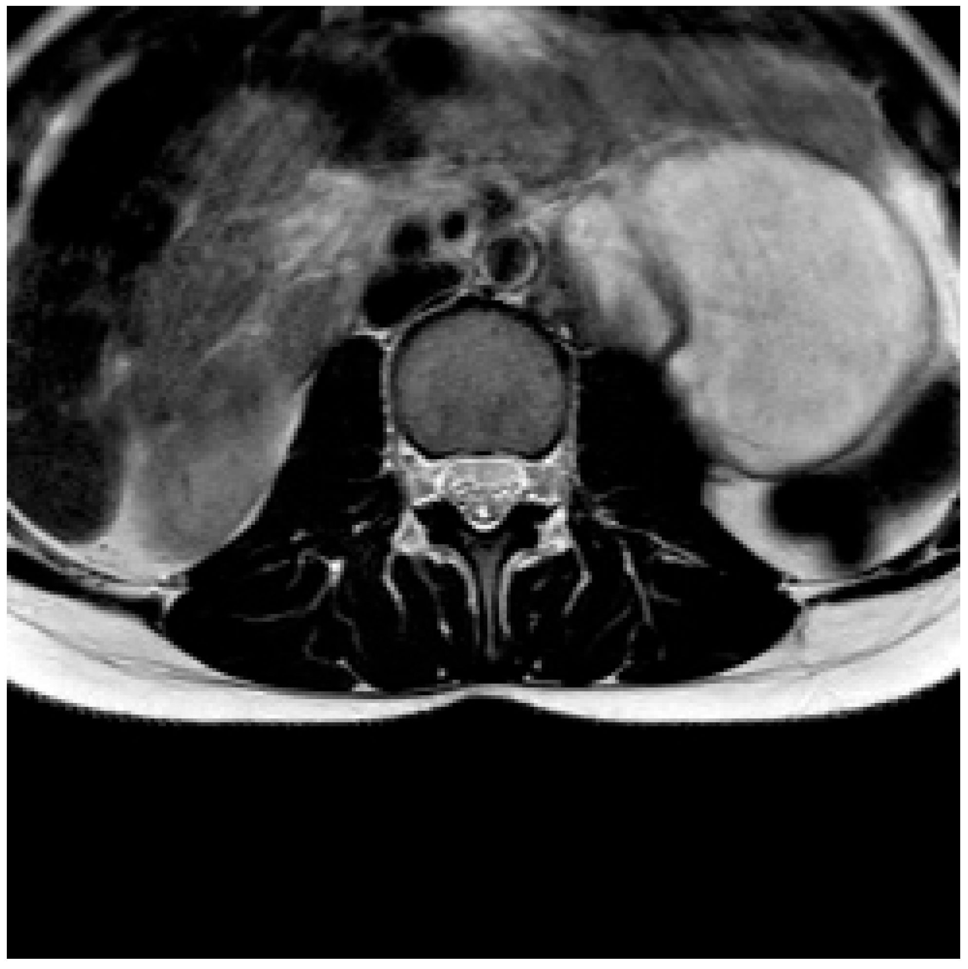
Abstract
:1. Introduction
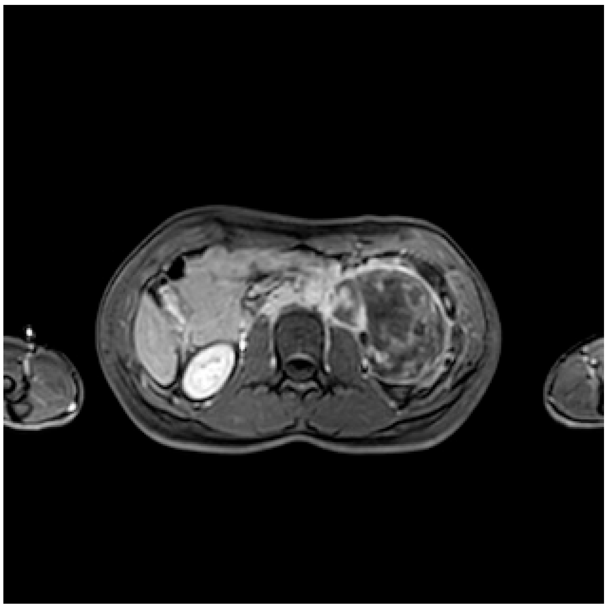
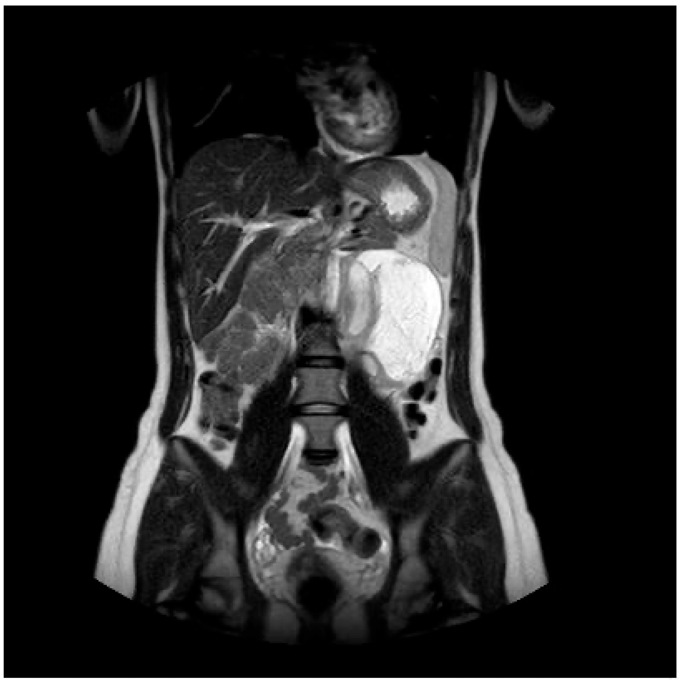
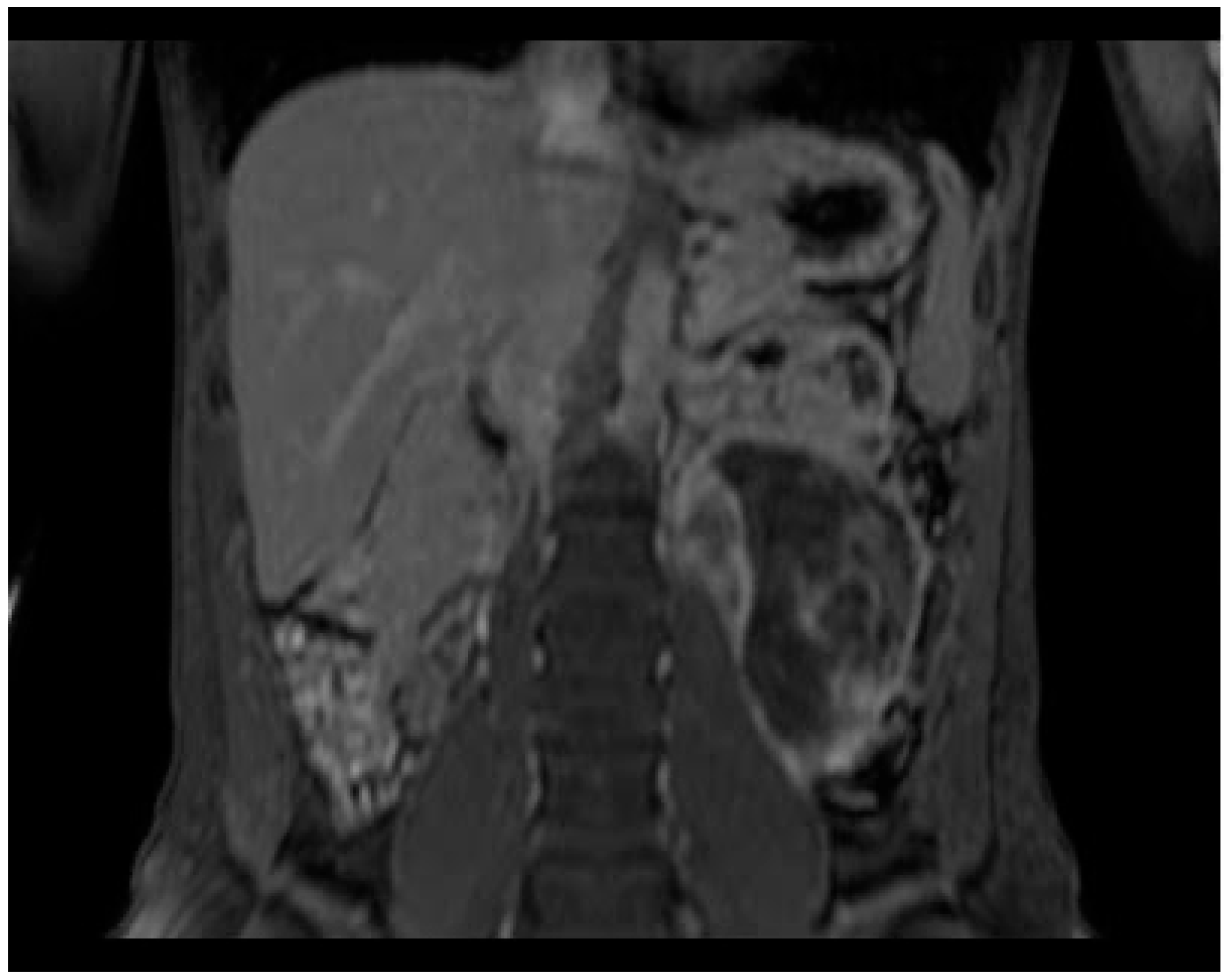
2. Case Presentation
3. Literature Review
4. Discussion
5. Conclusions
Author Contributions
Funding
Institutional Review Board Statement
Informed Consent Statement
Data Availability Statement
Conflicts of Interest
References
- Al-Dasuqi, K.; Irshaid, L.; Mathur, M. Radiologic-Pathologic Correlation of Primary Retroperitoneal Neoplasms. Radiographics 2020, 40, 1631–1657. [Google Scholar] [CrossRef] [PubMed]
- Shaaban, A.M.; Rezvani, M.; Tubay, M.; Elsayes, K.M.; Woodward, P.J.; Menias, C.O. Fat-containing Retroperitoneal Lesions: Imaging Characteristics, Localization, and Differential Diagnosis. Radiographics 2016, 36, 710–734. [Google Scholar] [CrossRef] [PubMed] [Green Version]
- Rajiah, P.; Sinha, R.; Cuevas, C.; Dubinsky, T.J.; Bush, W.H., Jr.; Kolokythas, O. Imaging of uncommon retroperitoneal masses. Radiographics 2011, 31, 949–976. [Google Scholar] [CrossRef]
- Guan, Y.B.; Zhang, W.D.; Zeng, Q.S.; Chen, G.Q.; He, J.X. CT and MRI findings of thoracic ganglioneuroma. Br. J. Radiol. 2012, 85, e365–e372. [Google Scholar] [CrossRef] [PubMed] [Green Version]
- Lonergan, G.J.; Schwab, C.M.; Suarez, E.S.; Carlson, C.L. Neuroblastoma, ganglioneuroblastoma, and ganglioneuroma: Radiologic-pathologic correlation. Radiographics 2002, 22, 911–934. [Google Scholar] [CrossRef] [PubMed]
- Zhang, Q.W.; Song, T.; Yang, P.P.; Hao, Q. Retroperitoneum ganglioneuroma: Imaging features and surgical outcomes of 35 cases at a Chinese Institution. BMC Med. Imaging 2021, 21, 114. [Google Scholar] [CrossRef] [PubMed]
- Noh, S.; Nessim, C.; Keung, E.Z.; Roland, C.L.; Strauss, D.; Sivarajah, G.; Fiore, M.; Biasoni, D.; Cioffi, S.P.B.; Mehtsun, W.; et al. Retrospective Analysis of Retroperitoneal-Abdominal-Pelvic Ganglioneuromas: An International Study by the Transatlantic Australasian Retroperitoneal Sarcoma Working Group. Ann. Surg. 2022. ahead of print. [Google Scholar] [CrossRef]
- Prakash, V.; Batanian, J.R.; Guzman, M.A.; Duncavage, E.J.; Geller, T.J. Malignant transformation of a desmoplastic infantile ganglioglioma in an infant carrier of a nonsynonymous TP53 mutation. Pediatr. Neurol. 2014, 51, 138–143. [Google Scholar] [CrossRef]
- Hayashi, Y.; Iwato, M.; Hasegawa, M.; Tachibana, O.; von Deimling, A.; Yamashita, J. Malignant transformation of a gangliocytoma/ganglioglioma into a glioblastoma multiforme: A molecular genetic analysis. Case report. J. Neurosurg. 2001, 95, 138–142. [Google Scholar] [CrossRef]
- Zhao, L.; Shi, L.; Huang, S.-G.; Cai, T.-N.; Guo, W.-L.; Gao, X.; Wang, J. Identification and validation of radiomic features from computed tomography for preoperative classification of neuroblastic tumors in children. BMC Pediatr. 2023, 23, 262. [Google Scholar] [CrossRef]
- Yılmaz, B.; Toktaş, Z.O.; Akakın, A.; Demir, M.K.; Yapıcıer, O.; Konya, D. Lumbar Spinal Immature Ganglioneuroma with Conus Medullaris Invasion: Case Report. Pediatr. Neurosurg. 2015, 50, 330–335. [Google Scholar] [CrossRef] [PubMed]
- Radin, R.; David, C.L.; Goldfarb, H.; Francis, I.R. Adrenal and extra-adrenal retroperitoneal ganglioneuroma: Imaging findings in 13 adults. Radiology 1997, 202, 703–707. [Google Scholar] [CrossRef]
- Xiao, J.; Zhao, Z.; Li, B.; Zhang, T. Primary Retroperitoneal Ganglioneuroma: A Retrospective Cohort Study of 32 Patients. Front. Surg. 2021, 8, 642451. [Google Scholar] [CrossRef] [PubMed]
- Do, S.-I.; Kim, G.Y.; Ki, K.-D.; Huh, C.-Y.; Kim, Y.-W.; Lee, J.; Park, Y.-K.; Lim, S.-J. Ganglioneuroma of the uterine cervix—A case report. Hum. Pathol. 2011, 42, 1573–1575. [Google Scholar] [CrossRef]
- Partecke, L.I.; Cziupka, K.; Thiele, A.; Diedrich, S.; von Bernstorff, W.; Heidecke, C.-D. Diagnostics and therapy for a ganglioneuroma of the retroperitoneal space—Case report. Zent. Chir. 2013, 138 (Suppl. S2), e127–e130. (In German) [Google Scholar] [CrossRef]
- Okamoto, T.; Suzuki, Y.; Sugiyama, N.; Kudo, S.; Yoneyama, T.; Hashimoto, Y.; Koie, T.; Kamimura, N.; Oyama, C. Ganglioneuroma with calcification mimicking adrenal tumor: A case report. Hinyokika Kiyo Acta Urol. Jpn. 2010, 56, 621–623. [Google Scholar]
- Cai, J.; Zeng, Y.; Zheng, H.; Qin, Y.; Kaiyong, T.; Zhao, J. Retroperitoneal ganglioneuroma in children: CT and MRI features with histologic correlation. Eur. J. Radiol. 2010, 75, 315–320. [Google Scholar] [CrossRef]
- Dyke, P.C.; Mulkey, D.A. Maturation of ganglioneuroblastoma to ganglioneuroma. Cancer 1967, 20, 1343–1349. [Google Scholar] [CrossRef]
- Zhang, Y.; Nishimura, H.; Kato, S.; Fujimoto, K.; Ohkuma, K.; Kojima, K.; Uchida, M.; Hayabuchi, N. MRI of ganglioneuroma: Histologic correlation study. J. Comput. Assist. Tomogr. 2001, 25, 617–623. [Google Scholar] [CrossRef]
- Otal, P.; Mezghani, S.; Hassissene, S.; Maleux, G.; Colombier, D.; Rousseau, H.; Joffre, F. Imaging of retroperitoneal ganglioneuroma. Eur. Radiol. 2001, 11, 940–945. [Google Scholar] [CrossRef]
- Scherer, A.; Niehues, T.; Engelbrecht, V.; Mödder, U. Imaging diagnosis of retroperitoneal ganglioneuroma in childhood. Pediatr. Radiol. 2001, 31, 106–110. [Google Scholar] [CrossRef]
- Nakashima, J.; Ueno, M.; Nakamura, K.; Tachibana, M.; Baba, S.; Guchi, N.; Tazaki, H.; Murai, M. Differential diagnosis of primary benign and malignant retroperitoneal tumors. Int. J. Urol. 1997, 4, 441–446. [Google Scholar] [CrossRef]
- Ichikawa, T.; Ohtomo, K.; Araki, T.; Fujimoto, H.; Nemoto, K.; Nanbu, A.; Onoue, M.; Aoki, K. Ganglioneuroma: Computed tomography and magnetic resonance features. Br. J. Radiol. 1996, 69, 114–121. [Google Scholar] [CrossRef]
- Yam, B.; Walczyk, K.; Mohanty, S.K.; Coren, C.V.; Katz, D.S. Radiology-pathology conference: Incidental posterior mediastinal ganglioneuroma. Clin. Imaging 2009, 33, 390–394. [Google Scholar] [CrossRef] [PubMed]
- Okamatsu, C.; London, W.B.; Naranjo, A.; Hogarty, M.D.; Gastier-Foster, J.M.; Look, A.T.; LaQuaglia, M.; Maris, J.M.; Cohn, S.L.; Matthay, K.K.; et al. Clinicopathological characteristics of ganglioneuroma and ganglioneuroblastoma: A report from the CCG and COG. Pediatr. Blood Cancer 2009, 53, 563–569. [Google Scholar] [CrossRef] [PubMed] [Green Version]
- Hayasaka, K.; Tanaka, Y.; Soeda, S.; Huppert, P.; Claussen, C.D. MR findings in primary retroperitoneal schwannoma. Acta Radiol. 1999, 40, 78–82. [Google Scholar] [CrossRef] [PubMed]
- Brueck, M.; Bandorski, D.; Rauber, K.; Kramer, W. Unilateral swollen leg caused by a retroperitoneal ganglioneuroma. Dtsch. Med. Wochenschr. 2006, 131, 2079–2082. (In German) [Google Scholar] [CrossRef] [PubMed]
- Amato, B.; Compagna, R.; Rocca, A.; Bianco, T.; Milone, M.; Sivero, L.; Vigliotti, G.; Amato, M.; Danzi, M.; Aprea, G.; et al. Fondaparinux vs warfarin for the treatment of unsuspected pulmonary embolism in cancer patients. Drug Des. Dev. Ther. 2016, 10, 2041–2046. [Google Scholar] [CrossRef] [Green Version]
- Nishino, M.; Hayakawa, K.; Minami, M.; Yamamoto, A.; Ueda, H.; Takasu, K. Primary retroperitoneal neoplasms: CT and MR imaging findings with anatomic and pathologic diagnostic clues. Radiographics 2003, 23, 45–57, Erratum in Radiographics 2003, 23, 1340. [Google Scholar] [CrossRef]
- Kirchweger, P.; Wundsam, H.V.; Fischer, I.; Rösch, C.S.; Böhm, G.; Tsybrovskyy, O.; Alibegovic, V.; Függer, R. Total resection of a giant retroperitoneal and mediastinal ganglioneuroma-case report and systematic review of the literature. World J. Surg. Oncol. 2020, 18, 248. [Google Scholar] [CrossRef]
- Lebby, E.; Kwan, D.; Bui, T.L.; O’Connell, R.; Seetharaman, M.; Houshyar, R. Retroperitoneal ganglioneuroma with nodal involvement in an adult patient with human immunodeficiency virus: A case report and review of the literature. J. Med. Case Rep. 2021, 15, 634. [Google Scholar] [CrossRef] [PubMed]
- Corssmit, E.P.; Romijn, J.A. Clinical management of paragangliomas. Eur. J. Endocrinol. 2014, 171, R231–R243. [Google Scholar] [CrossRef] [Green Version]
- Li, C.P.; Liu, B.N.; Wu, J.H.; Hao, C.Y. Contralateral internal iliac artery transposition for retroperitoneal sarcoma involving common iliac artery. Updates Surg. 2022, 74, 1157–1163. [Google Scholar] [CrossRef] [PubMed]
- Calise, F.; Giuliani, A.; Sodano, L.; Crolla, E.; Bianco, P.; Rocca, A.; Ceriello, A. Segmentectomy: Is minimally invasive surgery going to change a liver dogma? Updates Surg. 2015, 67, 111–115. [Google Scholar] [CrossRef] [PubMed]
- Boggi, U.; Vistoli, F.; Amorese, G. Twenty years of robotic surgery: A challenge for hu-man limits. Updates Surg. 2021, 73, 789–793. [Google Scholar] [CrossRef]
- Loffredo, D.; Marvaso, A.; Ceraso, S.; Cinelli, N.; Rocca, A.; Vitale, M.; Rossi, M.; Genovese, E.; Amato, B.; Cinelli, M. Minimal invasive surgery in treatment of liver metastases from colorectal carcinomas: Case studies and survival rates. BMC Surg. 2013, 13 (Suppl. S2), S45. [Google Scholar] [CrossRef] [Green Version]
- Jain, N.; Jain, V.; Chandi, A.; Srivastava, S.; Singh, S.; Vasundhara, N. Jain point: An alternate laparoscopic non-umbilical first blind entry port to avoid vessel, viscera, adhesions and bowel (VVAB). Updates Surg. 2021, 73, 2321–2329. [Google Scholar] [CrossRef]
- Rocca, A.; Cipriani, F.; Belli, G.; Berti, S.; Boggi, U.; Bottino, V.; Cillo, U.; Cescon, M.; Cimino, M.; Corcione, F.; et al. The Italian Consensus on minimally invasive simultaneous resections for synchronous liver metastasis and primary colorectal cancer: A Delphi methodology. Updates Surg. 2021, 73, 1247–1265. [Google Scholar] [CrossRef]
- Shimada, H.; Ambros, I.M.; Dehner, L.P.; Hata, J.; Joshi, V.V.; Roald, B. Terminology and morphologic criteria of neuroblastic tumors: Recommendations by the International Neuroblastoma Pathology Committee. Cancer 1999, 86, 349–363. [Google Scholar] [CrossRef]
- Rocca, A.; Scacchi, A.; Cappuccio, M.; Avella, P.; Bugiantella, W.; De Rosa, M.; Costa, G.; Polistena, A.; Codacci-Pisanelli, M.; Amato, B.; et al. Robotic surgery for colorectal liver metastases resection: A systematic review. Int. J. Med. Robot. Comput. Assist. Surg. 2021, 17, e2330. [Google Scholar] [CrossRef]
- Ceccarelli, G.; Andolfi, E.; Fontani, A.; Calise, F.; Rocca, A.; Giuliani, A. Robot-assisted liver surgery in a general surgery unit with a “Referral Centre Hub&Spoke Learning Program”. Early outcomes after our first 70 consecutive patients. Minerva Chir. 2018, 73, 460–468. [Google Scholar] [CrossRef]
- Medress, Z.A.; Bobrow, A.; Tigchelaar, S.S.; Henderson, T.; Parker, J.J.; Desai, A. Augmented Reality-Assisted Resection of a Large Presacral Ganglioneuroma: 2-Dimensional Operative Video. Oper. Neurosurg. 2022, 24, e284–e285. [Google Scholar] [CrossRef]
- Materazzi, G.; Rossi, L. Robot-assisted adrenalectomy: State of the art. Updates Surg. 2021, 73, 1131–1146. [Google Scholar] [CrossRef] [PubMed]
- He, Z.; Chen, S.; Lu, M.; Luo, Y.; Liu, T.; Xiao, Y.; Wang, X. A combination of laparoscopic approach and ERAS pathway optimizes outcomes and cost for adrenalectomy. Updat. Surg. 2021, 74, 519–525. [Google Scholar] [CrossRef]
- Piccoli, M.; Esposito, S.; Pecchini, F.; Francescato, A.; Colli, F.; Gozzo, D.; Trapani, V.; Alboni, C.; Rocco, B. Full robotic multivisceral resections: The Modena experience and literature review. Updates Surg. 2021, 73, 1177–1187. [Google Scholar] [CrossRef]
- Inoue, S.; Goto, K.; Ikeda, K.; Hieda, K.; Hayashi, T.; Teishima, J. Longitudinal analysis of retroperitoneoscopic adrenalectomy regarding cosmesis outcomes: Comparison of lateral transperitoneal and reduced port laparoscopic adrenalectomy. Updates Surg. 2021, 74, 757–764. [Google Scholar] [CrossRef] [PubMed]
- Zeh, R.D.; Konieczkowski, D.; Shen, C.; Tsilimigras, D.I.; Kim, A.; Grignol, V.P.; Contreras, C.M.; Welliver, M.; Tsung, A.; Pawlik, T.M.; et al. Prognostic factors in patients receiving surgery and radiation therapy for retroperitoneal sarcoma: A machine-learning analysis. Surgery 2023, 173, 640–644. [Google Scholar] [CrossRef] [PubMed]
- Thrussell, I.; Winfield, J.M.; Orton, M.R.; Miah, A.B.; Zaidi, S.H.; Arthur, A.; Thway, K.; Strauss, D.C.; Collins, D.J.; Koh, D.-M.; et al. Radiomic Features from Diffusion-Weighted MRI of Retroperitoneal Soft-Tissue Sarcomas Are Repeatable and Exhibit Change After Radiotherapy. Front. Oncol. 2022, 12, 899180. [Google Scholar] [CrossRef]
- Li, K.-P.; Duan, X.; Yang, X.-S.; Huang, J.; Wu, T. Partial versus total adrenalectomy for the treatment of unilateral aldosterone-producing adenoma: A systematic review and meta-analysis. Updat. Surg. 2021, 73, 2301–2313. [Google Scholar] [CrossRef]
- Rocca, A.; Brunese, M.C.; Cappuccio, M.; Scacchi, A.; Martucci, G.; Buondonno, A.; Perrotta, F.M.; Quarto, G.; Avella, P.; Amato, B. Impact of Physical Activity on Disability Risk in Elderly Patients Hospitalized for Mild Acute Diverticulitis and Diverticular Bleeding Undergone Conservative Management. Medicina 2021, 57, 360. [Google Scholar] [CrossRef]
- Komici, K.; Iacono, A.D.; De Luca, A.; Perrotta, F.; Bencivenga, L.; Rengo, G.; Rocca, A.; Guerra, G. Adiponectin and Sarcopenia: A Systematic Review with Meta-Analysis. Front. Endocrinol. 2021, 12, 576619. [Google Scholar] [CrossRef] [PubMed]
- Wang, H.; Chen, X.; Liu, H.; Yu, C.; He, L. Computed tomography-based radiomics for differential of retroperitoneal neuroblastoma and ganglioneuroblastoma in children. Nan Fang Yi Ke Da Xue Xue Bao 2021, 41, 1569–1576. (In Chinese) [Google Scholar] [CrossRef] [PubMed]
- Wang, H.; Chen, X.; Yu, W.; Xie, M.; Zhang, L.; Ding, H.; Li, T.; Qin, J.; He, L. Whole-tumor radiomics analysis of T2-weighted imaging in differentiating neuroblastoma from ganglioneuroblastoma/ganglioneuroma in children: An exploratory study. Abdom. Radiol. 2023, 48, 1372–1382. [Google Scholar] [CrossRef] [PubMed]
- Donato, I.; Velpula, K.K.; Tsung, A.J.; A Tuszynski, J.; Sergi, C.M. Demystifying neuroblastoma malignancy through fractal dimension, entropy, and lacunarity. Tumori J. 2023, 2023, 3008916221146208. [Google Scholar] [CrossRef]
- Ben-David, E.; Shochat, M.; Roth, I.; Nissenbaum, I.; Sosna, J.; Goldberg, S.N. Evaluation of a CT-Guided Robotic System for Precise Percutaneous Needle Insertion. J. Vasc. Interv. Radiol. 2018, 29, 1440–1446. [Google Scholar] [CrossRef]
- Fong, A.J.; Stewart, C.L.; Lafaro, K.; LaRocca, C.J.; Fong, Y.; Femino, J.D.; Crawford, B. Robotic assistance for quick and accurate image-guided needle placement. Updat. Surg. 2021, 73, 1197–1201. [Google Scholar] [CrossRef]
- Rocca, A.; Brunese, M.C.; Santone, A.; Avella, P.; Bianco, P.; Scacchi, A.; Scaglione, M.; Bellifemine, F.; Danzi, R.; Varriano, G.; et al. Early Diagnosis of Liver Metastases from Colorectal Cancer through CT Radiomics and Formal Methods: A Pilot Study. J. Clin. Med. 2021, 11, 31. [Google Scholar] [CrossRef]


Disclaimer/Publisher’s Note: The statements, opinions and data contained in all publications are solely those of the individual author(s) and contributor(s) and not of MDPI and/or the editor(s). MDPI and/or the editor(s) disclaim responsibility for any injury to people or property resulting from any ideas, methods, instructions or products referred to in the content. |
© 2023 by the authors. Licensee MDPI, Basel, Switzerland. This article is an open access article distributed under the terms and conditions of the Creative Commons Attribution (CC BY) license (https://creativecommons.org/licenses/by/4.0/).
Share and Cite
Pacella, G.; Brunese, M.C.; Donnarumma, F.; Barrassi, M.; Bellifemine, F.; Sciaudone, G.; Vallone, G.; Guerra, G.; Sallustio, G. Imaging of Ganglioneuroma: A Literature Review and a Rare Case of Cystic Presentation in an Adolescent Girl. Diagnostics 2023, 13, 2190. https://doi.org/10.3390/diagnostics13132190
Pacella G, Brunese MC, Donnarumma F, Barrassi M, Bellifemine F, Sciaudone G, Vallone G, Guerra G, Sallustio G. Imaging of Ganglioneuroma: A Literature Review and a Rare Case of Cystic Presentation in an Adolescent Girl. Diagnostics. 2023; 13(13):2190. https://doi.org/10.3390/diagnostics13132190
Chicago/Turabian StylePacella, Giulia, Maria Chiara Brunese, Federico Donnarumma, Michele Barrassi, Fabio Bellifemine, Guido Sciaudone, Gianfranco Vallone, Germano Guerra, and Giuseppina Sallustio. 2023. "Imaging of Ganglioneuroma: A Literature Review and a Rare Case of Cystic Presentation in an Adolescent Girl" Diagnostics 13, no. 13: 2190. https://doi.org/10.3390/diagnostics13132190