
New Corynebacterium Species with the Potential to Produce Diphtheria Toxin
by
 ,
,
Marta Prygiel
 ,
,
Maciej Polak
,
Ewa Mosiej
,
Karol Wdowiak
,
Kamila Formińska
and
Aleksandra Anna Zasada
* Department of Sera and Vaccines Evaluation, National Institute of Public Health NIH, National Research Institute, 00-791 Warsaw, Poland
*
Author to whom correspondence should be addressed.
Pathogens 2022, 11(11), 1264; https://doi.org/10.3390/pathogens11111264
Submission received: 29 September 2022
/
Revised: 20 October 2022
/
Accepted: 27 October 2022
/
Published: 30 October 2022
(This article belongs to the Section Bacterial Pathogens)
Abstract
:Only three Corynebacterium species are known to produce a lethal exotoxin called diphtheria toxin. These are C. diphtheriae, C. ulcerans and C. pseudotuberculosis. The diphtheria toxin gene (tox) is carried in a family of closely related corynebacteriophages and therefore the toxin can be produced only through lysogenisation, in which the corynephage encoding tox is stably inserted into the chromosome. However, ‘nontoxigenic tox gene-bearing’ (NTTB) strains, which are genotypically tox-positive but do not express the protein, have been described. The emergence of NTTB strains was first observed during the 1990s diphtheria epidemic in Eastern Europe and nowadays such isolates have been detected in many countries in the world. Recently, novel species of Corynebacterium genus have been described which might have the potential of producing the diphtheria toxin due to the possession of the diphtheria toxin gene but it has not produced toxin in laboratory tests. The circulation of NTTB strains could be related to the increased risk for diphtheria disease arising from the risk of re-emerging toxin expression. The article presents the mechanism of diphtheria toxin expression and action, recently described novel species of NTTB corynebacteria as well as the taxonomic changes within the C. diphtheriae group.
Keywords:
NTTB; diphtheria toxin; Corynebacterium; tox gene; C. rouxii; C. silvaticum; C. belfantii; C. diphtheriae; C. ulcerans1. Introduction
The genus Corynebacterium was first described in 1896 as a Gram-positive club-shaped bacillus with filamentous morphology [1]. The genus Corynebacterium belongs to the Phylum Actinobacteria characterized by high cytosine and guanine contents in DNA. Currently, this genus included about 145 different species [2]. More than half of the species were isolated from human and animal clinical samples which indicates their potential participation in pathogenesis [1,2]. In addition, strains of medical and veterinary importance, of the same species, such as Corynebacterium glutamicum and Corynebacterium efficiens, have biotechnological applications [1,2].
The most important human pathogen is Corynebacterium diphtheriae, which is the etiological agent of diphtheria, a serious, potentially fatal infection of the respiratory tract and occasionally the skin and other mucous membranes such as, e.g., eye, ear, and genital tract. The infection often causes complications in other body organs [3]. Since Friedrich Löffler’s isolation of toxin-secreting C. diphtheriae in 1884 [1], the species has been the best-known and probably most genetically diverse species of the genus [4,5]. Classical diphtheria is caused by the production of diphtheria toxin (DT) during infections by isolates holding the toxin gene. DT is the main virulence factor responsible for respiratory, neuro- or cardiopathological symptoms, causing pseudo-membranes, paralysis and cardiac failure [1]. In countries with high anti-diphtheria vaccination coverage, the disease is very rare, but in some regions of Africa and Asia diphtheria is still recognised, with thousands of cases reported annually [3]. The disease can emerge in case of the failed implementation of the recommended vaccination programs or lack of booster doses [3].
Corynebacterium ulcerans was described in 1926 by Gilbert and Steward [6]. The species is closely related to C. diphtheriae and also is able to produce DT. Nowadays, in European countries, C. ulcerans is recognized more frequently as an emerging pathogen associated with diphtheria-like symptoms [7,8]. Growing numbers of human infections caused by C. ulcerans are the result of zoonotic transmission by contact with animal hosts such as goats, cattle, domestic pigs, dogs, cats and even hedgehogs, monkeys, camels, foxes, squirrels, owls, orcas, otters and water rats [9].
Corynebacterium pseudotuberculosis is the etiological agent of ulcerative lymphangitis in equines, mastitis in dairy cattle, oedematous skin disease in buffalos, or abscesses and caseous lymphadenitis (CLA) in small ruminants, such as goat and sheep [10,11]. C. pseudotuberculosis has caused occasional infection in farm and animal health workers who remain in close contact with infected animals or their raw products, resulting in swellings of the lymph nodes in the neck or groin. C. pseudotuberculosis animal diseases cause severe economic losses [12]. The bacterium was first described in 1888 by Edmond Isidore Etienne Nocard and classified as C. pseudotuberculosis in 1918 by Eberson [13]. Historically, it is the third species known to be able to produce DT. However, toxin-producing C. pseudotuberculosis has been isolated extremely rarely.
Among the pathogenic species of the genus Corynebacterium, the C. diphtheriae, C. ulcerans and C. pseudotuberculosis, which can produce DT, were clustered together in the group of toxigenic corynebacteria named “C. diphtheriae complex” [14].
However, the infections caused by potentially toxigenic corynebacteria have recently changed. Toxigenic C. ulcerans has been isolated from clinical samples more often than in preceding years. Serious invasive infections caused by nontoxigenic C. diphtheriae have been noticed in many countries with high anti-diphtheria vaccination coverage [15]. What is more, new species capable of producing DT were described in 2020, called C. rouxii and C. silvaticum [16]. Based on the genomic sequencing and biochemical and chemotaxonomic analyses, the name of C. belfantii was proposed for strains previously considered C. diphtheriae biotype belfanti [17]. In this paper, we present current information concerning DT and potentially toxigenic Corynebacterium species.
2. The Structure of Diphtheria Toxin
Diphtheria toxin (DT) is encoded by a 1683-base-pair structural gene-tox (NCBI Reference Sequence: NC_002935.2), encoded not on the bacterial chromosome, but by a lysogenic phage called corynebacteriophage beta or corynephage β [18,19]. The integration of the corynephage β can occur at two specific sites: attB1 and attB2 [20]. Although tox is of bacteriophage origin, the regulation of its expression is reliant on bacteria [21]. The diphtheria toxin repressor gene (dtxR) is present on the bacterial chromosome. Its protein product (DtxR) is able to bind to the tox operator, blocking the transcription [22,23]. Interestingly, DtxR is activated by heavy metals, especially iron ions [23,24,25]. In the absence of iron ions apo-DtxR exists as an inactive monomer that is in weak equilibrium with a dimeric form. Once activated by the metal ions, DtxR forms stable dimers and two pairs of dimers have been shown to bind to almost opposite faces of the tox operator sequence. DtxR is composed of two major structural domains linked by a flexible tether containing a proline-rich region. The N-terminal domain contains the ancillary and primary metal ion-binding sites, a canonical helix-turn-helix DNA-recognition motif, and an extensive hydrophobic surface necessary for the formation of stable dimers. After DtxR dimers binding to tox promoter, the transcription of tox is repressed. When iron is limiting, the uncomplexed form of DtxR is unable to bind DNA, leading to the induction of diphtheria toxin [20,21]. The mature extracellular DT is a polypeptide consisting of 535 amino acid residues with a molecular mass of approximately 58 kDa [26,27,28]. The gene sequence analysis indicates that DT is preceded by 25 residues of leader peptide, which is most likely involved in toxin secretion [19]. DT is produced as a proenzyme that requires specific activation for its toxic function, either prior to or immediately after binding to a sensitive cell [29]. It is based on a proteolytic cleavage which cleaves the peptide bond located at the arginine (Arg) residue: Arg190, Arg192, or Arg193, resulting in the formation of two polypeptides: (1) a 193-residue amino-terminal fragment A (DT-A) which corresponds with the catalytic domain of DT, and (2) a 342-residue carboxyl-terminal fragment B (DT-B), corresponding with the translocation and receptor-binding domains of DT [26,30]. Both fragments remain covalently bound by the disulphide bonds between Cys186 and Cys201, and a reduction of this binding results in free forms of DT-A and DT-B, capable of infecting target cells [27]. DT has been described as the first example of group A–B toxins in which the catalytic and receptor-binding functions are separated into two different polypeptides [31]. Currently, the A–B motif is well-known and almost universal among intracellular toxins [32,33].
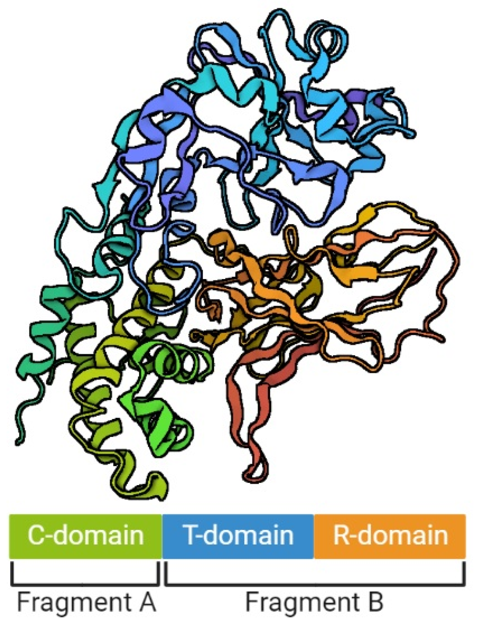
The model of DT structure has evolved over the years, along with the methods available for its determination [29,34,35]. It is assumed that a single DT molecule has three distinct folding domains – C, T and R (Figure 1), symbols of which are derived from the three main functions of this toxin, respectively: catalysis, translocation and receptor binding, respectively. They are arranged in the shape of the letter Y, with the lower part being the T domain, and the upper elements consisting of the C and R domains. The T domain is formed by the α-helical bundle, the R domain by the flattened β-barrel, and the C domain in turn is a combination of structures α and β. Functionally, the C domain forms fragment A of the mature DT, and the T and R domains – fragment B [29,35,36]. Concurrently, a crystallographic analysis showed that in the discussed Y shape, the cleft in the active site of the C domain is blocked by the R domain from accessing the substrate. This is the reason why those two fragments must be separated from each other in order to be toxic [29]. However, no trypsin-sensitive loop was found between the C and T domains that might be responsible for proenzyme proteolysis. Probably this place is created dynamically, which makes detecting it impossible [31]. Furthermore, there was an emphasized high similarity of the T domain to the hydrophobic N-terminal domain of the B chain of the non-toxic protein CRM45, that the ability to form pores in membranes under acidic conditions was attributed to [37,38].
3. The Mechanism of DT Toxicity
DT toxicity towards sensitive cells is based on the inactivation of a protein elongation factor-2 (EF-2), which is an essential element for protein synthesis, stimulating the GTP-dependent translocation of the ribosome [39,40,41]. This death process begins with the binding of DT by a receptor—a membrane-anchored form of the heparin binding EGF-like growth factor (HB-EGF precursor), on the cell surface of DT-sensitive cells. DT enters the cytosol via receptor-mediated endocytosis [28,42,43]. Inside endosomes, proteases partially cleave the bond between the A and B fragments of the toxin. The low pH inside the endosomes promotes a conformational change in DT by which the T domain is inserted into the endosomal membrane thereby allowing cytosolic DT-A exposure [44]. Then, the specific toxicity reaction begins—the A fragment transfers the ADP-ribose moiety of nicotinamide adenine dinucleotide (NAD+) to a modified histidine residue (diphthamide) on EF-2, deactivating it. Thus, the host is unable to produce protein and dies [39,45]. DT-A can exhibit its toxic activity against all eukaryotic EF-2s except mice and rats, and only one molecule of the toxin in the cytosol is sufficient to lead to cell death [37,46,47].
4. Nontoxigenic Toxin Gene-Bearing Strains
Historically, among the C. diphtheriae species, toxin-producing and non-toxin-producing strains have been distinguished. However, there has currently also been a third category: ‘nontoxigenic tox gene-bearing’ (NTTB). Non-toxin-producing (nontoxigenic) C. diphtheriae strains generally do not possess the tox gene, with the exception of some nontoxigenic strains which bear the tox gene. These strains called ‘nontoxigenic tox gene-bearing’ (NTTB) are genotypically tox-positive, but do not express the diphtheria toxin due to nucleotide mutations or deletions [48]. The circulation of NTTB C. diphtheriae strains was first detected during and after the diphtheria epidemic in the 1990s in Belarus [49] and in Russia during 1994–2002 [50]. NTTB C. diphtheriae strains have also been reported in other regions, for example in the United Kingdom [48,51], the United States [52] and Australia [53].
It is likely that mutations causing tox gene inactivation might be frequent in C. diphtheriae after epidemic waves, as a result of pathogen adaptation to circulation in the population with high anti-diphtheria antibody levels [54].
Nontoxigenic C. diphtheriae is recognized as a potential emerging pathogen, as it is with increasing frequency associated with severe invasive diseases. The growing number of detected nontoxigenic C. diphtheriae infections in the 1990s and in the early 2000s points out that the circulation of these strains is an escalating problem in Europe [55,56,57,58,59,60,61]. Nontoxigenic C. diphtheriae infections cannot be prevented by the contemporary vaccines targeting diphtheria toxin [62,63,64] and have quickly become prevalent in countries with high anti-diphtheria vaccination coverage.
Due to the fact that only toxigenic infections must be reported, the extent of the problem of nontoxigenic C. diphtheriae infections in Europe remains unknown since only toxigenic infections are registered. The lack of mandatory registration consequently leads to no prevention measures, which results in the spread of strains able to cause infections [65]. Nontoxigenic C. diphtheriae strains may become a public health threat in developed countries because they can persist and spread in the risk groups and then become the source of an outbreak [55].
The pathogenesis of nontoxigenic C. diphtheriae is not well elucidated. The most likely entry portals for nontoxigenic C. diphtheriae are skin lesions or dental caries [65]. In recent years, severe and often fatal systemic diseases (which were previously quite rare) caused by nontoxigenic C. diphtheriae have been registered in various countries. Nontoxigenic C. diphtheriae often were found to be associated with cutaneous lesions but can transform into severe clinical symptoms, such as myocarditis, polyneuritis, bacteraemia, septic arthritis and endocarditis, characterized by a high mortality rate reaching over 40% [58,60,61,63,65]. Among the factors that predispose to the invasive infections caused by nontoxigenic C. diphtheriae occurrence are homelessness, abuse of alcohol and injection drugs and diabetes mellitus, hepatic cirrhosis and dental caries [61]. Furthermore, refugees and foreign travellers constitute population groups that are particularly at risk of nontoxigenic C. diphtheriae infections [66,67].
The increasing number of cases of invasive infections caused by nontoxigenic isolates might suggest the acquisition of additional virulence factors [60]. It is theorized that the success in the prevention of toxin-mediated disease as an effect of anti-diphtheria vaccination has created selective pressure on C. diphtheriae strains to express or develop disease-causing mechanisms and virulence factors other than diphtheria toxin [60].
Nontoxigenic C. diphtheriae is a public health concern due to the lack of protection provided by current diphtheria vaccines and the potential for such circulating strains to readily become toxigenic through lysogenisation by toxin-encoding bacteriophages [68].
5. Corynebacterium silvaticum
Corynebacterium silvaticum sp. nov. is a novel species of the nontoxigenic tox-gene-bearing (NTTB) strains, firstly isolated from lymph nodes of wild boars showing severe lesions due to caseous lymphadenitis (CLA). The first case report on the isolation of atypical Corynebacterium strains from two wild boars causing CLA was published in 2011 [70]. The wild boars came from different provinces in southern Germany. Isolated strains (named KL0182T and KL0183) were positive for phospholipase D—the major virulence determinant of C. pseudotuberculosis, which plays a key role in the spread of bacteria from the site of infection to the lymph nodes. However, biochemical studies did not allow to classify them as C. pseudotuberculosis species. Sequencing of the 16S rRNA and rpoB genes allowed the classifying of the isolates to the C. ulcerans species. The tox gene for diphtheria toxin was detected in both isolates. DNA sequencing of the tox gene exhibited differences from sequences described for C. ulcerans strains and showed higher similarity to C. diphtheriae. The expression of diphtheria toxin was not be detected phenotypically. These results have indicated that wild boars could be a reservoir for zoonotic Corynebacterium. Since the description of these two isolates, bacteria causing caseous lymph node abscesses in wild boar have been consistently collected [71,72], causing the same difficulties in unambiguous classification as C. diphtheriae, C. belfantii, C. ulcerans or C. pseudotuberculosis, so far addressed as atypical C. ulcerans [70,71,72] or “wild boar cluster” (WBC) of C. ulcerans [73,74].
Based on the results presented below, Dangel et al. [75] classified those isolates into a novel species named C. silvaticum sp. nov. Thirty-four Corynebacterium sp. strains were collected from CLA of wild boar (33 isolates) and roe deer (1 isolate) from different regions of Germany between 1997 and 2018. Isolation procedures based on microbiological methods have demonstrated that bacteria grow under aerobic and microaerophilic conditions at 37 °C within 48 h on SBA (Sheep Blood Agar) and BHI (Brain Heart Infusion) plates (but slower than other Corynebacterium species). Colonies of the bacteria are small, creamy to waxy with a sleek area and a discrete β-haemolysis after incubation. The optimum pH range for growth is 7–8. The bacteria produce phospholipase D, catalase and urease and are inconstant for alpha-glucosidase and alkaline phosphatase production. Isolates show fermentation of ribose, glucose and maltose (same as C. ulcerans), but do not metabolize mannitol, D-xylose, lactose, glycogen and sucrose (same as C. pseudotuberculosis). They are sensitive to clindamycin, erythromycin and penicillin unlike C. ulcerans. The fatty acid analysis has assigned them to the C. diphtheriae group of genus Corynebacterium. The MALDI-TOF MS and rpoB gene sequencing have allocated them to the C. ulcerans, but the profile of polar major lipids and glycolipids differs from that of C. ulcerans. In the phylogenesis of the 16SrRNA and RpoB proteins, isolates have created separate branches with C. ulcerans as the closest relative. The quinone system has been almost identical as in C. ulcerans with the main menaquinone MK-8(H2). All analysed isolates (34) have been included in NTTB strains. The presence of the tox gene has been confirmed by real-time PCR, but the phenotypic expression of diphtheria toxin has not been detected in the Elek test. Whole genome sequencing has shown the specific sequence type 578 and a separate branch in MLST typing. The average nucleotide identity (ANI) values are <91, draft genome sizes are 2.55 Mbp and the G/C content is 54.4 mol%. Based on these results regarding phenotype, genotype and biochemistry, the said bacteria represent a separate species, for which the authors [75] have proposed the name C. silvaticum sp. nov. The full-length 16S rRNA gene sequence of C. silvaticum sp. nov is available at GenBank (accession number MK602323) and WGS draft genomes are available at the NCBI genome database (accession numbers: SDQO00000000, SDVC00000000 and SDVD00000000). The type strain is KL0182T (=CIP 111672T = DSM 109166T = LMG31313T) [75]. The study conducted by Möller et al. [76] might indicate a zoonotic potential of C. silvaticum. Cytotoxicity of this newly identified species has been demonstrated. This negative influence in in vitro conditions (different human epithelial cell lines) and in in vivo conditions (Galleria mellonella larvae) was comparable to diphtheria toxin-secreting C. ulcerans [76].
In the first proteome study conducted by scientists [77], 1305 proteins of C. silvaticum were identified. The potential known virulence factors such as phospholipase D and sialidase were also detected. Furthermore, an uncharacterized secreted protein trypsin-like protease having an impact on pathogenicity was found. In addition, the said proteome analyses confirmed the taxonomic relationship of C. silvaticum to be closely connected with the zoonotic species of the Corynebacterium genus.
The results of the study conducted by Viana et al. [78] showed that Corynebacterium PO100/5 strain (the first sequenced genome of a C. silvaticum) can also colonize livestock and not only wild forest animals. This strain was isolated from a skin abscess taken from a domestic pig in the southern region of Portugal. It was the first strain of C. silvaticum isolated outside Germany. The taxonomic analysis revealed that C. silvaticum species is genetically more homogeneous than C. ulcerans. Moreover, C. silvaticum has pilus subunit genes spaB (which play important role in adhesion on pharyngeal epithelial cells), conserved genomic islands and 172 genes that could be used as markers for molecular identification [78].
Initially recognized as the atypical C. ulcerans strains: isolate W25 and isolate KL1196 have been recently isolated from a case of CLA from wild boar and roe deer, respectively [79]. Phylogenetic analyses showed that those strains belong to a novel species C. silvaticum. The ANI values between the tested strains were 99% indicating a close relationship to the same species, the ANI values between the tested strains and the C. ulcerans genome were 92%, which proves belonging to a different species. The authors proved that one of the key biochemical differences separating C. silvaticum from C. ulcerans was the inability to ferment starch [79].
Wild boars and domestic pigs are reservoirs of C. silvaticum which are suspected to transmit the bacteria to other domestic animals and, eventually, humans. Therefore, finding the unique sequence and genes useful for this species classification is crucial for its detection and proper identification. Molecular characterization of C. pseudotuberculosis, C. auriscanis and C. silvaticum by ERIC 1+2-PCR genotyping could be useful as a diagnostic tool for the detection of the etiological agent of CLA [80]. The study conducted by Ramos et al. [80] showed that ERIC 1+2-PCR genotyping fingerprinted all tested eighty isolates into 24 genotypes: 22 genotypes corresponded to C. pseudotuberculosis, 1 genotype to C. auriscanis and 1 genotype to tested C. silvaticum strains. The maximum genetic similarity of 76% between C. pseudotuberculosis and C. silvaticum was observed [80]. The number of bands detected for all tested C. silvaticum isolates was 13 in the size range from 98 bp to 731 bp. Two of them (475 bp and 426 bp) were peculiar only to C. silvaticum profile. This study also confirmed that these bacteria were correctly classified into a new species called C. silvaticum.
The presence of mycolic acids in the outer membrane of the Corynebacterium genus is an important feature which may be related to virulence [81]. Dover et al. [81] analysed the genomes of 140 corynebacterial strains (representatives of 126 different species), the majority of which were isolated from humans and animals, and presented that these species had been organised into 19 phylogenetic groups proving their great diversity. Most of the important human and animal pathogens have been grouped into one group called Q. The group Q includes C. diphtheriae, C. pseudotuberculosis, C. ulcerans, C. silvaticum and C. rouxii. This similarity indicates the possibility that animal isolates can infect people [81].
6. Corynebacterium rouxii
Corynebacterium rouxii sp. nov. described as the new species of C. diphtheriae complex [16], was isolated between 2013 and 2017 in France. The isolates came from infected human skin tissues (ulceration) and peritoneum and from a dog’s skin and were initially identified as C. diphtheriae biovar belfanti. Previously, some Corynebacterium strains were isolated from domestic cats in the United States in 2010 with properties similar to the novel species C. rouxii [82]. Nowadays, after the proper species classification, those strains described in the United States have been reclassified as a novel species, Corynebacterium rouxii sp. nov.
C. rouxii is biochemically similar to C. diphtheriae biovar belfanti (currently reclassified as C. belfantii) except that the strains are negative for maltose fermentation. The investigated isolates were trehalose, urease, pyrazinamide, nitrate and glycogen negative. Moreover, all the investigated isolates of C. rouxii were positive in the PCR test for dtxR and rpoB genes and negative for C. ulcerans/C. pseudotuberculosis 16S rDNA and pld which indicated a similarity to C. diphtheriae but not to C. ulcerans and C. pseudotuberulosis. The tox gene was not detected [83,84].
The average genome size of C. rouxii is 2.4 Mb. The genomic sequencing results showed that the ANI value of C. rouxii was 92.4% with the C. diphtheriae clade and 91.4% with C. belfantii. The C. rouxii clade was genetically homogenous, which was revealed by ANI values ranging from 99.21% to 99.94% [83].
The type strain is FCR0190T (=CIP 111752T = DSM 110354T). The genome sequence is available in GenBank under access number MN535983.
7. Corynebacterium belfantii
C. diphtheriae was historically subdivided into four biovars: Mitis, Gravis, Intermedius and Belfanti, based on the colony morphology and biochemical properties (Table 1) [3,85]. The names of the first three biovars were supposed to refer to the illness severity they cause, however according to the current molecular epidemiology knowledge such a correlation does not occur [86,87]. Biovar Belfanti was later added to highlight the nontoxigenic C. diphtheriae strains isolated from ozaena patients that unlike strains from other biovars were nitrate negative [17,88]. This biovar was named after Serafino Belfanti who was the first to identify such strains [17,88].
Molecular typing studies have shown that the phenotypical differentiation of C. diphtheriae isolates into the biovars does not correspond to their genetic diversity [3,5,87,89,90].The isolates within the same biovar can be genetically more distant than isolates found in different biovars [90]. Recently, changes in C. diphtheriae taxonomy have been proposed on the basis of genomic sequencing findings [17,86]. In 2018, Dazas et al. [86] proposed to grant species status Corynebacterium belfantii sp. nov. for the group of isolates from the C. diphtheriae biovar Belfanti. As they proved, the isolates formed a clearly demarcated branch from C. diphtheriae biovars Mitis and Gravis [17]. The average nucleotide identity (ANI) of the C. belfantii isolates with C. diphtheriae type strain NCTC11397T was 94.85% and below the species threshold for bacteria (~95–96%) [17]. FRC0043T (CIP 111412T, DSM 105776T) was designated as C. belfantii type strain. In another phylogenetic concept, Tagini et al. [86] proposed a subdivision of C. diphtheriae into two subspecies—C. diphtheriae subsp. diphtheriae and C. diphtheriae subsp. lausannense [86]. However, as noted by Badell et al. [83], C. diphtheriae subsp. lausannense appears to be a later heterotypic synonym of C. belfantii, on the basis of the high genetic similarity between them and publication priority. Shortly thereafter, biovar Belfanti was the subject of another change in the taxonomy of C. diphtheriae; it was proposed to classify its atypical strains into a novel species named C. rouxii [83].
C. belfantii is a human pathogen that is commonly isolated from the respiratory tract, mostly from the nose or throat and often in association with ozaena [91,92]. Isolation of the bacteria from cutaneous infections is extremely rare [91,92,93]. A recent study revealed that C. belfantii can colonize and transmit between susceptible patients with cystic fibrosis [91]. Although isolation of C. belfantii from animals has been exceptionally rarely reported, such cases must be confirmed to avoid possible misclassification that may have occurred due to recent changes in the taxonomy of Corynebacterium genus [83,84,94,95]. In the past, C. belfantii species was rarely isolated, and generally in highly vaccinated countries where there is surveillance of Corynebacterium spp. infections [89,92]. Recently, with the shift from toxigenic to nontoxigenic C. diphtheriae population, the isolation frequency of C. belfantii has also increased [89,92].
Whole-genomic sequencing studies showed that C. belfantii isolates had an average genome size of 2.7 Mb, i.e., larger than that of C. rouxii isolates (2.4 Mb) and Mitis/Gravis isolates (2.45 Mb) [17,83]. Although C. belfantii is generally considered a nontoxigenic species, the tox gene was rarely reported in isolates of the former biovar Belfanti [87,96]. As in the case of C. diphtheriae, prophage insertions in the genome of C. belfantii are common [93]. For other virulence factors, the classical genes encoding pili (SpaA-, SpaD- and SpaH-type) were non-existent or only one of them (SrtB for SpaD-type pili) was present in C. belfantii isolates studied by Tagini et al. [86] and Li et al. [97], respectively. Moreover, in the isolate studied by Li et al. [97] more copies of genes involved in the ABC transporter were found compared to the reference strain of C. diphtheriae NCTC 13129, suggesting its potential increase of capacity to uptake iron and nutrition [97].
C. belfantii isolates similar to C. diphtheriae are generally susceptible to penicillin and erythromycin used for diphtheria treatment [89,92]. However, reduced susceptibility or resistance to one of these antibiotics was reported in some countries [93,97]. Moreover, reduced sensitivity or resistance to ciprofloxacin was described among recently collected isolates, suggesting asymptomatic carrier or undiagnosed Corynebacterium spp. infections [89,91,92].
8. Conclusions
The first Corynebacterium species was described almost 240 years ago. Since then, new species have been discovered and currently, the genus covers approximately 145 species. The development of new microbiological and molecular biology methods enables a more accurate analysis of isolated strains and, consequently, taxonomic changes. On the other hand, the development of sophisticated automated methods dedicated to medical diagnostic laboratories for routine work may not keep up with taxonomic changes and the new species described. The identification of new species at diagnostic laboratories is challenging but of crucial importance due to the fact that new species are revealed to be potentially harmful to humans as strains belonging to C. diphtheriae complex might change into toxin-producing pathogens and cause serious potentially fatal infections among non-vaccinated individuals, but also among vaccinated individuals who did not receive a booster dose of anti-diphtheria vaccine.
Funding
This study was supported by the National Institute of Public Health NIH—National Research Institute internal grant no. BS-2/2022.
Institutional Review Board Statement
Not applicable.
Informed Consent Statement
Not applicable.
Data Availability Statement
Not applicable.
Conflicts of Interest
The authors declare no conflict of interest.
References
- Plotkin, S.A.; Orenstein, W.A.; Offit, P.A. Plotkin’s Vaccines, 7th ed.; Elsevier: Philadelphia, PA, USA, 2017; pp. 262–263. [Google Scholar]
- Genus Corynebacterium. Available online: https://www.bacterio.net/genus/corynebacterium (accessed on 17 August 2022).
- Sharma, N.C.; Efstratiou, A.; Mokrousov, I.; Mutreja, A.; Das, B.; Ramamurthy, T. Diphtheria. Nat. Rev. Dis. Primers 2019, 5, 81. [Google Scholar] [CrossRef] [PubMed]
- Trost, E.; Blom, J.; Soares Sde, C.; Huang, I.H.; Al-Dilaimi, A.; Schröder, J.; Jaenicke, S.; Dorella, F.A.; Rocha, F.S.; Miyoshi, A.; et al. Pangenomic study of Corynebacterium diphtheriae that provides insights into the genomic diversity of pathogenic isolates from cases of classical diphtheria, endocarditis, and pneumonia. J. Bacteriol. 2012, 194, 3199–3215. [Google Scholar] [CrossRef] [PubMed] [Green Version]
- Sangal, V.; Hoskisson, P.A. Evolution, epidemiology and diversity of Corynebacterium diphtheriae: New perspectives on an old foe. Infect. Genet. Evol. 2016, 43, 364–370. [Google Scholar] [CrossRef] [PubMed] [Green Version]
- Gilbert, R.; Stewart, F.C. Corynebacterium ulcerans: A pathogenic microorganism resembling C. diphtheriae. J. Lab. Clin. Med. 1926, 12, 756–761. [Google Scholar]
- Hacker, E.; Antunes, C.A.; Mattos-Guaraldi, A.L.; Burkovski, A.; Tauch, A. Corynebacterium ulcerans an emerging human pathogen. Future Microbiol. 2016, 11, 1191–1208. [Google Scholar] [CrossRef]
- Zakikhany, K.; Efstratiou, A. Diphtheria in Europe: Current problems and new challenges. Future Microbiol. 2012, 7, 595–607. [Google Scholar] [CrossRef]
- Berger, A.; Dangel, A.; Peters, M.; Mühldorfer, K.; Braune, S.; Eisenberg, T.; Szentiks, C.A.; Rau, J.; Konrad, R.; Hörmansdorfer, S.; et al. Tox-positive Corynebacterium ulcerans in hedgehogs, Germany. Emerg. Microbes Infect. 2019, 8, 211–217. [Google Scholar] [CrossRef] [Green Version]
- Baird, G.J.; Fontaine, M.C. Corynebacterium pseudotuberculosis and its role in ovine caseous lymphadenitis. J. Comp. Pathol. 2007, 137, 179–210. [Google Scholar] [CrossRef]
- Fu, M.; Su, H.; Su, Z.; Yin, Z.; Jin, J.; Wang, L.; Zhang, Q.; Xu, X. Transcriptome analysis of Corynebacterium pseudotuberculosis-infected spleen of dairy goats. Microb. Pathog. 2020, 147, 104370. [Google Scholar] [CrossRef]
- Dorella, F.A.; Pacheco, L.G.; Oliveira, S.C.; Miyoshi, A.; Azevedo, V. Corynebacterium pseudotuberculosis: Microbiology, biochemical properties, pathogenesis and molecular studies of virulence. Vet. Res. 2006, 37, 201–218. [Google Scholar] [CrossRef] [Green Version]
- Stefańska, H.; Rzewuska, M.; Binek, M. Corynebacterium pseudotuberculosis—pathogenic processes in animals. Post Microbiol. 2007, 46, 101–112. [Google Scholar]
- Riegel, P.; Ruimy, R.; de Briel, D.; Prévost, G.; Jehl, F.; Christen, R.; Monteil, H. Taxonomy of Corynebacterium diphtheriae and related taxa, with recognition of Corynebacterium ulcerans sp. nov. nom. rev. FEMS Microbiol. Lett. 1995, 126, 271–276. [Google Scholar] [CrossRef] [PubMed]
- Brodzik, K.; Krysztopa-Grzybowska, K.; Polak, M.; Lach, J.; Strapagiel, D.; Zasada, A.A. Analysis of the Amino Acid Sequence Variation of the 67–72p Protein and the Structural Pili Proteins of Corynebacterium diphtheriae for their Suitability as Potential Vaccine Antigens. Pol. J. Microbiol. 2019, 68, 233–246. [Google Scholar] [CrossRef] [PubMed] [Green Version]
- Canário Viana, M.V.; Profeta, R.; Cerqueira, J.C.; Wattam, A.R.; Barh, D.; Silva, A.; Azevedo, V. Evidence of episodic positive selection in Corynebacterium diphtheriae complex of species and its implementations in identification of drug and vaccine targets. PeerJ 2022, 10, e12662. [Google Scholar] [CrossRef]
- Dazas, M.; Badell, E.; Carmi-Leroy, A.; Criscuolo, A.; Brisse, S. Taxonomic status of Corynebacterium diphtheriae biovar Belfanti and proposal of Corynebacterium belfantii sp. nov. Int. J. Syst. Evol. Microbiol. 2018, 68, 3826–3831. [Google Scholar] [CrossRef] [PubMed]
- Laird, W.; Groman, N. Orientation of the Tox Gene in the Prophage of Corynebacteriophage Beta. J. Virol. 1976, 19, 228–231. [Google Scholar] [CrossRef] [Green Version]
- Greenfield, L.; Bjorn, M.J.; Horn, G.; Fong, D.; Buck, G.A.; Collier, R.J.; Kaplan, D.A. Nucleotide Sequence of the Structural Gene for Diphtheria Toxin Carried by Corynebacteriophage Beta. Proc. Natl. Acad. Sci. USA 1983, 80, 6853–6857. [Google Scholar] [CrossRef] [Green Version]
- Tao, X.; Schiering, N.; Zeng, H.; Ringe, D.; Murphy, J.R. Iron, DtxR, and the Regulation of Diphtheria Toxin Expression. Mol. Microbiol. 1994, 14, 191–197. [Google Scholar] [CrossRef]
- Parveen, S.; Bishai, W.R.; Murphy, J.R. Corynebacterium diphtheriae: Diphtheria Toxin, the tox Operon, and Its Regulation by Fe2+ Activation of apo-DtxR. Microbiol. Spectr. 2019, 7. [Google Scholar] [CrossRef]
- Boyd, J.; Oza, M.N.; Murphy, J.R. Molecular Cloning and DNA Sequence Analysis of a Diphtheria Tox Iron-Dependent Regulatory Element (DtxR) from Corynebacterium Diphtheriae. Proc. Natl. Acad. Sci. USA 1990, 87, 5968–5972. [Google Scholar] [CrossRef] [Green Version]
- Schmitt, M.P.; Holmes, R.K. Characterization of a Defective Diphtheria Toxin Repressor (DtxR) Allele and Analysis of DtxR Transcription in Wild-Type and Mutant Strains of Corynebacterium Diphtheriae. Infect. Immun. 1991, 59, 3903–3908. [Google Scholar] [CrossRef] [PubMed] [Green Version]
- Pappenheimer, A.M.; Johnson, S.J. Studies in Diphtheria Toxin Production. I: The Effect of Iron and Copper. Br. J. Exp. Pathol. 1936, 17, 335–341. [Google Scholar]
- Tao, X.; Murphy, J.R. Binding of the Metalloregulatory Protein DtxR to the Diphtheria Tox Operator Requires a Divalent Heavy Metal Ion and Protects the Palindromic Sequence from DNase I Digestion. J. Biol. Chem. 1992, 267, 21761–21764. [Google Scholar] [CrossRef]
- Drazin, R.; Kandel, J.; Collier, R.J. Structure and Activity of Diphtheria Toxin. J. Biol. Chem. 1971, 246, 1504–1510. [Google Scholar] [CrossRef]
- Gill, D.M.; Pappenheimer, A.M. Structure-Activity Relationships in Diphtheria Toxin. J. Biol. Chem. 1971, 246, 1492–1495. [Google Scholar] [CrossRef]
- Sandvig, K.; Olsnes, S. Rapid Entry of Nicked Diphtheria Toxin into Cells at Low PH. Characterization of the Entry Process and Effects of Low PH on the Toxin Molecule. J. Biol. Chem. 1981, 256, 9068–9076. [Google Scholar] [CrossRef]
- Choe, S.; Bennett, M.J.; Fujii, G.; Curmi, P.M.G.; Kantardjieff, K.A.; Collier, R.J.; Eisenberg, D. The Crystal Structure of Diphtheria Toxin. Nature 1992, 357, 216–222. [Google Scholar] [CrossRef] [PubMed]
- Gill, D.M.; Dinius, L.L. Observations on the Structure of Diphtheria Toxin. J. Biol. Chem. 1971, 246, 1485–1491. [Google Scholar] [CrossRef]
- Collier, R.J. Understanding the Mode of Action of Diphtheria Toxin: A Perspective on Progress during the 20th Century. Toxicon 2001, 39, 1793–1803. [Google Scholar] [CrossRef]
- Lacy, D.B.; Stevens, R.C. Unraveling the Structures and Modes of Action of Bacterial Toxins. Curr. Opin. Struct. Biol. 1998, 8, 778–784. [Google Scholar] [CrossRef]
- Odumosu, O.; Nicholas, D.; Yano, H.; Langridge, W. AB Toxins: A Paradigm Switch from Deadly to Desirable. Toxins 2010, 2, 1612–1645. [Google Scholar] [CrossRef] [PubMed] [Green Version]
- Collier, R.J.; Westbrook, E.M.; McKay, D.B.; Eisenberg, D. X-ray Grade Crystals of Diphtheria Toxin. J. Biol. Chem. 1982, 257, 5283–5285. [Google Scholar] [CrossRef]
- Carroll, S.F.; Barbieri, J.T.; Collier, R.J. Dimeric Form of Diphtheria Toxin: Purification and Characterization. Biochemistry 1986, 25, 2425–2430. [Google Scholar] [CrossRef] [PubMed]
- Collier, R.J. Diphtheria Toxin: Mode of Action and Structure. Bacteriol. Rev. 1975, 39, 54–85. [Google Scholar] [CrossRef]
- Pappenheimer, A.M.; Harper, A.A.; Moynihan, M.; Brockes, J.P. Diphtheria Toxin and Related Proteins: Effect of Route of Injection on Toxicity and the Determination of Cytotoxicity for Various Cultured Cells. J. Infect. Dis. 1982, 145, 94–102. [Google Scholar] [CrossRef]
- Bacha, P.; Murphy, J.R.; Reichlin, S. Thyrotropin-Releasing Hormone-Diphtheria Toxin-Related Polypeptide Conjugates. Potential Role of the Hydrophobic Domain in Toxin Entry. J. Biol. Chem. 1983, 258, 1565–1570. [Google Scholar] [CrossRef]
- Honjo, T.; Nishizuka, Y.; Hayaishi, O. Diphtheria Toxin-Dependent Adenosine Diphosphate Ribosylation of Aminoacyl Transferase II and Inhibition of Protein Synthesis. J. Biol. Chem. 1968, 243, 3553–3555. [Google Scholar] [CrossRef]
- Van Ness, B.G.; Howard, J.B.; Bodley, J.W. ADP-Ribosylation of Elongation Factor 2 by Diphtheria Toxin. Isolation and Properties of the Novel Ribosyl-Amino Acid and Its Hydrolysis Products. J. Biol. Chem. 1980, 255, 10717–10720. [Google Scholar] [CrossRef]
- Holbourn, K.P.; Shone, C.C.; Acharya, K.R. A Family of Killer Toxins: Exploring the Mechanism of ADP-Ribosylating Toxins. FEBS J. 2006, 273, 4579–4593. [Google Scholar] [CrossRef]
- Draper, R.K.; Simon, M.I. The Entry of Diphtheria Toxin into the Mammalian Cell Cytoplasm: Evidence for Lysosomal Involvement. J. Cell Biol. 1980, 87, 849–854. [Google Scholar] [CrossRef]
- Naglich, J.G.; Metherall, J.E.; Russell, D.W.; Eidels, L. Expression Cloning of a Diphtheria Toxin Receptor: Identity with a Heparin-Binding EGF-like Growth Factor Precursor. Cell 1992, 69, 1051–1061. [Google Scholar] [CrossRef]
- D’Silva, P.R.; Lala, A.K. Organization of Diphtheria Toxin in Membranes. J. Biol. Chem. 2000, 275, 11771–11777. [Google Scholar] [CrossRef] [PubMed] [Green Version]
- Simon, N.C.; Aktories, K.; Barbieri, J.T. Novel Bacterial ADP-Ribosylating Toxins: Structure and Function. Nat. Rev. Microbiol. 2014, 12, 599–611. [Google Scholar] [CrossRef] [PubMed]
- Yamaizumi, M.; Mekada, E.; Uchida, T.; Okada, Y. One Molecule of Diphtheria Toxin Fragment a Introduced into a Cell Can Kill the Cell. Cell 1978, 15, 245–250. [Google Scholar] [CrossRef]
- Mekada, E.; Kohno, K.; Ishiura, M.; Uchida, T.; Okada, Y. Methylamine Facilitates Demonstration of Specific Uptake of Diphtheria Toxin by CHO Cell and Toxin-Resistant CHO Cell Mutants. Biochem. Biophys. Res. Commun. 1982, 109, 792–799. [Google Scholar] [CrossRef]
- Zakikhany, K.; Neal, S.; Efstratiou, A. Emergence and molecular characterisation of non-toxigenic tox gene-bearing Corynebacterium diphtheriae biovar mitis in the United Kingdom, 2003-2012. Euro Surveill. 2014, 19, 20819. [Google Scholar] [CrossRef] [Green Version]
- Grosse-Kock, S.; Kolodkina, V.; Schwalbe, E.; Blom, J.; Burkovski, A.; Hoskisson, P.A.; Brisse, S.; Smith, D.; Sutcliffe, I.C.; Titov, L.; et al. Genomic analysis of endemic clones of toxigenic and non-toxigenic Corynebacterium diphtheriae in Belarus during and after the major epidemic in 1990s. BMC Genom. 2017, 18, 873. [Google Scholar] [CrossRef] [Green Version]
- Mel’nikov, V.G.; Kombarova, S.; Borisova, O.; Volozhantsev, N.V.; Verevkin, V.V.; Volkovoĭ, K.I.; Mazurova, I.K. Kharakteristika netoksigennykh shtammov Corynebacterium diphtheriae, nesushchikh gen difteriĭnogo toksina [Corynebacterium diphtheriae nontoxigenic strain carrying the gene of diphtheria toxin]. Zh. Mikrobiol. Epidemiol. Immunobiol. 2004, 1, 3–7. [Google Scholar]
- Gower, C.M.; Scobie, A.; Fry, N.K.; Litt, D.J.; Cameron, J.C.; Chand, M.A.; Brown, C.; Collins, S.; White, J.M.; Ramsay, M.E.; et al. The changing epidemiology of diphtheria in the United Kingdom, 2009 to 2017. Euro Surveill. 2020, 25, 1900462. [Google Scholar] [CrossRef] [Green Version]
- Williams, M.M.; Waller, J.L.; Aneke, J.S.; Weigand, M.R.; Diaz, M.; Bowden, K.E.; Simon, A.K.; Peng, Y.; Xiaoli, L.; Cassiday, P.K.; et al. Detection and Characterization of Diphtheria Toxin Gene-Bearing Corynebacterium Species through a New Real-Time PCR Assay. J. Clin. Microbiol. 2020, 58, e00639-20. [Google Scholar] [CrossRef]
- Doyle, C.J.; Mazins, A.; Graham, R.M.; Fang, N.X.; Smith, H.V.; Jennison, A.V. Sequence Analysis of Toxin Gene-Bearing Corynebacterium diphtheriae Strains, Australia. Emerg. Infect. Dis. 2017, 23, 105–107. [Google Scholar] [CrossRef] [PubMed] [Green Version]
- Czajka, U.; Wiatrzyk, A.; Mosiej, E.; Formińska, K.; Zasada, A.A. Changes in MLST profiles and biotypes of Corynebacterium diphtheriae isolates from the diphtheria outbreak period to the period of invasive infections caused by nontoxigenic strains in Poland (1950-2016). BMC Infect. Dis. 2018, 18, 121. [Google Scholar] [CrossRef] [PubMed] [Green Version]
- Dangel, A.; Berger, A.; Konrad, R.; Bischoff, H.; Sing, A. Geographically Diverse Clusters of Nontoxigenic Corynebacterium diphtheriae Infection, Germany, 2016–2017. Emerg. Infect. Dis. 2018, 24, 1239–1245. [Google Scholar] [CrossRef] [PubMed] [Green Version]
- Farfour, E.; Badell, E.; Zasada, A.; Hotzel, H.; Tomaso, H.; Guillot, S.; Guiso, N. Characterization and comparison of invasive Corynebacterium diphtheriae isolates from France and Poland. J. Clin. Microbiol. 2012, 50, 173–175. [Google Scholar] [CrossRef]
- Gubler, J.; Huber-Schneider, C.; Gruner, E.; Altwegg, M. An outbreak of nontoxigenic Corynebacterium diphtheriae infection: Single bacterial clone causing invasive infection among Swiss drug users. Clin. Infect. Dis. 1998, 27, 1295–1298. [Google Scholar] [CrossRef] [Green Version]
- Reacher, M.; Ramsay, M.; White, J.; de Zoysa, A.; Efstratiou, A.; Mann, G.; Mackay, A.; George, R.C. Nontoxigenic Corynebacterium diphtheriae: An emerging pathogen in England and Wales? Emerg. Infect. Dis. 2000, 6, 640–645. [Google Scholar] [CrossRef] [Green Version]
- von Hunolstein, C.; Alfarone, G.; Scopetti, F.; Pataracchia, M.; La Valle, R.; Franchi, F.; Pacciani, L.; Manera, A.; Giammanco, A.; Farinelli, S.; et al. Molecular epidemiology and characteristics of Corynebacterium diphtheriae and Corynebacterium ulcerans strains isolated in Italy during the 1990s. J. Med. Microbiol. 2003, 52, 181–188. [Google Scholar] [CrossRef] [Green Version]
- Zasada, A.A.; Baczewska-Rej, M.; Wardak, S. An increase in non-toxigenic Corynebacterium diphtheriae infections in Poland--molecular epidemiology and antimicrobial susceptibility of strains isolated from past outbreaks and those currently circulating in Poland. Int. J. Infect. Dis. 2010, 14, e907-12. [Google Scholar] [CrossRef] [Green Version]
- Zasada, A.A. Nontoxigenic highly pathogenic clone of Corynebacterium diphtheriae, Poland, 2004–2012. Emerg. Infect. Dis. 2013, 19, 1870–1872. [Google Scholar] [CrossRef]
- Lowe, C.F.; Bernard, K.A.; Romney, M.G. Cutaneous diphtheria in the urban poor population of Vancouver, British Columbia, Canada: A 10-year review. J. Clin. Microbiol. 2011, 49, 2664–2666. [Google Scholar] [CrossRef] [Green Version]
- Romney, M.G.; Roscoe, D.L.; Bernard, K.; Lai, S.; Efstratiou, A.; Clarke, A.M. Emergence of an invasive clone of nontoxigenic Corynebacterium diphtheriae in the urban poor population of Vancouver, Canada. J. Clin. Microbiol. 2006, 44, 1625–1629. [Google Scholar] [CrossRef] [PubMed] [Green Version]
- Zasada, A.A.; Rzeczkowska, M. Nontoxigenic Corynebacterium diphtheriae Infections, Europe. Emerg. Infect. Dis. 2019, 25, 1437–1438. [Google Scholar] [CrossRef] [PubMed] [Green Version]
- Zasada, A.A.; Zaleska, M.; Podlasin, R.B.; Seferynska, I. The first case of septicemia due to nontoxigenic Corynebacterium diphtheriae in Poland: Case report. Ann. Clin. Microbiol. Antimicrob. 2005, 5, 4–8. [Google Scholar] [CrossRef] [PubMed] [Green Version]
- FitzGerald, R.P.; Rosser, A.J.; Perera, D.N. Non-toxigenic penicillin-resistant cutaneous C. diphtheriae infection: A case report and review of the literature. J. Infect. Public Health 2015, 8, 98–100. [Google Scholar] [CrossRef]
- Meinel, D.M.; Kuehl, R.; Zbinden, R.; Boskova, V.; Garzoni, C.; Fadini, D.; Dolina, M.; Blümel, B.; Weibel, T.; Tschudin-Sutter, S.; et al. Outbreak investigation for toxigenic Corynebacterium diphtheriae wound infections in refugees from Northeast Africa and Syria in Switzerland and Germany by whole genome sequencing. Clin. Microbiol. Infect. 2016, 22, 1003.e1–1003.e8. [Google Scholar] [CrossRef]
- Freeman, V.J. Studies on the virulence of bacteriophage-infected strains of Corynebacterium diphtheriae. J. Bacteriol. 1951, 61, 675–688. [Google Scholar] [CrossRef] [Green Version]
- Dinu, S.; Damian, M.; Badell, E.; Dragomirescu, C.C.; Guiso, N. New diphtheria toxin repressor types depicted in a Romanian collection of Corynebacterium diphtheriae isolates. J. Basic Microbiol. 2014, 54, 1136–1139. [Google Scholar] [CrossRef]
- Contzen, M.; Sting, R.; Blazey, B.; Rau, J. Corynebacterium ulcerans from diseased wild boars. Zoonoses Public Health 2011, 58, 479–488. [Google Scholar] [CrossRef]
- Eisenberg, T.; Kutzer, P.; Peters, M.; Sing, A.; Contzen, M.; Rau, J. Nontoxigenic tox-bearing Corynebacterium ulcerans infection among game animals, Germany. Emerg. Infect. Dis. 2014, 20, 448–452. [Google Scholar] [CrossRef]
- Rau, J.; Blazey, B.; Contzen, M.; Sting, R. Corynebacterium ulcerans-Infektion bei einem Reh (Capreolus capreolus) [Corynebacterium ulcerans infection in roe deer (Capreolus capreolus)]. Berl. Munch. Tierarztl. Wochenschr. 2012, 125, 159–162. [Google Scholar]
- Rau, J.; Eisenberg, T.; Peters, M.; Berger, A.; Kutzer, P.; Lassnig, H.; Hotzel, H.; Sing, A.; Sting, R.; Contzen, M. Reliable differentiation of a non-toxigenic tox gene-bearing Corynebacterium ulcerans variant frequently isolated from game animals using MALDI-TOF MS. Vet. Microbiol. 2019, 237, 108399. [Google Scholar] [CrossRef] [PubMed]
- Dangel, A.; Berger, A.; Konrad, R.; Sing, A. NGS-based phylogeny of diphtheria-related pathogenicity factors in different Corynebacterium spp. implies species-specific virulence transmission. BMC Microbiol. 2019, 19, 28. [Google Scholar] [CrossRef] [PubMed] [Green Version]
- Dangel, A.; Berger, A.; Rau, J.; Eisenberg, T.; Kämpfer, P.; Margos, G.; Contzen, M.; Busse, H.J.; Konrad, R.; Peters, M.; et al. Corynebacterium silvaticum sp. nov., a unique group of NTTB corynebacteria in wild boar and roe deer. Int. J. Syst. Evol. Microbiol. 2020, 70, 3614–3624. [Google Scholar] [CrossRef] [PubMed]
- Möller, J.; Busch, A.; Berens, C.; Hotzel, H.; Burkovski, A. Newly Isolated Animal Pathogen Corynebacterium silvaticum Is Cytotoxic to Human Epithelial Cells. Int. J. Mol. Sci. 2021, 22, 3549. [Google Scholar] [CrossRef]
- Möller, J.; Schorlemmer, S.; Hofmann, J.; Burkovski, A. Cellular and Extracellular Proteome of the Animal Pathogen Corynebacterium silvaticum, a Close Relative of Zoonotic Corynebacterium ulcerans and Corynebacterium pseudotuberculosis. Proteomes 2020, 8, 19. [Google Scholar] [CrossRef]
- Viana, M.V.C.; Profeta, R.; da Silva, A.L.; Hurtado, R.; Cerqueira, J.C.; Ribeiro, B.F.S.; Almeida, M.O.; Morais-Rodrigues, F.; Soares, S.C.; Oliveira, M.; et al. Taxonomic classification of strain PO100/5 shows a broader geographic distribution and genetic markers of the recently described Corynebacterium silvaticum. PLoS ONE 2020, 15, e0244210. [Google Scholar] [CrossRef]
- Möller, J.; Musella, L.; Melnikov, V.; Geißdörfer, W.; Burkovski, A.; Sangal, V. Phylogenomic characterisation of a novel corynebacterial species pathogenic to animals. Antonie Van Leeuwenhoek 2020, 113, 1225–1239. [Google Scholar] [CrossRef]
- Ramos, C.P.; Dorneles, E.M.; Haas, D.J.; Veschi, J.L.; Loureiro, D.; Portela, R.D.; Azevedo, V.; Heinemann, M.B.; Lage, A.P. Molecular characterization of Corynebacterium pseudotuberculosis, C. silvaticum, and C. auriscanis by ERIC-PCR. Ciência Rural 2022, 52, e2021032. [Google Scholar] [CrossRef]
- Dover, L.G.; Thompson, A.R.; Sutcliffe, I.C.; Sangal, V. Phylogenomic Reappraisal of Fatty Acid Biosynthesis, Mycolic Acid Biosynthesis and Clinical Relevance Among Members of the Genus Corynebacterium. Front. Microbiol. 2021, 12, 802532. [Google Scholar] [CrossRef]
- Hall, A.J.; Cassiday, P.K.; Bernard, K.A.; Bolt, F.; Steigerwalt, A.G.; Bixler, D.; Pawloski, L.C.; Whitney, A.M.; Iwaki, M.; Baldwin, A.; et al. Novel Corynebacterium diphtheriae in Domestic Cats. Emerg. Infect. Dis. 2010, 16, 688–691. [Google Scholar] [CrossRef]
- Badell, E.; Hennart, M.; Rodrigues, C.; Passet, V.; Dazas, M.; Panunzi, L.; Bouchez, V.; Carmi-Leroy, A.; Toubiana, J.; Brisse, S. Corynebacterium rouxii sp. nov., a novel member of the diphtheriae species complex. Res. Microbiol. 2020, 171, 122–127. [Google Scholar] [CrossRef] [PubMed]
- Schlez, K.; Eisenberg, T.; Rau, J.; Dubielzig, S.; Kornmayer, M.; Wolf, G.; Berger, A.; Dangel, A.; Hoffmann, C.; Ewers, C.; et al. Corynebacterium rouxii, a recently described member of the C. diphtheriae group isolated from three dogs with ulcerative skin lesions. Antonie van Leeuwenhoek 2021, 114, 1361–1371. [Google Scholar] [CrossRef] [PubMed]
- WHO. WHO Laboratory Manual for the Diagnosis of Diphtheria and Other Related Infections; World Health Organization: Geneva, Switzerland, 2021. [Google Scholar]
- Tagini, F.; Pillonel, T.; Croxatto, A.; Bertelli, C.; Koutsokera, A.; Lovis, A.; Greub, G. Distinct Genomic Features Characterize Two Clades of Corynebacterium diphtheriae: Proposal of Corynebacterium diphtheriae Subsp. diphtheriae Subsp. nov. and Corynebacterium diphtheriae Subsp. lausannense Subsp. nov. Front. Microbiol. 2018, 9, 1743. [Google Scholar] [CrossRef] [PubMed]
- Bolt, F.; Cassiday, P.; Tondella, M.L.; Dezoysa, A.; Efstratiou, A.; Sing, A.; Zasada, A.; Bernard, K.; Guiso, N.; Badell, E.; et al. Multilocus sequence typing identifies evidence for recombination and two distinct lineages of Corynebacterium diphtheriae. J. Clin. Microbiol. 2010, 48, 4177–4185. [Google Scholar] [CrossRef] [PubMed] [Green Version]
- Bezjak, V. Differentiation of Corynebacterium diphtheriae of the mitis type found in diphtheria and ozaena. I. Biochemical properties. Antonie Van Leeuwenhoek 1954, 20, 269–272. [Google Scholar] [CrossRef]
- Farfour, E.; Badell, E.; Dinu, S.; Guillot, S.; Guiso, N. Microbiological changes and diversity in autochthonous non-toxigenic Corynebacterium diphtheriae isolated in France. Clin. Microbiol. Infect. 2013, 19, 980–987. [Google Scholar] [CrossRef] [Green Version]
- Sangal, V.; Burkovski, A.; Hunt, A.C.; Edwards, B.; Blom, J.; Hoskisson, P.A. A lack of genetic basis for biovar differentiation in clinically important Corynebacterium diphtheriae from whole genome sequencing. Infect. Genet. Evol. 2014, 21, 54–57. [Google Scholar] [CrossRef]
- Pivot, D.; Fanton, A.; Badell-Ocando, E.; Benouachkou, M.; Astruc, K.; Huet, F.; Amoureux, L.; Neuwirth, C.; Criscuolo, A.; Aho, S.; et al. Carriage of a Single Strain of Nontoxigenic Corynebacterium diphtheriae bv. Belfanti (Corynebacterium belfantii) in Four Patients with Cystic Fibrosis. J. Clin. Microbiol. 2019, 57, e00042-19. [Google Scholar] [CrossRef] [Green Version]
- Benamrouche, N.; Hasnaoui, S.; Badell, E.; Guettou, B.; Lazri, M.; Guiso, N.; Rahal, K. Microbiological and molecular characterization of Corynebacterium diphtheriae isolated in Algeria between 1992 and 2015. Clin. Microbiol. Infect. 2016, 22, 1005.e1–1005.e7. [Google Scholar] [CrossRef]
- Hoefer, A.; Pampaka, D.; Herrera-León, S.; Peiró, S.; Varona, S.; López-Perea, N.; Masa-Calles, J.; Herrera-León, L. Molecular and Epidemiological Characterization of Toxigenic and Nontoxigenic Corynebacterium diphtheriae, Corynebacterium belfantii, Corynebacterium rouxii, and Corynebacterium ulcerans Isolates Identified in Spain from 2014 to 2019. J. Clin. Microbiol. 2021, 59, e02410-20. [Google Scholar] [CrossRef]
- Sing, A.; Konrad, R.; Meinel, D.M.; Mauder, N.; Schwabe, I.; Sting, R. Corynebacterium diphtheriae in a free-roaming red fox: Case report and historical review on diphtheria in animals. Infection 2016, 44, 441–445. [Google Scholar] [CrossRef] [PubMed]
- Corboz, L.; Thoma, R.; Braun, U.; Zbinden, R. Isolierung von of Corynebacterium diphtheriae subsp. belfanti bei einer Kuh mit chronisch-aktiver Dermatitis [Isolation of Corynebacterium diphtheriae subsp. belfanti from a cow with chronic active dermatitis]. Schweiz. Arch. Tierheilkd. 1996, 138, 596–599. [Google Scholar] [PubMed]
- Pimenta, F.P.; Matias, G.A.; Pereira, G.A.; Camello, T.C.; Alves, G.B.; Rosa, A.C.; Hirata, R., Jr.; Mattos-Guaraldi, A.L. A PCR for dtxR gene: Application to diagnosis of non-toxigenic and toxigenic Corynebacterium diphtheriae. Mol. Cell Probes 2008, 22, 189–192. [Google Scholar] [CrossRef] [PubMed]
- Li, G.; Wang, S.; Zhao, S.; Zhou, Y.; Pan, X. Whole genome sequence of a non-toxigenic Corynebacterium diphtheriae strain from a hospital in southeastern China. BMC Genom. Data 2021, 22, 42. [Google Scholar] [CrossRef]
Figure 1.
The structure of diphtheria toxin. The figure was created with BioRender.com, accessed on 1 September 2022.
Figure 1.
The structure of diphtheria toxin. The figure was created with BioRender.com, accessed on 1 September 2022.


Table 1.
Differentiation of the C. diphtheriae biovars on the basis of colony morphology on primary media and biochemical properties [85].
Table 1.
Differentiation of the C. diphtheriae biovars on the basis of colony morphology on primary media and biochemical properties [85].
| C. diphtheriae Biovar | Blood Agar | Hoyle’s Tellurite Agar | Lipophilism | Nitrate Reduction | Ability to Utilize Glycogen |
|---|---|---|---|---|---|
| Gravis | non-hemolytic | dull, grey/black, opaque colonies, 1.5–2.0 mm in diameter, matt surface, friable, tending to break into small segments when touched with a straight wire | − | + | + |
| Mitis | colonies may exhibit a small zone of β-haemolysis | grey/black, opaque colonies, 1.5–2.0 mm in diameter, entire edge and glossy smooth surface; size variation is common | − | + | − |
| Intermedius | colonies exhibit a small zone of β-haemolysis | small, grey/black, shiny surface, discrete, translucent colonies, 0.5–1.0 mm in diameter | + | + | − |
| Belfanti | colonies may exhibit a small zone of β-haemolysis | grey/black, opaque colonies, 1.5–2.0 mm in diameter, entire edge and glossy smooth surface, size variation is common | − | − | − |
Colony morphology after 24 h of aerobic incubation at 35–37 °C. + positive; − negative.
Publisher’s Note: MDPI stays neutral with regard to jurisdictional claims in published maps and institutional affiliations. |
© 2022 by the authors. Licensee MDPI, Basel, Switzerland. This article is an open access article distributed under the terms and conditions of the Creative Commons Attribution (CC BY) license (https://creativecommons.org/licenses/by/4.0/).
Share and Cite
MDPI and ACS Style
Prygiel, M.; Polak, M.; Mosiej, E.; Wdowiak, K.; Formińska, K.; Zasada, A.A. New Corynebacterium Species with the Potential to Produce Diphtheria Toxin. Pathogens 2022, 11, 1264. https://doi.org/10.3390/pathogens11111264
AMA Style
Prygiel M, Polak M, Mosiej E, Wdowiak K, Formińska K, Zasada AA. New Corynebacterium Species with the Potential to Produce Diphtheria Toxin. Pathogens. 2022; 11(11):1264. https://doi.org/10.3390/pathogens11111264
Chicago/Turabian StylePrygiel, Marta, Maciej Polak, Ewa Mosiej, Karol Wdowiak, Kamila Formińska, and Aleksandra Anna Zasada. 2022. "New Corynebacterium Species with the Potential to Produce Diphtheria Toxin" Pathogens 11, no. 11: 1264. https://doi.org/10.3390/pathogens11111264
Note that from the first issue of 2016, this journal uses article numbers instead of page numbers. See further details here.