Article

Scanning Electron Microscopy and First Molecular Data of Two Species of Lamproglena (Copepoda: Lernaeidae) from Labeo victorianus (Cyprinidae) and Clarias gariepinus (Clariidae) in Kenya

1
DSI-NRF SARChI Chair (Ecosystem Health), Department of Biodiversity, University of Limpopo, Private Bag X1106, Sovenga 0727, South Africa
2
Department of Biological Sciences, Kisii University, P.O. Box 408, Kisii 40200, Kenya
3
Department of Environment, Natural Resources and Aquatic Sciences, Kisii University, P.O. Box 408, Kisii 40200, Kenya
4
Kenya Marine and Fisheries Research Institute, P.O. Box 837, Naivasha 20117, Kenya
5
Water Research Group, Unit for Environmental Sciences and Management, North-West University, Private Bag X6001, Potchefstroom 2520, South Africa
6
South African Institute for Aquatic Biodiversity, Makhanda 6140, South Africa
*
Author to whom correspondence should be addressed.
Pathogens 2023, 12(8), 980; https://doi.org/10.3390/pathogens12080980
Submission received: 1 June 2023 / Revised: 9 July 2023 / Accepted: 13 July 2023 / Published: 27 July 2023

Abstract

:
A parasitological study carried out in May 2022 and March 2023 in the Nyando River of Lake Victoria Basin, Kenya, disclosed two parasitic lernaeid copepods: Lamproglena cleopatra Humes, 1957, from the gills of a cyprinid, the Ningu Labeo victorianus Boulenger, 1901, endemic to the Lake Victoria drainage system, and Lamproglena clariae Fryer, 1957, from a clariid, the North African catfish Clarias gariepinus (Burchell, 1822). The copepods were studied and supplementary taxonomic information was presented using scanning electron micrographs and genetic data. Scanning electron microscopy (SEM) provided information on the morphology of L. cleopatra’s antennae, oral region, thoracic legs (2–5), and furcal rami not previously reported. Analyses of the partial fragments of 18S and 28S rDNA and cytochrome c oxidase subunit 1 (cox1) of the two parasites showed them to be distinct from all other Lamproglena taxa retrieved from GenBank. This study presents new taxonomic information on morphology using SEM and provides the first ribosomal (18S and 28S rDNA) and mitochondrial (mtDNA) data for these two parasite species. The cox1 data provided are the first for all 38 nominal species of Lamproglena. Notably, the study also provides a new host record for L. cleopatra and extends the geographical information of this species to Kenya.

Graphical Abstract

1. Introduction

Lernaeidae Cobbold, 1879 comprises among other things the cosmopolitan parasitic freshwater copepods Lamproglena von Nordmann, 1832. This genus, comprising 38 nominal species, is regarded as the oldest and second-largest member of this family [1,2,3]. Out of the 38 valid species only 12 (31.59%) have been reported from Africa (Lamproglena hemprichii von Nordmann, 1832 (Zambia, Zimbabwe, Sudan, Egypt, Nigeria, Niger, and South Africa); Lamproglena werneri Zimmermann, 1923 (Sudan); Lamproglena angusta Wilson, 1924 (Egypt and Sudan); Lamproglena monodi Capart, 1944 (Malawi, Kenya, Zimbabwe, and Egypt); Lamproglena wilsoni Capart, 1956 (Sudan); Lamproglena clariae Fryer, 1956 (Malawi, Sudan, Zimbabwe, and South Africa); Lamproglena elongata Capart, 1956 (Sudan); Lamproglena cleopatra Humes, 1957 (Egypt and South Africa); Lamproglena barbicola Fryer, 1961; (South Africa and Kenya); Lamproglena cornuta Fryer, 1965 (South Sudan and South Africa); Lamproglena hoi Dippenaar, Luus-Powell & Roux, 2001 (South Africa); and Lamproglena hepseti Van As & Van As, 2007 (Botswana)). Three of the African lamproglenoids have been recorded from Lake Victoria Basin, Kenya, namely L. barbicola, L. monodi, and L. clariae [4,5,6,7].
Humes [8] described L. cleopatra from the cyprinid Labeo forskalii Rüppell, 1835 obtained from the Giza market in Cairo, Egypt, but this fish was presumed to have come from the Nile River in Egypt. The description of Humes [8] employed the use of light microscopy (LM) with detailed line drawings of every taxonomic structure. Six decades later, Kunutu et al. [2] gave an expanded description of this lernaeid copepod from two cyprinids from South Africa (Labeo rosae Steindachner, 1894 from Flag Boshielo Dam and the Leaden labeo, Labeo molybdinus du Plessis, 1963 from Nwanedi-Luphephe Dam) and one cyprinid Silver labeo Labeo ruddi Boulenger, 1907 from the River Bubye in Zimbabwe. Line drawings, scanning electron micrographs, morphometric measurements of the taxonomic features of this parasite, and a key to adult females of Lamproglena species were also provided [2].
Fryer [5] provided the first description of L. clariae, a species endemic to Africa from Mudfish Clarias anguillaris (Linnaeus, 1758) collected from Lake Malawi. Fryer [6,9,10] recorded the same parasite from Lake Victoria, the White Nile, Lake Albert, and Lake Malawi and provided additional taxonomic features on the number of setae on the legs and furcal rami. Thurston [11], Shötter [12], and Euler and Avenant-Oldewage [13] recorded this parasite from clariid fishes in Lake George-Edward (Uganda), the Galma River (Nigeria), and the Olifants River (South Africa), respectively. Later, Marx and Avenant-Oldewage [14] provided a comprehensive redescription of morphological features using LM and scanning electron microscopy (SEM) on specimens collected from the gills of C. gariepinus sampled in the Olifants River in Kruger National Park, South Africa, and the Cuando River in the Caprivi Strip, Namibia.
The present study, carried out in May 2022 and March 2023 along the Nyando River of Lake Victoria Basin in Kenya, resulted in the collection of two Lamproglena species, L. cleopatra and L. clariae, from the gills of the cyprinid Ningu L. victorianus and the clariid C. gariepinus (the North African catfish), respectively. The study used SEM to add new taxonomic information on the morphology of L. cleoptra and provided the first ribosomal DNA (18S and 28S) and mitochondrial (mtDNA) genetic data for these two parasitic copepods. The study also provided a new host record and extended the geographical report for L. cleopatra to Kenya.

2. Materials and Methods

2.1. Sample Collection, Examination, and Identification

In May 2022 and March 2023, 34 L. victorianus and 2 C. gariepinus were collected from the Nyando River near Ahero town [15] using an Electrofisher (SAMUS 1000, Samus Special Electronics, RX 28371, China). The fish were identified using Okeyo and Ojwang’s photographic guide [16]. The common names and nomenclature of fishes in this study followed FishBase [17].
Fish were killed by cervical dislocation [18] and gills were parasitologically examined in situ using a Leica Zoom 2000 Stereo microscope (model no. Z30V Shanghai, China). All female lernaeids found were removed using a Camel’s hair paintbrush and identified as species of Lamproglena using the Boxshall and Halsey [19] key. The specific species identities were determined using the Kunutu et al. [2] key. The recovered Lamproglena species were transferred to 70% ethanol for morphological and 96% ethanol for molecular studies. The samples were transported to the parasitology laboratory in the Department of Biodiversity, University of Limpopo, South Africa, for further examination and analysis.

2.2. Morphological Analyses

Five specimens preserved in 70% ethanol were prepared for LM. The specimens were cleared in lactic acid for 24 h and examined with an Olympus U-DA 0C13617 compound microscope (model BX50F no. 4C05604 Olympus Optical Co., Ltd., Tokyo, Japan) fitted with a digital camera and a drawing tube. Measurements of the body regions of the parasite were recorded (Table 1) for comparisons with previous descriptions. All measurements were expressed in millimetres (mm) unless otherwise indicated and presented as a mean with range in parentheses.
For SEM, four specimens fixed in 70% ethanol were prepared by dehydrating through graded ascending ethanol concentrations. The dehydration process consisted of 20 min sequential exchanges in increasing ethanol concentrations of 80%, 90%, 96%, 96%, 99.98%, and 99.98%. The samples were then dried for a 20 min sequential exchange using graded ascending series of Bis(trimethylsilyl)amine 30%, 40%, 50%, 60%, 70%, 80%, 90%, 100%, 100%, and 100% based on the procedures outlined by Nation [20] and Dos Santos et al. [21] with adjustments on the concentrations of ethanol and Bis(trimethylsilyl)amine and timing. Following this, the copepods were transferred into a glass desiccator for 24 h at room temperature and gold coated using a Quorum TM Q150T Emscope sputter coater (Quorum Technologies Ltd., Newhaven, U.K.). The copepods were then examined using a Zeiss Sigma 500VP scanning electron microscope (Jena, Germany) at 4 kV acceleration voltages at the University of Limpopo. Photomicrographs from LM and SEM aided in the morphometric redescription of the copepods.

2.3. DNA Extraction, PCR, and Sequencing

Total genomic DNA was extracted from the isolated egg strings of two L. cleopatra and two L. clariae specimens. This was conducted using a NucleoSpin® Tissue Genomic DNA Tissue Kit (Macherey-Nagel, Düren, Germany) following the manufacturer’s instructions. Two partial fragments of the 18S rDNA and 28S rDNA genes were amplified using the primer combinations 18SF (5′–AAGGTGTGMCCTATCAACT–3′) with 18SR (5′–TTACTTCCTCTAAACGCTC–3′) and 28SF (5′–ACAACTGTGATGCCCTTAG–3′) with 28SR (5′– TGGTCCGTGTTTCAAGACG–3′). The partial fragment of the cytochrome c oxidase subunit 1 (cox1) mitochondrial gene region (mtDNA) was amplified using the primer sets LCO1490 (5′–GGTCAACAAATCATAAAGTATTGG–3′) and HCO2198 (5′ TAAACTTCAGGGTGACAAAAAATCA–3′) [22]. PCR reactions were performed in a total volume of 25 µL containing 1.25 µL of each primer (10 µM), 7 µL of molecular-grade water, 12.5 µL of DreamTaqTM Hot Start Green PCR Master Mix (2X) (ThermoFisher Scientific, Waltham, Massachusetts, USA), and 3 µL of the DNA template, following the thermocycler conditions described in Song et al. [23] for the 18S and 28S rDNA genes. The thermal cycling profile for cox1 mtDNA had an initial denaturation of 95 °C for 5 min, followed by 37 cycles of 95 °C for 30 s, 47 °C for 30 s, 72 °C for 1 min, and final extension at 72 °C for 7 min. Successful amplification products were verified using a 1% agarose gel electrophoresis and sent for purification and sequencing to Inqaba Biotechnical Industries (Pty) Ltd. (Pretoria, South Africa).

2.4. Phylogenetic Analyses

The novel sequence data obtained were assembled and inspected using the built-in De Novo Assembly tool in Geneious Prime v2022.2. (https://www.geneious.com). The resulting consensus sequences, 18S, 28S rDNA, and cox1, were subjected to a Basic Local Alignment Search Tool (BLAST, https://blast.ncbi.nlm.nih.gov/Blast.cgi, accessed on 10 July 2023) [24] to identify the closest congeners. Alignments for each gene/region fragment were constructed under the default parameters of MAFFT in Geneious and trimming of the 28S alignment was performed in trimAL v.1.2. using the “gappyout” parameter selection under default settings to remove gaps in the alignment [25]. There were no comparable sequences in GenBank for Lamproglena for the cox1 sequences generated in this study. The species used in the phylogenetic trees are outlined in Table 2. For all the alignments the parasitic copepod Lernea cyprinacea Linnaeus, 1758 was selected as the outgroup. The best fitting model selected for 18S and 28S rDNA alignments according to the Akaike Information Criterion (AIC) from jModelTest v2.1.4. [26] was the GTR + I + G (general time-reversible model with invariant sites and gamma distribution) model. Maximum Likelihood (ML) analyses were computed in phyML using ATGC Montpellier Bioinformatics Platform specifying AIC criterion, model selection, and a bootstrap value of 100 (http://www.atgc-montpellier.fr/, accessed on 10 July 2023) [27]. Bayesian Inference (BI) analyses were performed in MrBayes using the CIPRES [28] computational resource. The BI analyses were generated by implementing a data block criterion running two independent Markov Chain Monte Carlo (MCMC) chains of four chains for 1 million generations. A sampling of the MCMC chain was set at every 1000th generation and a burn-in was set to the first 25% of the sample generations. Phylogenetic trees generated were visualised in FigTree v1.4.4. [29]. The uncorrected pairwise distances (p-distances) were estimated in MEGA 7.0 [30] and the number of base pair differences was calculated in Geneious.

3. Results

A total of 20 female L. cleopatra occurred on the gills of 34 Labeo victorianus.

3.1. Taxonomic Summary

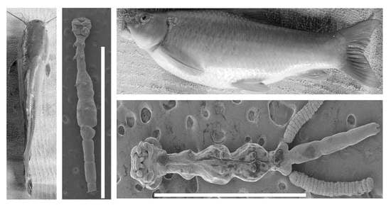
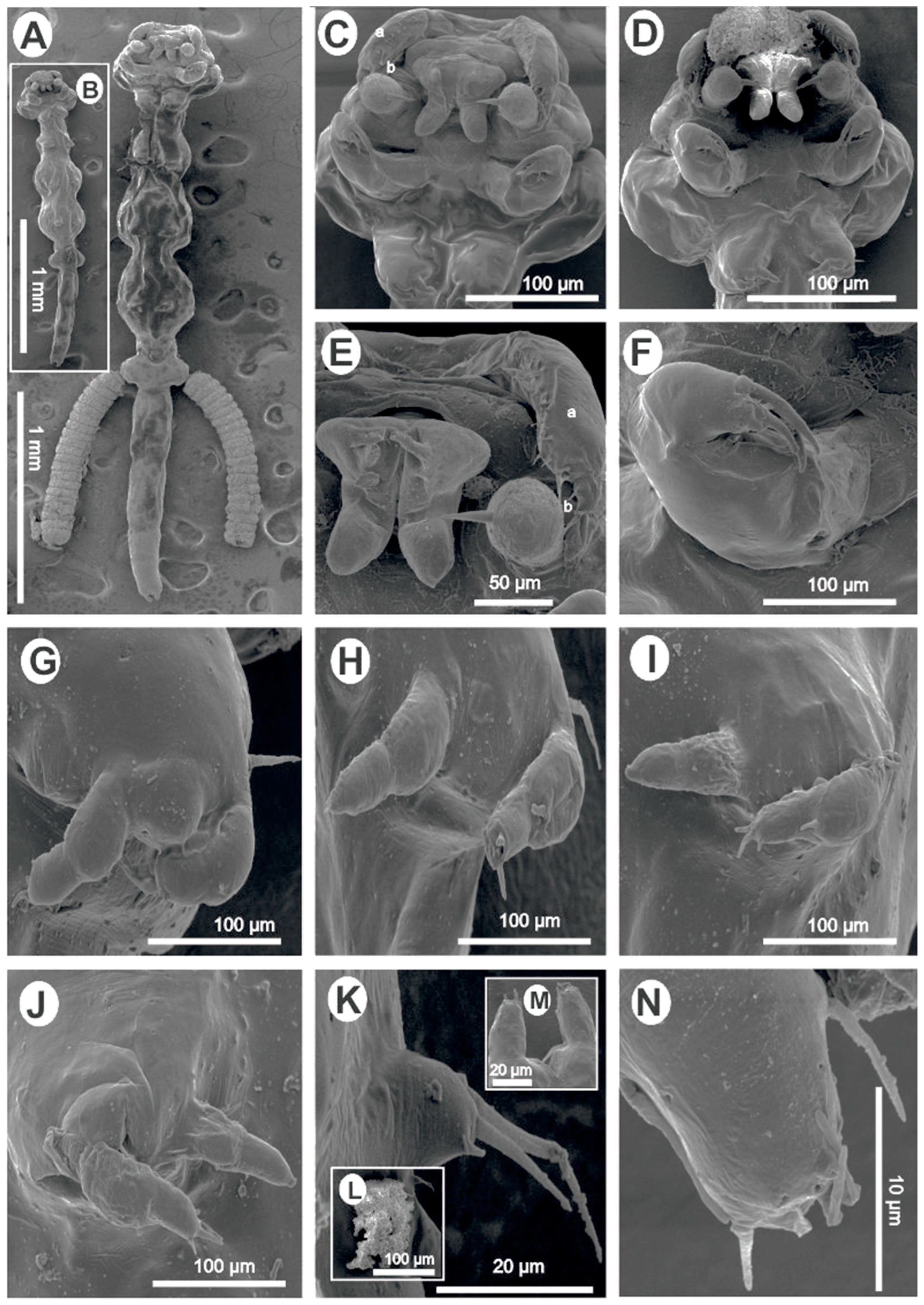
  • Lamproglena cleopatra Humes, 1957.
  • Host: Labeo victorianus Boulenger, 1901 (Cypriniformes: Cyprinidae).
  • Site of infection: Gills.
  • Locality/collection date: Nyando River-Ahero (Lake Victoria drainage system), KisumuCounty, Kenya (0°0′ 0°22′ S, 34°51′ E 35°11′ E), collected 10 May 2022 and 10 March 2023 by Drs. Nehemiah M. Rindoria and George N. Morara.
  • Materials studied: 14 specimens (5 for morphometrics, 4 for SEM, and 5 for molecular analysis).
  • Deposition of voucher specimens: A total of six voucher female specimens were deposited in the Helminthological Collection of the Institute of Parasitology, the Biology Centre of the Czech Academy of Sciences, České Budějovice, Czech Republic (IPCAS Cr-38).
  • Deposition of sequences: Sequence data obtained were deposited in GenBank: 18S rDNA (OR242501, OR242502), 28S rDNA (OR338169, OR338170), and cox1 (OR232207).
  • Redescription (Figure 1) Female (based on nine specimens, five morphometrics (all measurements in millimetres), and four SEM): Body elongated, slender, cylindrical, total length (excluding caudal rami) 2.71 (2.41–3.20) (Figure 1A,B). Body divided into cephalothorax, thorax, and abdomen (Figure 1A,B). Cephalothorax length 0.43 (0.36–0.54), width 0.56 (0.51–0.62), width represents 20.20% of total length, laterally indented; wider posterior part than thorax; U-shaped ridge on dorsal surface (Figure 1A,B). First thoracic segment fused with the head (Figure 1A–D). Second, third, and fourth thoracic segments free, with pedigerous segments distinct and well separated with indentations laterally (Figure 1A,B). Second segment 0.35 (0.24–0.42) wide, 0.26 (0.19–0.31) long. Third and fourth segments 0.42 (0.35–0.53) and 0.50 (0.37–0.54) long, respectively; width subequal 0.51 (0.39–0.59) and 0.50 (0.41–0.56), respectively, wider than the second segment (Figure 1A,B). Fifth thoracic segment narrower 0.24 (0.15–0.27), shorter 0.096 (0.07–0.13), bearing tiny fifth legs (Figure 1A,B,K). Genital segment free, wider 0.354 (0.31–0.40) than fifth thoracic segment, 0.19 (0.13–0.24) long, with egg sacs attached laterally (Figure 1A); other specimens with chitinous, kidney-shaped spermatophores attached ventrally (Figure 1A,B,L). Abdomen length 0.94 (0.79–1.10) (about 34.23% (29.43–37.69) of the total body length) composed of three approximately equal, poorly demarcated segments (Figure 1A,B). Furcal rami (Figure 1A,B,M,N) minute, 0.028 (0.02–0.03) wide, 0.037 (0.03–0.04) long. Each ramus with one long seta, one pore on inner and outer margins, and terminally with four setae, one blunt process, and two pores (Figure 1N). Antennules uniramous, indistinctly two-jointed with long swollen basal podomere bearing 11–14 naked setae and small distal podomere with 5 naked setae, 1 lateral and 4 terminal. Dorsal side of antennule with circular pores (Figure 1C–E). Antenna uniramous, indistinctly four-jointed, distal segment with five small terminal setae (Figure 1C–E). Oral region consisting of distinct projecting sucker-like with two lateral lobes from which arises two long setae and two finger-like posterior lobes (Figure 1C–E). Mandible not observed. Maxilla uniramous, rigid, covered with a thin layer through which distinct terminal spine projects, basal region finely granulated (Figure 1A–E). Maxilliped equipped with three roughly equal, curved claws, with a minute spine-like protrusion on the proximal part (Figure 1F). Legs 1–4 biramous, rami of legs indistinctly two-jointed. Endopodites of legs 1–4 all similar, terminating in a minute, rather blunt seta. Protopodite of legs 1–4 with one lateral long seta at the base before exopodite (Figure 1G–J). Exopodite of first leg first podomere with one smaller seta and four long terminal setae on the second podomere (Figure 1G). Second leg first exopodite podomere with one basal seta, second exopodite podomere with two small setae and a minute knob, an opening between setae and knob (Figure 1H). Second exopodite podomere of third and fourth legs with four setae: two long, one medium, one min (Figure 1I,J). Fifth leg made of small lobe with two long distal and one lateral seta (Figure 1K). Spermatophore observed (Figure 1I,L). Egg sac 0.98 × 0.24, containing about 20 eggs (19–22) (Figure 1A).
Remarks: The parasitic copepods studied here were indistinguishable from L. cleopatra as per the available morphological information published by Humes [8] and Kunutu et al. [2] and clearly distinct from other species of this genus. The indistinguishable features were as follows: body elongated, cylindrical and divided into a cephalothorax, thorax, and abdomen; cephalothorax broader than neck; first thoracic legs fused with the head; thoracic segments marked by lateral constrictions; indistinctly segmented abdomen; three clawed maxilliped; genital somite laterally protruding and distinctly demarcated from the rest of the thorax by a deep indent; antennule larger than antenna; biramous legs; and furcal rami with long lateral processes and terminal setae. Slight variations were noted between the present material and previous records of Humes [8] and Kunutu et al. [2], but the additional taxonomic features observed in the present material were as follows: two long setae on lateral lobes of the oral region (Figure 1C–E) and four circular pores on the furcal rami (Figure 1N).
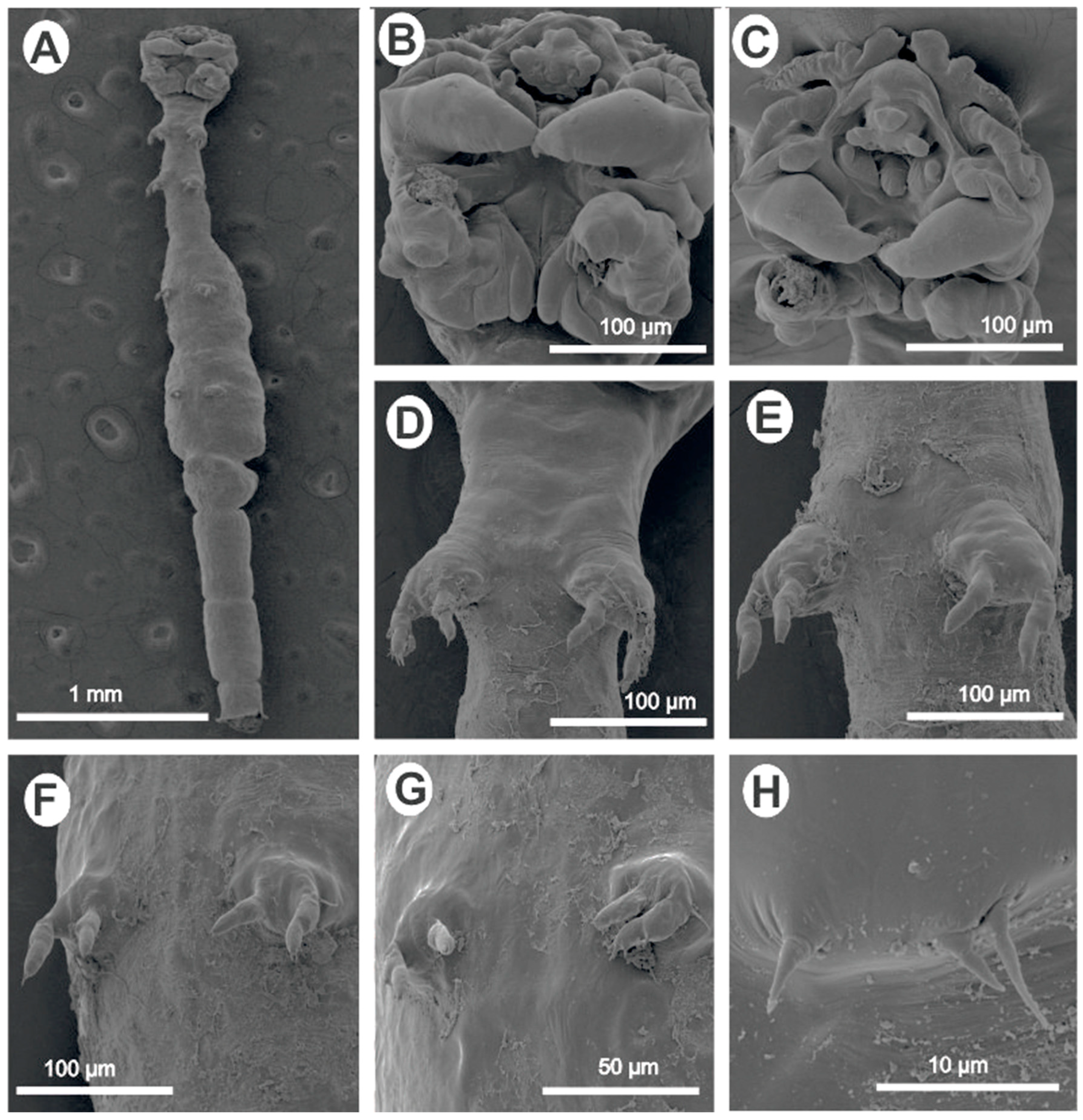
  • Lamproglena clariae Fryer, 1956 (Figure 2).
  • Host: Clarias gariepinus (Burchell, 1822) (Siluriformes, Clariidae).
  • Site of infection: Gills.
  • Locality/collection date: Nyando River-Ahero (Lake Victoria drainage system), Kisumu County, Kenya (0°0′ 0°22′S, 34°51′E 35°11′E), collected 10 May 2022 and 10 March 2023 by Drs. Nehemiah M. Rindoria and George N. Morara.
  • Materials examined: Two specimens, one for SEM and one for molecular analysis.
  • Deposition of voucher specimens: Not deposited.
  • Deposition of sequences: Sequence data obtained were deposited in GenBank: 18S rRNA (OR242503, OR242504), 28S rRNA (OR338195, OR338196), and cox1 (OR232208, OR232209).
  • Remarks: Based on the morphological data available from the reports of Fryer [5] and Marx and Avenant-Oldewage [14], the present material was identical to L. clariae. Following a detailed redescription of this parasite using LM and SEM by Marx and Avenant-Oldewage [14], the present study only provided the SEM images to confirm the identity of our specimen and most importantly provided genetic sequences using 18S, 28S, and cox1 markers.

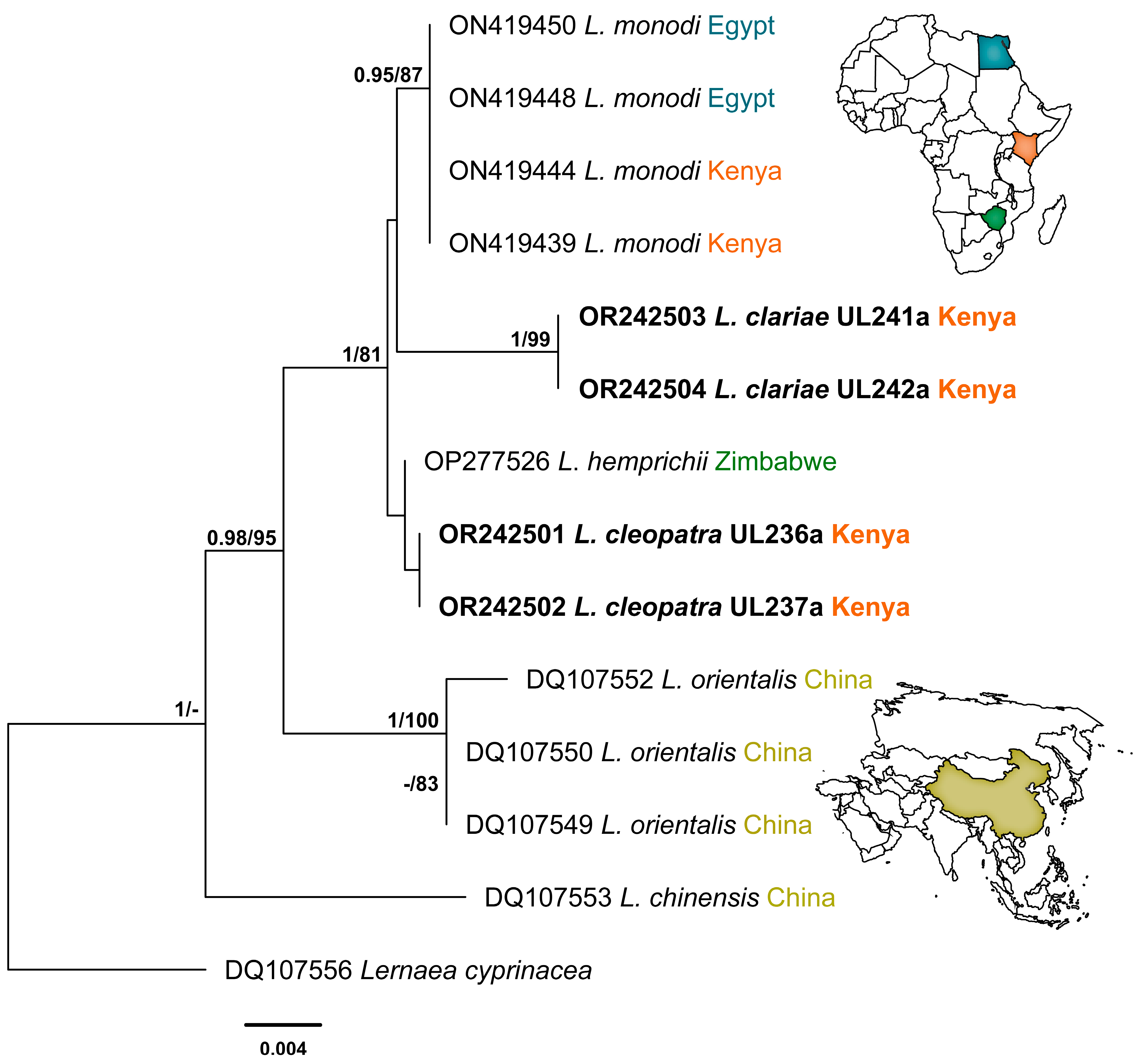
3.2. Molecular Identification

This study generated a total of 11 novel sequences of the three genetic markers: 5 sequences for L. cleopatra and 6 sequences for L. clariae. The Bayesian Inference and Maximum Likelihood analyses of the 18S alignment yielded similar hypotheses (nt = 1325) (Figure 3). The newly generated sequences for L. clariae and L. cleopatra fell into the clade of Lamproglena species previously reported from Africa with strong support. The sequences for L. clariae clustered together with high nodal support and formed a separate branch to the L. monodi clade with no nodal support. The novel sequences for L. cleopatra clustered together and formed a separate clade with L. hemprichii (OP277526) at the basal position of the African clade with no nodal support. The BI and ML analyses for the 28SrDNA dataset showed similar topologies (nt = 696) (Figure 4). A clear distinction between Lamproglena species from Africa and Asia clades were observed. The sequences for L. clariae fell at the basal position of the African clade with strong nodal support. The L. cleopatra sequences clustered with the L. hemprichii (OP277527) previously reported from Zimbabwe with strong nodal support.
The results from the analysis of the 18S and 28S rDNA haplotypes showed a distinct match with all sequences of the four Lamproglena species present in GenBank. There were no cox1 mtDNA sequences available in GenBank for this genus for species comparisons. The pairwise distances (p-distances) and number of base pair differences of L. cleopatra and L. clariae for small (18S) and large (28S) subunit rDNA and all sequences belonging to the Lernaeidae used in this analysis are presented in Table 3 and Table 4, respectively.
The two copepods in the present study, L. clariae and L. cleopatra, were distinct from other Lamproglena species by p-distances of 0.9–2.1% (13–29 bp) and 0.1–2.0% (1–30 bp) based on 18S rDNA (Table 3). For the 28S rDNA, the results showed p-distances of 16.8–23.7% (120–167 bp) and 7.1–23.3% (46–156 bp), respectively (Table 4). The two ribosomal DNA (18S and 28S) markers produced nearly similar topologies with insignificant intraspecific branching. The unavailability of mitochondrial (cox1) marker sequences in GenBank made it impossible to construct any phylogeny tree; therefore, the p-distance and number of base pair differences are provided for cox1 sequences (nt = 683) generated from the present study (Table 5).

4. Discussion

In the present study, lamploglenoids collected in the Nyando River, Kenya, from L. victorianus and C. gariepinus were identified as L. cleopatra and L. clariae, respectively. To a large extent, the parasites bore resemblance to the original descriptions of L. cleopatra by Humes [8] and L. clariae by Fryer [5], respectively.
For L. cleopatra, the original description by Humes [8] and the redescription by Kunutu et al. [2] gave illustrations with morphological and morphometric information which forms a basis for comparison with the current study. The morphometrics given in the present study (see Table 1) are within the ranges provided by Humes [8] and Kunutu et al. [2]. It is worth noting that the present study failed to compare the SEM images provided by Kunutu et al. [2] as the images provided do not conform with the original description of Humes [8] especially on the position of the first thoracic segment. The SEM images of Kunutu et al. [2] show the first thoracic segment just after the cephalothorax, which differs from the same authors’ line micrographs. The line micrographs presented by these authors are in agreement with the original description of L. cleopatra (see Humes [8]), which also corresponds with the morphology of the present study material (Figure 1A–D). Kunutu et al. [2] collected their study materials from three cyprinid species, L. rosae, L. ruddi, and L. molybdinus, from Flag Boshielo Dam, Nwanedi-Luphephe Dam, and River Bubye, respectively, the first two from South Africa and the latter from Zimbabwe, both in the Limpopo River System. We assume that the authors might have had more than one Lamproglena species hence the discrepancy in their line drawings and SEM images. Kunutu et al. [2] failed to provide SEM images of thoracic legs 1–4 but only provided this in the form of line micrographs, and interestingly the descriptions of the thoracic legs correspond well with the present study specimens, in which the four thoracic legs have been well illustrated (Figure 1G–J). Based on morphology, the present study recorded additional taxonomic features which were conspicuous and had not been previously recorded by Humes [8] and Kunutu et al. [2], including two long setae on lateral lobes of the oral region (Figure 1C–E) and four circular pores on the caudal region (Figure 1N).
The morphological study of the second species identified as L. clariae (Figure 2A–H) received little attention in the current study because Marx and Avenant-Oldewage [14] provided detailed morphological studies giving both line drawings and SEM images in addition to the original description by Fryer [5]. In this material, the present study provided an SEM image (for morphological identification) and a genetic description.
The analyses of both 18S and 28S rDNA sequence data for L. clariae and L. cleopatra proved to be distinct from all comparable Lernaeidae and the four Lamproglena sequences available in GenBank. Despite this, the pairwise distances calculated for all the Lamproglena species used in our analysis are from African 18S rDNA (0.9–1.0% 13 bp for L. clariae and 0.1–1.0% 1–14 bp for L. cleopatra) and 28S rDNA (1.3–18.4% 9–131 bp for L. clariae and 7.1–20.4% 46–135 bp for L. cleopatra). These pairwise distances from Africa suggest the conspecificity of L. cleopatra and L. hemprichii. Mabika et al. [31] noted that such a suggestion is improbable because of the distinctive morphology and host specificity of these two species (Cyprinidae and Alestidae, respectively). Rindoria et al. [7] found no variation in the 18S rDNA gene region for L. monodi collected from Egypt and Kenya, confirming the marker’s stability in distinguishing the taxa as also suggested by Mabika et al. [31]. For the mitochondrial marker (cox1), the present study was not able to construct any phylogeny tree due to the unavailability of sequences in GenBank for comparison. However, the study was able to give a comparison of L. clariae and L. cleopatra with p-distances (19.9–20.1%) and the number of base pair differences (136–137 bp) (Table 5), which confirms the distinctness of the two species.
Based on the results found in this study, the importance of global genetic data from the highly variant cox1 gene is highlighted, and more sequences need to be generated to help resolve the taxonomic position of all Lamproglena species. This study shows molecular advances in our knowledge of the diversity of Lamproglena and represents a significant milestone, as it is the first study to provide supplementary genetic data for L. clariae and L. cleopatra (the first ribosomal (18S and 28S rDNA) and the first mitochondrial (cox1 mtDNA) data for any of the 38 nominal species of Lamproglena). It also adds new taxonomic information on morphology using SEM for L. cleopatra. Furthermore, the study provides a new host record for L. cleopatra and extends the geographical information of this species to Kenya. We believe that both the morphological and molecular approaches during the classification of Lamproglena species are vital in expanding our understanding of their taxonomic position.

Author Contributions

Conceptualization, N.M.R. and W.J.L.-P.; methodology, N.M.R. and C.v.W.; software, N.M.R. and C.v.W.; validation, N.M.R., Z.G., G.N.M., W.J.S. and W.J.L.-P.; formal analysis, N.M.R., G.N.M. and C.v.W.; investigation, N.M.R., Z.G. and G.N.M.; resources, W.J.L.-P. and N.J.S.; data curation, N.M.R., C.v.W. and Z.G.; writing—original draft preparation, N.M.R.; writing—review and editing, N.M.R., Z.G., G.N.M., C.v.W., W.J.S., N.J.S. and W.J.L.-P.; visualization, N.M.R., Z.G. and C.v.W.; supervision, W.J.L.-P.; project administration, N.M.R. and W.J.L.-P.; funding acquisition, W.J.L.-P. All authors have read and agreed to the published version of the manuscript.

Funding

This work is based on the research supported in part by the Department of Science and Innovation and the National Research Foundation of South Africa (Grant Numbers 101054 and 146162). The funder had no role in the manuscript writing, editing, approval, or decision to publish.

Institutional Review Board Statement

The authors confirm that the ethical policies of the journal, as noted on the journal’s author guidelines page, have been adhered to. No permit and ethical approval were required for this specific study as fish were collected as part of the routine aquatic research surveys of the Kenya Marine and Fisheries Research Institute (KMFRI), the government agency mandated to conduct research in fisheries and aquatic ecology of all water bodies in Kenya.

Informed Consent Statement

Not applicable.

Data Availability Statement

The data presented in this study are openly available in GenBank (accession number OR232207-OR232209; OR242501-OR242504; OR338169 and OR338170; OR338195 and OR338196) and the Helminthological Collection of the Institute of Parasitology, the Biology Centre of the Czech Academy of Sciences, České Budějovice, Czech Republic (voucher numbers IPCAS Cr-38).

Acknowledgments

The authors would like to thank Elijah M. Kembenya and his team from KMFRI-Sangoro station for their assistance during sample collections. Special thanks are given to Joan M. Maraganga and Gladys N. J. Rindoria from the Kisii University Department of Fisheries and Aquaculture and Judy K. Rindoria (Wildlife and Fisheries Research Institute—Naivasha campus) for their support in the collection and examination of the specimens in the laboratory.

Conflicts of Interest

The authors declare no conflict of interest.

References

  1. Jirsa, F.; Zitek, A.; Schachner, O. First record of Lamproglena pulchella Nordmann, 1832 in the Pielach and Melk rivers, Austria. J. Appl. Ichthyol. 2006, 22, 404–406. [Google Scholar] [CrossRef]
  2. Kunutu, K.D.; Tavakol, S.; Halajian, A.; Baker, C.; Paoletti, M.; Heckmann, R.A.; Luus-Powell, W.J. Expanded description of Lamproglena cleopatra Humes, 1957 (Lernaeidae: Copepoda) from Labeo spp. (Cyprinidae) with a key to species of Lamproglena von Nordmann, 1832. Syst. Parasitol. 2018, 95, 91–103. [Google Scholar] [CrossRef]
  3. Piasecki, W. Comparative morphology of the three species of Lamproglena (Copepoda, Cyclopoida, Lernaeidae) described by von Nordmann, based on re-examination of the types. Mitt. Zool. Mus. Berl. 1993, 69, 307–315. [Google Scholar] [CrossRef]
  4. Capart, A. Notes sur les Copépodes parasites. III. Copépodes parasites des poissons d’eau douce du Congo Belge. Bull. Mus. R. Hist. Nat. Belg. 1944, 20, 1–24. [Google Scholar]
  5. Fryer, G. A report on the parasitic Copepoda and Branchiura of the fishes of Lake Nyasa. Proc. Zool. Soc. Lond. 1956, 127, 29–44. [Google Scholar] [CrossRef]
  6. Fryer, G. The parasitic Copepoda and Branchiura of the fishes of Lake Victoria and the Victoria Nile. Proc. Zool. Soc. Lond. 1961, 137, 41–60. [Google Scholar] [CrossRef]
  7. Rindoria, N.M.; Dos Santos, Q.M.; Ali, S.E.; Ibraheem, M.H.; Avenant Oldewage, A. Lamproglena monodi Capart, 1944 infecting Oreochromis niloticus (Linnaeus, 1758): Additional information on infection, morphology and genetic data. Afr. Zool. 2022, 57, 98–110. [Google Scholar] [CrossRef]
  8. Humes, A.G. Two new caligoid copepods from Egyptian fishes. J. Parasitol. 1957, 43, 201–208. [Google Scholar] [CrossRef]
  9. Fryer, G. Further studies on the parasitic Crustacea of African freshwater fishes. Proc. Zool. Soc. Lond. 1964, 143, 79–102. [Google Scholar]
  10. Fryer, G. The parasitic Crustacea of African freshwater fishes; their biology and distribution. J. Zool. 1968, 156, 45–95. [Google Scholar] [CrossRef]
  11. Thurston, P. The incidence of Monogenea and parasitic Crustacea on the gills of fish in Uganda. Rev. Zool. Bot. Afric. 1970, 82, 111–130. [Google Scholar]
  12. Shotter, R.A. Copepod parasites of fishes from northern Nigeria. Bull. I.F.A.N. 1977, 39, 583–600. [Google Scholar]
  13. Uler, C.; Avenant-Oldewage, A. Die biologie van verteenwoordiges van die genus Lamproglena. Suid-Afrik. Tydskr. Nat. Wet. Tegnol. 1992, 11, 72–73. [Google Scholar]
  14. Marx, H.M.; Avenant-Oldewage, A. Redescription of Lamproglena clariae Fryer, 1956 (Copepoda: Lernaeidae) with notes on its occurrence and distribution. Crustaceana 1996, 69, 509–523. [Google Scholar]
  15. Rindoria, N.M.; Moraa, G.N.; Smit, W.J.; Truter, M.; Smit, N.J.; Luu-Powell, W.J. Integrated morphological and molecular characterization of the fish parasitic nematode Rhabdochona (Rhabdochona) gendrei Campana-Rouget, 1961 infecting Labeobarbus altianalis (Boulenger, 1900) in Kenya. IJP-PAW 2023, 21, 201–209. [Google Scholar] [CrossRef] [PubMed]
  16. Okeyo, D.O.; Ojwang, W.O. A Photographic Guide to Freshwater Fishes of Kenya; 2015; p. 66. Available online: www.seriouslyfish.com/publications/ (accessed on 1 March 2022).
  17. Froese, R.; Pauly, D. (Eds.) FishBase. World Wide Web Electronic Publication; 2013; Available online: www.fishbase.org (accessed on 3 August 2022).
  18. Schäperclaus, W. Fischkrankheiten; Akademie Verlag: Berlin, Germany, 1990. [Google Scholar]
  19. Boxshall, G.A.; Halsey, S.H. An Introduction to Copepod Diversity; Part II; Ray Society: London, UK, 2004. [Google Scholar]
  20. Nation, J.L. A new method using hexamethyldisilazane for preparation of soft insect tissues for scanning electron microscopy. Stain Technol. 1983, 58, 347–351. [Google Scholar] [CrossRef]
  21. Dos Santos, Q.M.; Avenant-Oldewage, A. Soft tissue digestion of Paradiplozoon vaalense for SEM of sclerites and simultaneous molecular analysis. J. Parasitol. 2015, 101, 94–97. [Google Scholar] [CrossRef]
  22. Folmer, O.; Black, M.; Hoeh, W.; Lutz, R.; Vrijenhoek, R. DNA primers for amplification of mitochondrial cytochrome c oxidase subunit I from diverse metazoan invertebrates. Mol. Mar. Biol. Biotechnol. 1994, 3, 294–299. [Google Scholar]
  23. Song, Y.; Wang, G.T.; Yao, W.J.; Gao, Q.; Nei, P. Phylogeny of freshwater parasitic copepods in the Ergasilidae (Copepoda: Poecilostomatoida) based on 18S and 28S rDNA sequences. Parasitol. Res. 2008, 102, 299–306. [Google Scholar] [CrossRef] [Green Version]
  24. Altschul, S.F.; Gish, W.; Miller, W.; Myers, E.W.; Lipman, D.J. Basic Local Alignment Search Tool. J. Mol. Biol. 1990, 215, 403–410. [Google Scholar] [CrossRef]
  25. Capella-Gutiérrez, S.; Silla-Martínez, J.M.; Gabaldón, T. trimAl: A tool for automated alignment trimming in large-scale phylogenetic analyses. Bioinformatics 2009, 25, 1972–1973. [Google Scholar] [CrossRef] [PubMed] [Green Version]
  26. Darriba, D.; Taboada, G.L.; Doallo, R.; Posada, D. jModelTest 2: More models, new heuristics and parallel computing. Nat. Methods. 2012, 9, 772. [Google Scholar] [CrossRef] [PubMed] [Green Version]
  27. Guindon, S.; Gascuel, O. A simple, fast, and accurate algorithm to estimate large phylogenies by maximum likelihood. Syst. Biol. 2003, 52, 696–704. [Google Scholar] [CrossRef] [PubMed] [Green Version]
  28. Miller, M.A.; Pfeiffer, W.; Schwartz, T. Creating the CIPRES Science Gateway for inference of large phylogenetic trees. In Proceedings of the 2010 Gateway Computing Environments Workshop (GCE), New Orleans, LA, USA, 14 November 2010; pp. 1–8. [Google Scholar]
  29. Rambaut, A. FigTree v. 1.4. In Molecular Evolution, Phylogenetics and Epidemiology; University of Edinburgh, Institute of Evolutionary Biology: Edinburgh, UK, 2012; Available online: http://tree.bio.ed.ac.uk/software/figtree/ (accessed on 1 August 2022).
  30. Kumar, S.; Stecher, G.; Tamura, K. MEGA7: Molecular evolutionary genetics analysis version 7.0 for bigger datasets. Mol. Biol. Evol. 2016, 33, 1870–1874. [Google Scholar] [CrossRef] [Green Version]
  31. Mabika, N.; Barson, M.; Dos Santos, Q.M.; van Dyk, C.; Avenant-Oldewage, A. Additional Taxonomic Information for Lamproglena hemprichii (Copepoda: Lernaeidae) Based on Scanning Electron Microscopy and Genetic Characterization, Alongside Some Aspects of its Ecology. Zool. Sci. 2023, 40. [Google Scholar] [CrossRef]
Figure 1. Scanning electron micrographs of a Lamproglena cleopatra Humes, 1957 female: (A,B) ventral view of the adult; (CE) ventral view of cephalothorax showing antennules, antennae, oral region, and maxillae; (F) maxilliped; (G) first leg; (H) second leg; (I) third leg; (J) fourth leg; (K) fifth leg; (L) spermatophore; (M) furcal rami, showing the anal opening; (N) furcal rami. Abbreviations: a, antennules; b, antenna.
Pathogens 12 00980 g001
Figure 2. Lamproglena clariae Fryer, 1956 female: (A) ventral view of a mature adult; (B,C) ventral view of cephalothorax showing antennules, antennae, oral region, maxillae, and maxillipeds; (D) first leg; (E) second leg; (F) third leg; (G) fourth leg; (H) fifth leg.
Pathogens 12 00980 g002
Figure 3. Phylogenetic relationship of Lamproglena cleopatra Humes, 1957 and Lamproglena clariae Fryer, 1956 to other Lernaeidae based on 18S rDNA. Phylogenies were reconstructed using Bayesian Inference (BI) and Maximum Likelihood (ML) with Lernaea cyprinacea designated as the outgroup. Sequences of the present study are highlighted in bold. Nodal support for BI and ML is indicated along the branch nodes (BI/ML); values < 0.90 (BI) and < 70 (ML) are not shown.
Pathogens 12 00980 g003
Figure 4. Phylogenetic relationship of Lamproglena cleopatra Humes, 1957 and Lamproglena clariae Fryer, 1956 to other Lernaeidae based on 28S rDNA. Phylogenies were reconstructed using Bayesian Inference (BI) and Maximum Likelihood (ML) with Lernaea cyprinacea designated as the outgroup. Sequences of the present study are highlighted in bold. Nodal support for BI and ML is indicated along the branch nodes (BI/ML); values < 0.90 (BI) and < 70 (ML) are not shown.
Pathogens 12 00980 g004
Table 1. Measurements in millimetres with mean followed by standard deviation and range in parentheses of various taxonomic features of Lamproglena cleopatra Humes, 1957 for the present study and comparisons with previous studies.
Humes [8]Kunutu et al. [2]Present Study
Country/fish species/no. measuredEgypt: L. forskalii n = 5SA: L. rosae and L. molybdinus
ZIM: L. ruddi n = 40
KEN: L. victorianus n = 5
Taxonomic feature
Total length2.60 (2.43–2.77)2.79 ± 0,39 (1.66–3.38)2.71 ± 0.30 (2.41–3.20)
Cephalothorax L0.504-0.43 ± 0.07 (0.36–0.54)
W0.3750.58 ± 0.07 (0.41–0.71)0.56 ± 0.05 (0.51–0.62)
Second thoracic segmentL-0.28 ± 0.07 (0.16–0.41)0.26 ± 0.05 (0.19–0.31)
W0.2910.32 ± 0.05 (0.19–0.40)0.35 ± 0.07 (0.24–0.42)
Third thoracic segmentL-0.38 ± 0.06 (0.15–0.48)0.42 ± 0.07 (0.35–0.53)
W0.4220.43 ± 0.08 (0.20–0.59)0.52 ± 0.08 (0.39–0.59)
Fourth thoracic segmentL-0.41 ± 0.07 (0.16–0.51)0.50 ± 0.07 (0.37–0.54
W0.4130.43 ± 0.08 (0.20–0.590.50 ± 0.06 (0.41–0.56)
Fifth leg-bearing segmentL-0.09 ± 0.02 (0.06–0.14)0.096 ± 0.02 (0.07–0.13)
W0.2120.22 ± 0.03 (0.16–0.30)0.242 ± 0.05 (0.15–0.29)
Genital segmentL-0.17 ± 0.03 (0.13–0.22)0.194 ± 0.04 (0.13–0.24)
W0.3430.35 ± 0.06 (0.16–0.43)0.354 ± 0.02 (0.31–0.40)
Egg sacL1.321.22 ± 0.23 (0.92–1.46)0.976 (n = 1)
W0.171-0.24 (n = 1)
AbdomenL0.9750.96 ± 0.16 (0.56–1.22)0.94 ± 0.13 (0.79–1.10)
W-0.19 ± 0.02 (0.14–0.25)-
% of the abdomen to total body length 373434
Furcal ramiL0.0390.04 ± 0.01 (0.03–0.06)0.037 (0.03–0.04)
W0.026 0.028 (0.02–0.03)
Abbreviations: SA, South Africa; KEN, Kenya; ZIM, Zimbabwe; -, not reported.
Table 2. Information for the species, hosts, families, geographical localities, and accession numbers of 18S, 28S, and cox1 used from Lernaeidae used in molecular analyses.
SpeciesHostFamilyLocality18S28Scox1Reference
Lamproglena orientalisSqualiobarbus curriculusXenocyprididaeDangjiangkou Reservoir, ChinaDQ107552DQ107544Song et al. [2]
Lamproglena orientalisChanodichthys erythropterusXenocyprididaeTangxun Lake, China DQ107551DQ107541 Song et al. [2]
Lamproglena orientalisChanodichthys mongolicusXenocyprididaeE-zhou farm, ChinaDQ107550DQ107543 Song et al. [2]
Lamproglena orientalisChanodichthys dabryiXenocyprididaeTangxun Lake, ChinaDQ107549DQ107542 Song et al. [2]
Lamproglena hemprichiiHydrocynus vittatusAlestidaeLake Kariba, ZimbabweOP277526OP277527Mabika et al. [28]
Lamproglena cleopatra
Isolate UL236
Labeo victorianusCyprinidaeNyando River, KenyaOR242501OR338169Present study
Lamproglena cleopatra
Isolate UL237
Labeo victorianusCyprinidaeNyando River, KenyaOR242502OR338170OR232207Present study
Lamproglena clariae
Isolate UL241
Clarias gariepinusClariidaeNyando River, KenyaOR242503OR338195
OR338196
OR232208Present study
Lamproglena clariae
Isolate UL242
Clarias gariepinusClariidaeNyando River, KenyaOR242504OR232209Present study
Lamproglena monodiOreochromis niloticusCichlidaeKibos Fish Farm, Kenya ON419439ON419422Rindoria et al. [7]
Lamproglena monodiOreochromis niloticusCichlidaeKibos Fish Farm, KenyaON419444ON419428Rindoria et al. [7]
Lamproglena monodiOreochromis niloticusCichlidaeSharqia, EgyptON419450ON419435Rindoria et al. [7]
Lamproglena monodiOreochromis niloticusCichlidaeEl-Minia, EgyptON419448ON419432Rindoria et al. [7]
Lamproglena chinensisChanna argusChannidaeDangjiangkou ReservoirDQ107553DQ107545Song et al. [2]
Lernea cyprinaceaChanodichthys erythropterusXenocyprididaeLake Dongxi, ChinaDQ107556DQ107548Song et al. [2]
― not available.
Table 3. Pairwise distances (%, unshaded diagonal) and the number of base pair differences (shaded diagonal) between Lamproglena cleopatra Humes, 1957, Lamproglena clariae Fryer, 1957, other Lamproglena species, and Lernaea cyprinacea Linnaeus, 1758 based on 18S rDNA (present study species % and base pairs are in bold).
Accession
Number
1234567891011121314
1L. cleopatra UL236OR242501 0.01.01.00.10.20.20.20.21.21.21.42.02.3
2L. cleopatra UL237OR2425020 1.01.00.10.20.20.20.21.21.21.42.02.3
3L. clariae UL241OR2425031414 0.00.90.90.90.90.91.71.71.92.02.4
4L. clariae UL242OR24250414140 1.01.01.01.01.01.91.91.92.12.6
5L. hemprichiiOP277526111313 0.30.30.30.31.11.11.42.02.4
6L. monodiON4194393313134 0.00.00.01.31.31.52.02.4
7L. monodiON41944433131340 0.00.01.31.31.52.02.4
8L. monodiON419448331313400 0.01.31.31.52.02.4
9L. monodiON4194503313134000 1.31.31.52.02.4
10L. orientalisDQ107549161624241517171717 0.00.32.22.6
11L. orientalisDQ1075501616242415171717170 0.32.22.6
12L. orientalisDQ10755219192625192020202044 2.42.8
13L. chinensisDQ107553303029282929292929323235 2.5
14Lernaea cyprinaceaDQ10755633333535343434343437373938
Table 4. Pairwise distances (%, unshaded diagonal) and the number of base pair differences (shaded diagonal) between Lamproglena cleopatra Humes, 1957, Lamproglena clariae Fryer, 1957, other Lamproglena species, and Lernaea cyprinacea Linnaeus, 1758 based on 28S rDNA (present study species % and base pairs are in bold).
Accession
Number
123456789101112131415
1L. cleopatra UL236OR338169 0.019.219.47.79.99.99.910.120.821.021.220.921.722.2
2L. cleopatra UL237OR3381700 20.420.47.19.19.19.19.222.422.622.722.523.323.0
3L. clariae UL241 c3OR338195115135 1.318.416.816.816.816.923.523.723.523.321.124.0
4L. clariae UL242 c25OR3381961161359 17.916.816.816.816.923.223.423.223.020.724.0
5L. hemprichiiOP277527 4647131128 6.6 6.6 6.6 6.8 19.9 20.0 20.1 19.9 19.9 22.5
6L. monodiON419422 596012012048 0.00.00.018.718.819.018.919.422.4
7L. monodiON419428 5960120120480 0.00.018.718.819.018.919.422.4
8L. monodiON419432 59601201204800 0.018.718.819.018.919.422.4
9L. monodiON419435 596012012048111 18.718.819.018.919.422.5
10L. orientalisDQ107541 122146166164139131131131130 0.10.32.521.022.2
11L. orientalisDQ1075431231471671651401321321321311 0.42.621.222.4
12L. orientalisDQ10754212414816616414113313313313223 2.721.322.5
13L. orientalisDQ107544 125149167165142135135135134202122 20.722.0
14L. chinensisDQ107545 132156154151144141141141140151152153151 22.7
15Lernaea cyprinaceaDQ107548 155176195195182183183183183181182183182180
Table 5. Pairwise distances (%, unshaded diagonal) and the number of base pair differences (shaded diagonal) between Lamproglena cleopatra Humes, 1957, Lamproglena clariae Fryer, 1957, other Lamproglena species, and Lernaea cyprinacea Linnaeus, 1758 based on cox1 (present study species % and base pairs are in bold).
Accession
Number
OR232207
L. cleopatra
OR232208
L. clariae
OR232209
L. clariae
NC 025239
Lernaea cyprinacea
L. cleopatra UL237OR232207 20.119.926.8
L. clariae UL241OR232208137 0.126.2
L. clariae UL242OR2322091361 26.4
Lernaea cyprinaceaNC 025239183179180
Disclaimer/Publisher’s Note: The statements, opinions and data contained in all publications are solely those of the individual author(s) and contributor(s) and not of MDPI and/or the editor(s). MDPI and/or the editor(s) disclaim responsibility for any injury to people or property resulting from any ideas, methods, instructions or products referred to in the content.

