
Viral Diagnosis of Hepatitis B and Delta: What We Know and What Is Still Required? Specific Focus on Low- and Middle-Income Countries
 ,
, 
Abstract
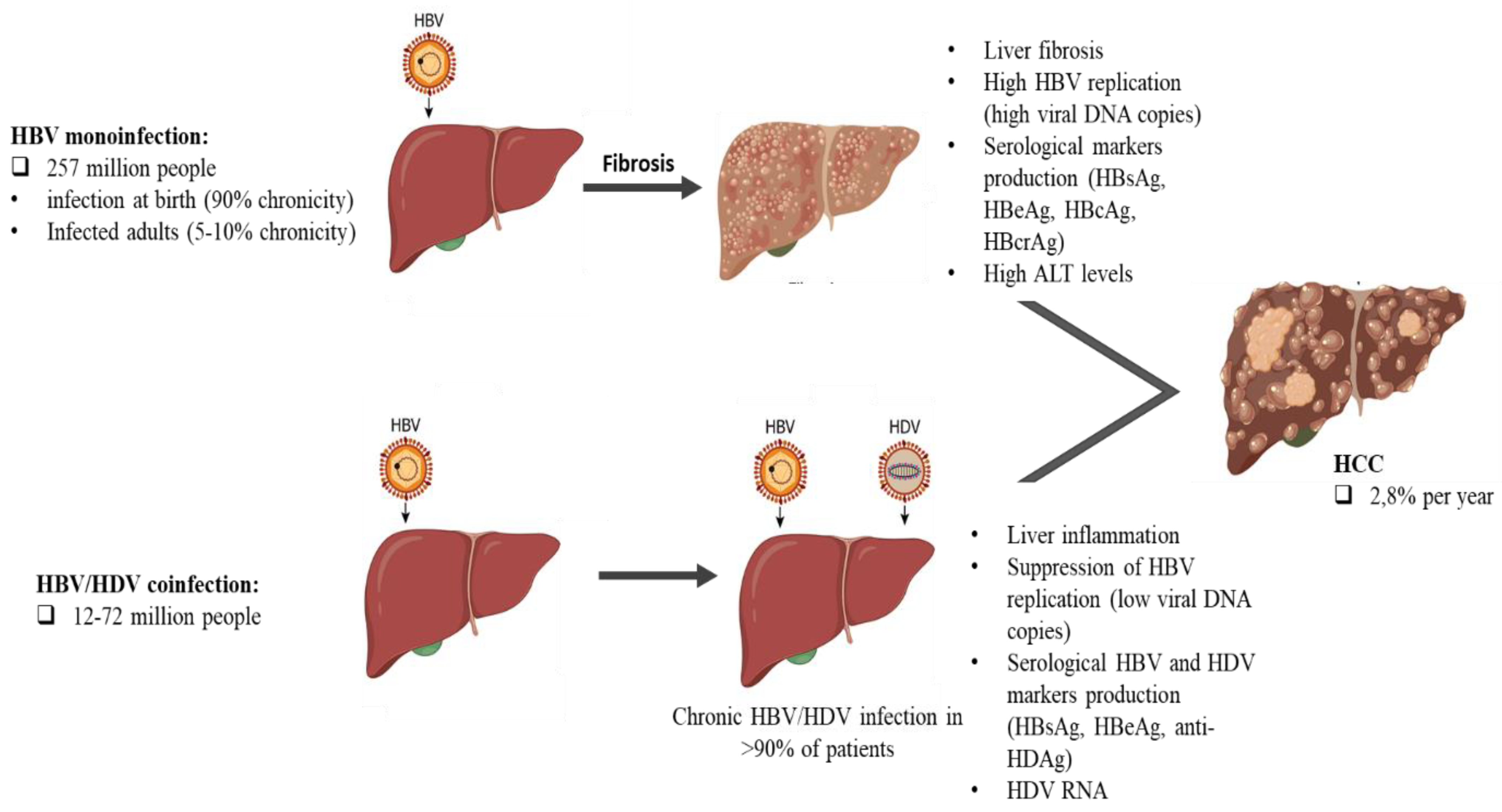
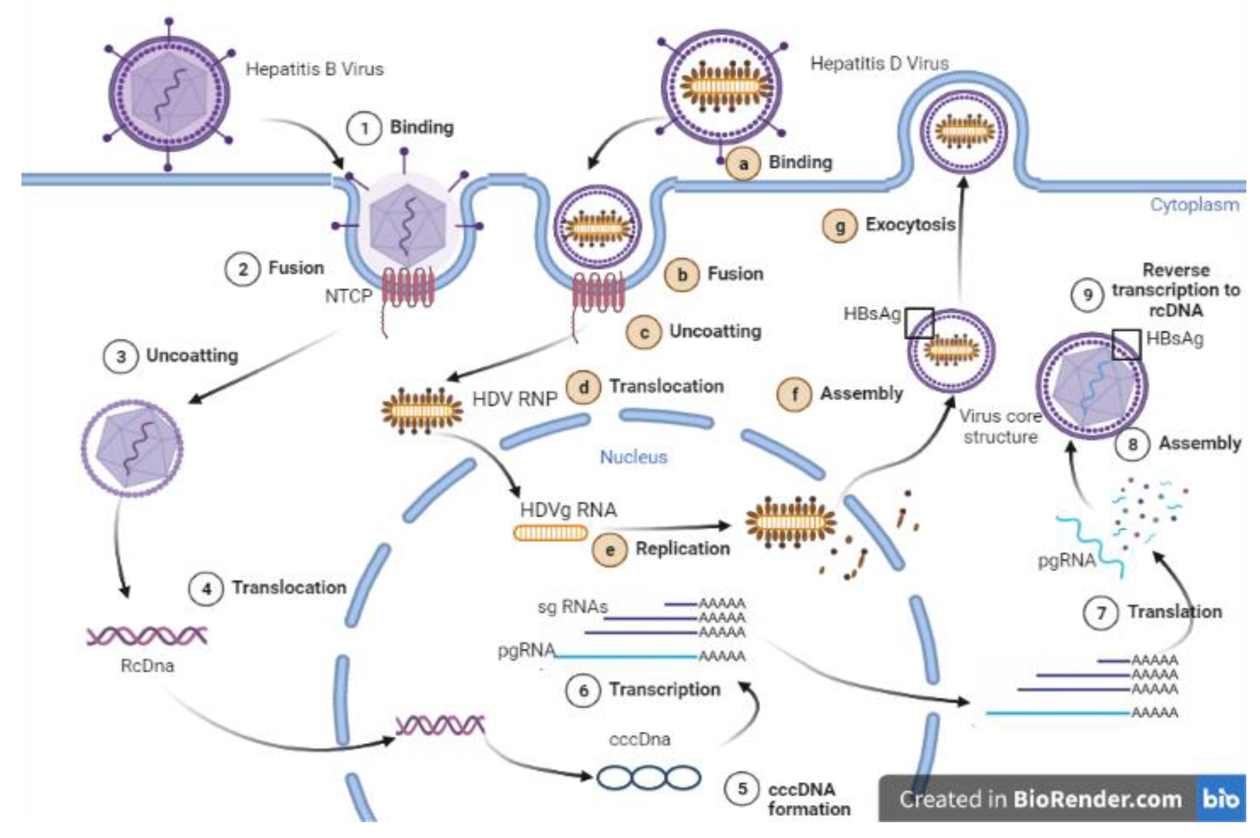
:1. Introduction
2. Serological Markers and Related Diagnosis Tools
2.1. HDV/HBV Coinfection First-Line Markers
2.2. Detection of HBsAg and Anti-HBs Antibodies
2.3. Detection of Hepatitis B Core Antibody (HBcAb)
2.4. Detection of Anti-HDV Antibodies
2.5. Hepatitis Be Antigen (HBeAg) and Its Associated Antibodies (Anti-HBe)
2.6. Hepatitis B Core-Related Antigen (HBcrAg)
3. Rapid Detection Tests (RDTs)
4. Biochemical Markers and Related Diagnosis Tools
4.1. Aspartate Aminotransferase (AST)/Alanine Aminotransferase (ALT)
4.2. Alpha-Fetoprotein (AFP)
5. Molecular Markers and Related Diagnosis Tools
5.1. HBV and HDV Viral Load
5.2. Detection and Quantification of HBV DNA
5.3. New Tools under Development for Diagnosis of HBV/HDV Coinfection
5.4. Detection and Quantification of HDV RNA
5.5. HDV Genotyping
5.6. HBV Pre-Genomic RNA
5.7. A Point-of-Care Test (POCT) for HDV Anti-IgM and IgG
6. Summary
7. Conclusions
Author Contributions
Funding
Institutional Review Board Statement
Conflicts of Interest
References
- Lanini, S.; Pisapia, R.; Capobianchi, M.R.; Ippolito, G. Global epidemiology of viral hepatitis and national needs for complete control. Expert Rev. Anti-infective Ther. 2018, 16, 625–639. [Google Scholar] [CrossRef] [PubMed]
- Botelho-Souza, L.F.; Vasconcelos, M.P.A.; Santos, A.D.O.D.; Salcedo, J.M.V.; Vieira, D.S. Hepatitis delta: Virological and clinical aspects. Virol. J. 2017, 14, 177. [Google Scholar] [CrossRef] [PubMed] [Green Version]
- Lee, A.U.; Lee, C. Hepatitis D Review: Challenges for the Resource-Poor Setting. Viruses 2021, 13, 1912. [Google Scholar] [CrossRef]
- Stockdale, A.J.; Kreuels, B.; Henrion, M.Y.; Giorgi, E.; Kyomuhangi, I.; de Martel, C.; Hutin, Y.; Geretti, A.M. The global prevalence of hepatitis D virus infection: Systematic review and meta-analysis. J. Hepatol. 2020, 73, 523–532. [Google Scholar] [CrossRef]
- Fattovich, G.; Stroffolini, T.; Zagni, I.; Donato, F. Hepatocellular carcinoma in cirrhosis: Incidence and risk factors. Gastroenterology 2004, 127, S35–S50. [Google Scholar] [CrossRef]
- Magnius, L.O. Characterization of a New Antigen-Antibody System Associated with Hepatitis B. Clin. Exp. Immunol. 1975, 20, 209–216. [Google Scholar]
- Shah, P.A.; Choudhry, S.; Reyes, K.J.C.; Lau, D.T.Y. An update on the management of chronic hepatitis D. Gastroenterol. Rep. 2019, 7, 396–402. [Google Scholar] [CrossRef]
- Lampertico, P.; Agarwal, K.; Berg, T.; Buti, M.; Janssen, H.L.A.; Papatheodoridis, G.; Zoulim, F.; Tacke, F. EASL 2017 Clinical Practice Guidelines on the management of hepatitis B virus infection. J. Hepatol. 2017, 67, 370–398. [Google Scholar] [CrossRef] [Green Version]
- Tiwari, A.K.; Upadhyay, A.P.; Arora, D.; Wadhwa, T.; Aggarwal, G.; Pabbi, S.; Luthra, A.S.; Rawat, S.S. Head-to-head comparison of Enzyme Linked Immunosorbent Assay (ELISA) and Enhanced Chemiluminescence Immunoassay (ECLIA) for the detection of Transfusion Transmitted Disease (TTD) Markers; HIV, HCV and HBV in blood donors, in India. J. Virol. Methods 2020, 285, 113962. [Google Scholar] [CrossRef] [PubMed]
- Rocco, C.; Bonavolta, R.; Vallefuoco, L.; Braschi, U.; Sorrentino, R.; Terracciano, D.; Portella, G. Comparison of anti–hepatitis D virus (HDV) ETI-AB-DELTAK-2 assay and the novel LIAISON® XL MUREX anti-HDV assay in the diagnosis of HDV infection. Diagn. Microbiol. Infect. Dis. 2019, 95, 114873. [Google Scholar] [CrossRef]
- Blumberg, B.S. Australia Antigen and the Biology of Hepatitis B. Science 1977, 197, 17–25. [Google Scholar] [CrossRef] [PubMed]
- Rodella, A.; Galli, C.; Terlenghi, L.; Perandin, F.; Bonfanti, C.; Manca, N. Quantitative analysis of HBsAg, IgM anti-HBc and anti-HBc avidity in acute and chronic hepatitis B. J. Clin. Virol. 2006, 37, 206–212. [Google Scholar] [CrossRef] [PubMed]
- Hansson, B.G. Evaluation of Three Reverse Passive Haemagglutination Methods and Two Radioimmunoassay Tests to Be Used for the Detection of Hepatitis B Surface Antigen. Acta Pathol. Microbiol. Scand. Sect. C Immunol. 2009, 84, 53–58. [Google Scholar] [CrossRef] [PubMed]
- Chan, H.L.; Wong, V.W.; Tse, A.M.; Tse, C.; Chim, A.M.; Chan, H.; Wong, G.L.; Sung, J.J. Serum Hepatitis B Surface Antigen Quantitation Can Reflect Hepatitis B Virus in the Liver and Predict Treatment Response. Clin. Gastroenterol. Hepatol. 2007, 5, 1462–1468. [Google Scholar] [CrossRef] [PubMed]
- Madiyal, M.; Sagar, S.; Vishwanath, S.; Banerjee, B.; Eshwara, V.K.; Chawla, K. Comparing Assay Performance of ELISA and Chemiluminescence Immunoassay in Detecting Antibodies to Hepatitis B Surface Antigen. J. Clin. Diagn. Res. 2016, 10, DC22–DC25. [Google Scholar] [CrossRef] [PubMed]
- Yuan, Q.; Song, L.-W.; Cavallone, D.; Moriconi, F.; Cherubini, B.; Colombatto, P.; Oliveri, F.; Coco, B.A.; Ricco, G.; Bonino, F.; et al. Total Hepatitis B Core Antigen Antibody, a Quantitative Non-Invasive Marker of Hepatitis B Virus Induced Liver Disease. PLoS ONE 2015, 10, e0130209. [Google Scholar] [CrossRef] [PubMed]
- Izumida, K.; Kaneko, A.; Takahashi, K.; Kusumoto, S.; Narita, T.; Takami, A.; Iida, S.; Aoyagi, K.; Tanaka, Y. Clinical evaluation of a novel and highly sensitive immunoassay for anti-hepatitis B core antigen using a fully automated immunochemical analyzer. Hepatol. Res. 2018, 48, 1081–1091. [Google Scholar] [CrossRef] [PubMed] [Green Version]
- Wranke, A.; Borzacov, L.M.P.; Parana, R.; Lobato, C.; Hamid, S.; Ceausu, E.; Dalekos, G.N.; Rizzetto, M.; Turcanu, A.; Niro, G.A.; et al. Clinical and virological heterogeneity of hepatitis delta in different regions world-wide: The Hepatitis Delta International Network (HDIN). Liver Int. 2017, 38, 842–850. [Google Scholar] [CrossRef]
- Ricco, G.; Popa, D.C.; Cavallone, D.; Iacob, S.; Salvati, A.; Tabacelia, D.; Oliveri, F.; Mascolo, G.; Bonino, F.; Yuan, Q.; et al. Quantification of serum markers of hepatitis B (HBV) and Delta virus (HDV) infections in patients with chronic HDV infection. J. Viral Hepat. 2018, 25, 911–919. [Google Scholar] [CrossRef]
- Wranke, A.; Heidrich, B.; Ernst, S.; Calle Serrano, B.; Caruntu, F.A.; Curescu, M.G.; Yalcin, K.; Gürel, S.; Zeuzem, S.; Erhardt, A.; et al. Anti-HDV IgM as a Marker of Disease Activity in Hepatitis Delta. PLoS ONE 2014, 9, e101002. [Google Scholar] [CrossRef]
- Kennedy, P.T.; Sandalova, E.; Jo, J.; Gill, U.; Ushiro–Lumb, I.; Tan, A.T.; Naik, S.; Foster, G.R.; Bertoletti, A. Preserved T-Cell Function in Children and Young Adults with Immune-Tolerant Chronic Hepatitis B. Gastroenterology 2012, 143, 637–645. [Google Scholar] [CrossRef] [PubMed]
- Croagh, C.M.; Lubel, J.S. Natural history of chronic hepatitis B: Phases in a complex relationship. World J. Gastroenterol. 2014, 20, 10395–10404. [Google Scholar] [CrossRef] [PubMed]
- Charre, C.; Levrero, M.; Zoulim, F.; Scholtès, C. Non-invasive biomarkers for chronic hepatitis B virus infection management. Antivir. Res. 2019, 169, 104553. [Google Scholar] [CrossRef]
- Yang, H.I.; Lu, S.N.; Liaw, Y.F.; You, S.L.; Sun, C.A.; Wang, L.Y.; Hsiao, C.K.; Chen, P.J.; Chen, D.S.; Chen, C.J. Hepatitis B e Antigen and the Risk of Hepatocellular Carcinoma. N. Engl. J. Med. 2002, 347, 168–174. [Google Scholar] [CrossRef] [PubMed] [Green Version]
- Thompson, A.J.; Nguyen, T.; Iser, D.; Ayres, A.; Jackson, K.; Littlejohn, M.; Slavin, J.; Bowden, S.; Gane, E.J.; Abbott, W.; et al. Serum hepatitis B surface antigen and hepatitis B e antigen titers: Disease phase influences correlation with viral load and intrahepatic hepatitis B virus markers. Hepatology 2010, 51, 1933–1944. [Google Scholar] [CrossRef] [PubMed]
- Liu, X.; Chen, J.; Lou, J.; Huang, Y.; Yan, Y.; Sun, G.; Li, N. Correlation between hepatitis B virus DNA levels and diagnostic tests for HBsAg, HBeAg, and PreS1-Ag in chronic hepatitis B. Genet. Mol. Res. 2016, 15, 1–9. [Google Scholar] [CrossRef] [PubMed]
- Shimakawa, Y.; Yan, H.-J.; Tsuchiya, N.; Bottomley, C.; Hall, A.J. Association of Early Age at Establishment of Chronic Hepatitis B Infection with Persistent Viral Replication, Liver Cirrhosis and Hepatocellular Carcinoma: A Systematic Review. PLoS ONE 2013, 8, e69430. [Google Scholar] [CrossRef]
- Seck, A.; Maylin, S.; Akbar, S.M.F.; Funk, A.L.; Bercion, R.; Mishiro, S.; Ndiaye, B.; Fontanet, A.; Vray, M.; Simon, F.; et al. Poor Sensitivity of Commercial Rapid Diagnostic Tests for Hepatitis B e Antigen in Senegal, West Africa. Am. J. Trop. Med. Hyg. 2018, 99, 428–434. [Google Scholar] [CrossRef]
- Wang, S.-J.; Chen, Z.-M.; Wei, M.; Liu, J.-Q.; Li, Z.-L.; Shi, T.-S.; Nian, S.; Fu, R.; Wu, Y.-T.; Zhang, Y.-L.; et al. Specific determination of hepatitis B e antigen by antibodies targeting precore unique epitope facilitates clinical diagnosis and drug evaluation against hepatitis B virus infection. Emerg. Microbes Infect. 2021, 10, 37–50. [Google Scholar] [CrossRef]
- Gani, A.W.; Wei, W.; Shi, R.-Z.; Ng, E.; Nguyen, M.; Chua, M.-S.; So, S.; Wang, S.X. An Automated, Quantitative, and Multiplexed Assay Suitable for Point-of-Care Hepatitis B Virus Diagnostics. Sci. Rep. 2019, 9, 15615. [Google Scholar] [CrossRef] [Green Version]
- Shimakawa, Y.; Ndow, G.; Njie, R.; Njai, H.F.; Takahashi, K.; Akbar, S.M.F.; Cohen, D.; Nayagam, A.S.; Jeng, A.; Ceesay, A.; et al. Hepatitis B core-related antigen (HBcrAg): An alternative to HBV DNA to assess treatment eligibility in Africa. Clin. Infect. Dis. 2019, 70, 1442–1452. [Google Scholar] [CrossRef]
- Suzuki, F.; Miyakoshi, H.; Kobayashi, M.; Kumada, H. Correlation between serum hepatitis B virus core-related antigen and intrahepatic covalently closed circular DNA in chronic hepatitis B patients. J. Med Virol. 2008, 81, 27–33. [Google Scholar] [CrossRef] [PubMed]
- Wong, D.K.-H.; Seto, W.-K.; Cheung, K.-S.; Chong, C.-K.; Huang, F.-Y.; Fung, J.; Lai, C.-L.; Yuen, M.-F. Hepatitis B virus core-related antigen as a surrogate marker for covalently closed circular DNA. Liver Int. 2017, 37, 995–1001. [Google Scholar] [CrossRef] [PubMed]
- Testoni, B.; Lebossé, F.; Scholtes, C.; Berby, F.; Miaglia, C.; Subic, M.; Loglio, A.; Facchetti, F.; Lampertico, P.; Levrero, M.; et al. Serum hepatitis B core-related antigen (HBcrAg) correlates with covalently closed circular DNA transcriptional activity in chronic hepatitis B patients. J. Hepatol. 2019, 70, 615–625. [Google Scholar] [CrossRef] [PubMed]
- Suzuki, F.; Hosaka, T.; Imaizumi, M.; Kobayashi, M.; Ohue, C.; Suzuki, Y.; Fujiyama, S.; Kawamura, Y.; Sezaki, H.; Akuta, N.; et al. Potential of ultra-highly sensitive immunoassays for hepatitis B surface and core-related antigens in patients with or without development of hepatocellular carcinoma after hepatitis B surface antigen seroclearance. Hepatol. Res. 2021, 51, 426–435. [Google Scholar] [CrossRef]
- Inoue, T.; Kusumoto, S.; Iio, E.; Ogawa, S.; Suzuki, T.; Yagi, S.; Kaneko, A.; Matsuura, K.; Aoyagi, K.; Tanaka, Y. Clinical efficacy of a novel, high-sensitivity HBcrAg assay in the management of chronic hepatitis B and HBV reactivation. J. Hepatol. 2021, 75, 302–310. [Google Scholar] [CrossRef]
- Hosaka, T.; Suzuki, F.; Kobayashi, M.; Fujiyama, S.; Kawamura, Y.; Sezaki, H.; Akuta, N.; Kobayashi, M.; Suzuki, Y.; Saitoh, S.; et al. Ultrasensitive Assay for Hepatitis B Core-Related Antigen Predicts Hepatocellular Carcinoma Incidences During Entecavir. Hepatol. Commun. 2022, 6, 36–49. [Google Scholar] [CrossRef]
- Shinkai, N.; Kusumoto, S.; Murakami, S.; Ogawa, S.; Ri, M.; Matsui, T.; Tamori, A.; Toyoda, H.; Ishida, T.; Iida, S.; et al. Novel monitoring of hepatitis B reactivation based on ultra-high sensitive hepatitis B surface antigen assay. Liver Int. 2017, 37, 1138–1147. [Google Scholar] [CrossRef]
- Shimakawa, Y.; Ndow, G.; Kaneko, A.; Aoyagi, K.; Lemoine, M.; Tanaka, Y. Rapid Point-of-Care Test for Hepatitis B Core-Related Antigen to Diagnose High Viral Load in Resource-Limited Settings. Clin. Gastroenterol. Hepatol. 2022, 27, R713–R715. [Google Scholar] [CrossRef]
- Naseri, M.; Ziora, Z.M.; Simon, G.P.; Batchelor, W. ASSURED-compliant point-of-care diagnostics for the detection of human viral infections. Rev. Med Virol. 2022, 32, e2263. [Google Scholar] [CrossRef]
- Ceesay, A.; Lemoine, M.; Cohen, D.; Chemin, I.; Ndow, G. Clinical utility of the ‘Determine HBsAg’ Point-of-Care Test for Diagnosis of Hepatitis B Surface Antigen in Africa. Expert Rev. Mol. Diagn. 2022, 22, 497–505. [Google Scholar] [CrossRef] [PubMed]
- Biselli, R.; Nisini, R.; Lista, F.; Autore, A.; Lastilla, M.; De Lorenzo, G.; Peragallo, M.S.; Stroffolini, T.; D’Amelio, R. A Historical Review of Military Medical Strategies for Fighting Infectious Diseases: From Battlefields to Global Health. Biomedicines 2022, 10, 2050. [Google Scholar] [CrossRef] [PubMed]
- Zhang, W.; Aryan, M.; Qian, S.; Cabrera, R.; Liu, X. A Focused Review on Recent Advances in the Diagnosis and Treatment of Viral Hepatitis. Gastroenterol. Res. 2021, 14, 139–156. [Google Scholar] [CrossRef] [PubMed]
- Xiao, Y.; Thompson, A.J.; Howell, J. Point-of-Care Tests for Hepatitis B: An Overview. Cells 2020, 9, 2233. [Google Scholar] [CrossRef] [PubMed]
- Chevaliez, S.; Challine, D.; Naija, H.; Luu, T.C.; Laperche, S.; Nadala, L.; Allain, J.-P.; Lee, H.H.; Pawlotsky, J.-M. Performance of a new rapid test for the detection of hepatitis B surface antigen in various patient populations. J. Clin. Virol. 2014, 59, 89–93. [Google Scholar] [CrossRef] [Green Version]
- Servant-Delmas, A.; Ly, T.D.; Hamon, C.; Houdah, A.K.; Laperche, S. Comparative Performance of Three Rapid HBsAg Assays for Detection of HBs Diagnostic Escape Mutants in Clinical Samples. J. Clin. Microbiol. 2015, 53, 3954–3955. [Google Scholar] [CrossRef] [Green Version]
- Amini, A.; Varsaneux, O.; Kelly, H.; Tang, W.; Chen, W.; Boeras, D.I.; Falconer, J.; Tucker, J.D.; Chou, R.; Ishizaki, A.; et al. Diagnostic accuracy of tests to detect hepatitis B surface antigen: A systematic review of the literature and meta-analysis. BMC Infect. Dis. 2017, 17, 19–37. [Google Scholar] [CrossRef]
- Picchio, C.A.; Nomah, D.K.; Araujo, S.G.; Rando-Segura, A.; Fernández, E.; Buti, M.; Rodríguez-Tajes, S.; Lens, S.; Rodríguez-Frías, F.; Lazarus, J.V. A novel model of care for simplified testing of HBV in African communities during the COVID-19 pandemic in Spain. Sci. Rep. 2021, 11, 17063. [Google Scholar] [CrossRef]
- Papatheodoridis, G.V.; Manolakopoulos, S.; Liaw, Y.-F.; Lok, A. Follow-up and indications for liver biopsy in HBeAg-negative chronic hepatitis B virus infection with persistently normal ALT: A systematic review. J. Hepatol. 2012, 57, 196–202. [Google Scholar] [CrossRef] [Green Version]
- Gong, X.; Yang, J.; Tang, J.; Gu, C.; Huang, L.; Zheng, Y.; Liang, H.; Wang, M.; Wu, C.; Chen, Y.; et al. A Mechanistic Assessment of the Discordance between Normal Serum Alanine Aminotransferase Levels and Altered Liver Histology in Chronic Hepatitis B. PLoS ONE 2015, 10, e0134532. [Google Scholar] [CrossRef]
- Song, L.-W.; Liu, P.-G.; Liu, C.-J.; Zhang, T.-Y.; Cheng, X.-D.; Wu, H.-L.; Yang, H.-C.; Hao, X.-K.; Yuan, Q.; Zhang, J.; et al. Quantitative hepatitis B core antibody levels in the natural history of hepatitis B virus infection. Clin. Microbiol. Infect. 2015, 21, 197–203. [Google Scholar] [CrossRef] [PubMed]
- Shen, J.; Dai, J.; Zhang, Y.; Xie, F.; Yu, Y.; Li, C.; Wen, T. Baseline HBV-DNA load plus AST/ALT ratio predicts prognosis of HBV-related hepatocellular carcinoma after hepatectomy: A multicentre study. J. Viral Hepat. 2021, 28, 1587–1596. [Google Scholar] [CrossRef] [PubMed]
- Mbaye, P.S.; Sarr, A.; Sire, J.-M.; Evra, M.-L.; Ba, A.; Daveiga, J.; Diallo, A.; Fall, F.; Chartier, L.; Simon, F.; et al. Liver Stiffness Measurement and Biochemical Markers in Senegalese Chronic Hepatitis B Patients with Normal ALT and High Viral Load. PLoS ONE 2011, 6, e22291. [Google Scholar] [CrossRef] [PubMed] [Green Version]
- Shimakawa, Y.; Njie, R.; Ndow, G.; Vray, M.; Mbaye, P.S.; Bonnard, P.; Sombié, R.; Nana, J.; Leroy, V.; Bottero, J.; et al. Development of a simple score based on HBeAg and ALT for selecting patients for HBV treatment in Africa. J. Hepatol. 2018, 69, 776–784. [Google Scholar] [CrossRef] [Green Version]
- Segeral, O.; Dim, B.; Durier, C.; Prak, S.; Chhim, K.; Vong, C.; Pech, S.; Tiv, S.; Nem, B.; Hout, K.; et al. Hepatitis B e Antigen (HBeAg) Rapid Test and Alanine Aminotransferase Level–Based Algorithm to Identify Pregnant Women at Risk of HBV Mother-to-Child Transmission: The ANRS 12345 TA PROHM Study. Clin. Infect. Dis. 2020, 71, e587–e593. [Google Scholar] [CrossRef] [Green Version]
- Kafeero, H.M.; Ndagire, D.; Ocama, P.; Kato, C.D.; Wampande, E.; Kajumbula, H.; Kateete, D.P.; Walusansa, A.; Kudamba, A.; Ssenku, J.E.; et al. TREAT-B Algorithm for Treatment Eligibility Among Chronically Infected Hepatitis B Virus Persons in a Low and a High Endemic Region: A Potential Strategy Towards Virus Elimination by 2030. Front. Virol. 2022, 2, 754711. [Google Scholar] [CrossRef]
- Lemoine, M.; Shimakawa, Y.; Nayagam, S.; Khalil, M.; Suso, P.; Lloyd, J.; Goldin, R.; Njai, H.-F.; Ndow, G.; Taal, M.; et al. The gamma-glutamyl transpeptidase to platelet ratio (GPR) predicts significant liver fibrosis and cirrhosis in patients with chronic HBV infection in West Africa. Gut 2016, 65, 1369–1376. [Google Scholar] [CrossRef] [Green Version]
- Force, M.; Park, G.; Chalikonda, D.; Roth, C.; Cohen, M.; Halegoua-DeMarzio, D.; Hann, H.-W. Alpha-Fetoprotein (AFP) and AFP-L3 Is Most Useful in Detection of Recurrence of Hepatocellular Carcinoma in Patients after Tumor Ablation and with Low AFP Level. Viruses 2022, 14, 775. [Google Scholar] [CrossRef]
- Bruix, J.; Sherman, M. Management of hepatocellular carcinoma: An update. Hepatology 2011, 53, 1020–1022. [Google Scholar] [CrossRef] [Green Version]
- Ding, Y.; Liu, K.; Xu, Y.; Zhao, Q.; Lou, S.; Xiang, X.; Yan, L.; Cao, Z.; Xie, Q.; Zhu, C.; et al. Combination of inflammatory score/liver function and AFP improves the diagnostic accuracy of HBV-related hepatocellular carcinoma. Cancer Med. 2020, 9, 3057–3069. [Google Scholar] [CrossRef] [Green Version]
- Ren, A.; Li, Z.; Zhou, X.; Zhang, X.; Huang, X.; Deng, R.; Ma, Y. Evaluation of the Alpha-Fetoprotein Model for Predicting Recurrence and Survival in Patients with Hepatitis B Virus (HBV)–Related Cirrhosis Who Received Liver Transplantation for Hepatocellular Carcinoma. Front. Surg. 2020, 7, 52. [Google Scholar] [CrossRef] [PubMed]
- Song, T.; Wang, L.; Su, B.; Zeng, W.; Jiang, T.; Zhang, T.; Sun, G.; Wu, H. Diagnostic value of alpha-fetoprotein, Lens culinaris agglutinin-reactive alpha-fetoprotein, and des-gamma-carboxyprothrombin in hepatitis B virus-related hepatocellular carcinoma. J. Int. Med Res. 2019, 48, 0300060519889270. [Google Scholar] [CrossRef] [Green Version]
- Liu, K.; Ding, Y.; Wang, Y.; Zhao, Q.; Yan, L.; Xie, J.; Liu, Y.; Xie, Q.; Cai, W.; Bao, S.; et al. Combination of IL-34 and AFP improves the diagnostic value during the development of HBV related hepatocellular carcinoma. Clin. Exp. Med. 2022. [Google Scholar] [CrossRef] [PubMed]
- Hochberger, S.; Althof, D.; Deschrott, R.; Nachbaur, N.; Rock, H.; Leying, H. Fully automated quantitation of Hepatitis B virus (HBV) DNA in human plasma by the COBAS® AmpliPrep/COBAS® TaqMan® System. J. Clin. Virol. 2006, 35, 373–380. [Google Scholar] [CrossRef] [PubMed]
- Abe, A.; Inoue, K.; Tanaka, T.; Kato, J.; Kajiyama, N.; Kawaguchi, R.; Tanaka, S.; Yoshiba, M.; Kohara, M. Quantitation of Hepatitis B Virus Genomic DNA by Real-Time Detection PCR. J. Clin. Microbiol. 1999, 37, 2899–2903. Available online: https://journals-asm-org.docelec.univ-lyon1.fr/doi/epub/10.1128/JCM.37.9.2899-2903.1999 (accessed on 24 January 2022). [CrossRef] [PubMed] [Green Version]
- Liu, Y.; Hussain, M.; Wong, S.; Fung, S.K.; Yim, H.J.; Lok, A.S.F. A Genotype-Independent Real-Time PCR Assay for Quantification of Hepatitis B Virus DNA. J. Clin. Microbiol. 2007, 45, 553–558. [Google Scholar] [CrossRef] [PubMed] [Green Version]
- Ghosh, S.; Sow, A.; Guillot, C.; Jeng, A.; Ndow, G.; Njie, R.; Toure, S.; Diop, M.; Mboup, S.; Kane, C.T.; et al. Implementation of an in-house quantitative real-time polymerase chain reaction method for Hepatitis B virus quantification in West African countries. J. Viral Hepat. 2016, 23, 897–904. [Google Scholar] [CrossRef]
- Assih, M.; Ouattara, A.K.; Diarra, B.; Yonli, A.T.; Compaore, T.R.; Obiri-Yeboah, D.; Djigma, F.W.; Karou, S.; Simpore, J. Genetic diversity of hepatitis viruses in West-African countries from 1996 to 2018. World J. Hepatol. 2018, 10, 807–821. [Google Scholar] [CrossRef]
- Vanhomwegen, J.; Kwasiborski, A.; Diop, A.; Boizeau, L.; Hoinard, D.; Vray, M.; Bercion, R.; Ndiaye, B.; Dublineau, A.; Michiyuki, S.; et al. Development and clinical validation of loop-mediated isothermal amplification (LAMP) assay to diagnose high HBV DNA levels in resource-limited settings. Clin. Microbiol. Infect. 2021, 27, 1858.e9–1858.e15. [Google Scholar] [CrossRef]
- Marcuccilli, F.; Chevaliez, S.; Muller, T.; Colagrossi, L.; Abbondanza, G.; Beyser, K.; Wlassow, M.; Ortonne, V.; Perno, C.; Ciotti, M. Multicenter Evaluation of the Cepheid Xpert® HBV Viral Load Test. Diagnostics 2021, 11, 297. [Google Scholar] [CrossRef]
- Auzin, A.M.; Slavenburg, S.; Peters, C.; Boland, G.; Rahamat-Langendoen, J.; Melchers, W.J.; Schuurman, R. Rapid, random-access, and quantification of hepatitis B virus using the Cepheid Xpert HBV viral load assay. J. Med Virol. 2021, 93, 3999–4003. [Google Scholar] [CrossRef] [PubMed]
- Poiteau, L.; Wlassow, M.; Hézode, C.; Pawlotsky, J.-M.; Chevaliez, S. Evaluation of the Xpert HBV Viral Load for hepatitis B virus molecular testing. J. Clin. Virol. 2020, 129, 104481. [Google Scholar] [CrossRef]
- Abravanel, F.; Lhomme, S.; Trémeaux, P.; Migueres, M.; Harter, A.; Haslé, C.; Bruel, P.; Alric, L.; Métivier, S.; Raymond, S.; et al. Performance of the Xpert HBV Viral Load assay versus the Aptima Quant assay for quantifying hepatitis B virus DNA. Diagn. Microbiol. Infect. Dis. 2020, 96, 114946. [Google Scholar] [CrossRef]
- Buti, M.; Esteban, R.; Roggendorf, M.; Fernandez, J.; Jardi, R.; Rashofer, R.; Allende, H.; Genesca, J.; Esteban, J.I.; Guardia, J. Hepatitis D virus RNA in acute delta infection: Serological profile and correlation with other markers of hepatitis D virus infection. Hepatology 1988, 8, 1125–1129. [Google Scholar] [CrossRef] [PubMed]
- Jardi, R.; Buti, M.; Cotrina, M.; Rodriguez, F.; Allende, H.; Esteban, R.; Guardia, J. Determination of hepatitis delta virus RNA by polymerase chain reaction in acute and chronic delta infection. Hepatology 1995, 21, 25–29. [Google Scholar] [CrossRef] [PubMed]
- Karataylı, E.; Altunoğlu, Y.; Karataylı, S.C.; Alagöz, S.G.K.; Çınar, K.; Yalçın, K.; Idilman, R.; Yurdaydın, C.; Bozdayı, A.M. A one step real time PCR method for the quantification of hepatitis delta virus RNA using an external armored RNA standard and intrinsic internal control. J. Clin. Virol. 2014, 60, 11–15. [Google Scholar] [CrossRef] [PubMed]
- Smedile, A.; Niro, M.G.; Rizzetto, M.; Hamatake, R.K.; Lau, J.Y.N. Detection of Serum HDV RNA by RT-PCR. Methods Mol. Med. 2004, 95, 85–94. [Google Scholar] [CrossRef] [PubMed]
- Scholtes, C.; Icard, V.; Amiri, M.; Chevallier-Queyron, P.; Trabaud, M.-A.; Ramière, C.; Zoulim, F.; André, P.; Dény, P. Standardized One-Step Real-Time Reverse Transcription-PCR Assay for Universal Detection and Quantification of Hepatitis Delta Virus from Clinical Samples in the Presence of a Heterologous Internal-Control RNA. J. Clin. Microbiol. 2012, 50, 2126–2128. [Google Scholar] [CrossRef] [PubMed] [Green Version]
- Ferns, R.; Nastouli, E.; Garson, J. Quantitation of hepatitis delta virus using a single-step internally controlled real-time RT-qPCR and a full-length genomic RNA calibration standard. J. Virol. Methods 2012, 179, 189–194. [Google Scholar] [CrossRef]
- Shang, D.; Hughes, S.A.; Horner, M.; Bruce, M.J.; Dong, Y.; Carey, I.; Suddle, A.R.; Agarwal, K.; Harrison, P.M.; Atkins, M. Development and validation of an efficient in-house real-time reverse transcription polymerase chain reaction assay for the quantitative detection of serum hepatitis delta virus RNA in a diverse South London population. J. Virol. Methods 2012, 184, 55–62. [Google Scholar] [CrossRef]
- Le Gal, F.; Dziri, S.; Gerber, A.; Alloui, C.; Ben Abdesselam, Z.; Roulot, D.; Brichler, S.; Gordien, E. Performance Characteristics of a New Consensus Commercial Kit for Hepatitis D Virus RNA Viral Load Quantification. J. Clin. Microbiol. 2017, 55, 431–441. [Google Scholar] [CrossRef] [PubMed]
- Stelzl, E.; Ciesek, S.; Cornberg, M.; Maasoumy, B.; Heim, A.; Chudy, M.; Olivero, A.; Miklau, F.N.; Nickel, A.; Reinhardt, A.; et al. Reliable quantification of plasma HDV RNA is of paramount importance for treatment monitoring: A European multicenter study. J. Clin. Virol. 2021, 142, 104932. [Google Scholar] [CrossRef] [PubMed]
- Mederacke, I.; Filmann, N.; Yurdaydin, C.; Bremer, B.; Puls, F.; Zacher, B.J.; Heidrich, B.; Tillmann, H.L.; Rosenau, J.; Bock, C.-T.; et al. Rapid early HDV RNA decline in the peripheral blood but prolonged intrahepatic hepatitis delta antigen persistence after liver transplantation. J. Hepatol. 2012, 56, 115–122. [Google Scholar] [CrossRef]
- Hofmann, J.; Frenzel, K.; Minh, B.Q.; von Haeseler, A.; Edelmann, A.; Ross, S.R.; Berg, T.; Krüger, D.H.; Meisel, H. Quantitative detection and typing of hepatitis D virus in human serum by real-time polymerase chain reaction and melting curve analysis. Diagn. Microbiol. Infect. Dis. 2010, 67, 172–179. [Google Scholar] [CrossRef]
- Kodani, M.; Martin, A.; Mixson-Hayden, T.; Drobeniuc, J.; Gish, R.R.; Kamili, S. One-step real-time PCR assay for detection and quantitation of hepatitis D virus RNA. J. Virol. Methods 2013, 193, 531–535. [Google Scholar] [CrossRef] [PubMed]
- Katsoulidou, A.; Manesis, E.; Rokka, C.; Issaris, C.; Pagoni, A.; Sypsa, V.; Hatzakis, A. Development and assessment of a novel real-time PCR assay for quantitation of hepatitis D virus RNA to study viral kinetics in chronic hepatitis D. J. Viral Hepat. 2013, 20, 256–262. [Google Scholar] [CrossRef]
- Madejón, A.; Romero, M.; Hernández, Á.; García-Sánchez, A.; Sánchez-Carrillo, M.; Olveira, A.; García-Samaniego, J. Hepatitis B and D viruses replication interference: Influence of hepatitis B genotype. World J. Gastroenterol. 2016, 22, 3165–3174. [Google Scholar] [CrossRef]
- Shirazi, R.; Ram, D.; Rakovsky, A.; Bucris, E.; Gozlan, Y.; Lustig, Y.; Shaked-Mishan, P.; Picard, O.; Shemer-Avni, Y.; Ben-Zvi, H.; et al. Characterization of hepatitis B and delta coinfection in Israel. BMC Infect. Dis. 2018, 18, 97. [Google Scholar] [CrossRef]
- Casey, J.L.; Brown, T.L.; Colan, E.J.; Wignall, F.S.; Gerin, J.L. A Genotype of Hepatitis D Virus that Occurs in Northern South America. Proc. Natl. Acad. Sci. USA 1993, 90, 9016–9020. [Google Scholar] [CrossRef] [PubMed] [Green Version]
- Hughes, S.A.; Wedemeyer, H.; Harrison, P.M. Hepatitis delta virus. Lancet 2011, 378, 73–85. [Google Scholar] [CrossRef]
- Coller, K.E.; Butler, E.K.; Luk, K.-C.; Rodgers, M.; Cassidy, M.; Gersch, J.; McNamara, A.L.; Kuhns, M.C.; Dawson, G.J.; Kaptue, L.; et al. Development and performance of prototype serologic and molecular tests for hepatitis delta infection. Sci. Rep. 2018, 8, 2095. [Google Scholar] [CrossRef] [PubMed]
- Wu, J.C.; Chiang, T.Y.; Sheen, I.J. Characterization and phylogenetic analysis of a novel hepatitis D virus strain discovered by restriction fragment length polymorphism analysis. J. Gen. Virol. 1998, 79, 1105–1113. [Google Scholar] [CrossRef] [PubMed] [Green Version]
- Wu, S.; Zhang, Y.; Tang, Y.; Yao, T.; Lv, M.; Tang, Z.; Zang, G.; Yu, Y.; Chen, X. Molecular epidemiology and clinical characteristics of hepatitis delta virus (HDV) infected patients with elevated transaminases in Shanghai, China. BMC Infect. Dis. 2020, 20, 565. [Google Scholar] [CrossRef] [PubMed]
- Green, M.R.; Sambrook, J. Nested Polymerase Chain Reaction (PCR). Cold Spring Harb. Protoc. 2019, 2019, pdb-prot095182. [Google Scholar] [CrossRef]
- Khair, O.M.M.; Enan, K.A.A.; Hussien, M.O.; Mohammed, A.A.A.; Bozdayi, M.A.; Karatayli, E.; Elhussein, A.A.M.; Elkhider, I.M.; Yurdaydin, C. Seroprevalence and Molecular Detection of Hepatitis Delta Virus (HDV) Among Hemodialysis Patients and Blood Donors in a Cross-Sectional Study in Khartoum State, Sudan. Int. J. Infect. 2016, 3, e35391. [Google Scholar] [CrossRef] [Green Version]
- Miller, R.H.; Marion, P.L.; Robinson, W.S. Hepatitis B viral DNA-RNA hybrid molecules in particles from infected liver are converted to viral DNA molecules during an endogenous dna polymerase reaction. Virology 1984, 139, 64–72. [Google Scholar] [CrossRef]
- Köck, J.; Theilmann, L.; Galle, P.; Schlicht, H.J. Hepatitis B virus nucleic acids associated with human peripheral blood mononuclear cells do not originate from replicating virus. Hepatology 1996, 23, 405–413. [Google Scholar] [CrossRef]
- Kramvis, A.; Chang, K.-M.; Dandri, M.; Farci, P.; Glebe, D.; Hu, J.; Janssen, H.L.A.; Lau, D.T.Y.; Penicaud, C.; Pollicino, T.; et al. A roadmap for serum biomarkers for hepatitis B virus: Current status and future outlook. Nat. Rev. Gastroenterol. Hepatol. 2022, 19, 727–745. [Google Scholar] [CrossRef] [PubMed]
- Martinez, M.G.; Boyd, A.; Combe, E.; Testoni, B.; Zoulim, F. Covalently closed circular DNA: The ultimate therapeutic target for curing HBV infections. J. Hepatol. 2021, 75, 706–717. [Google Scholar] [CrossRef]
- Wang, J.; Shen, T.; Huang, X.; Kumar, G.R.; Chen, X.; Zeng, Z.; Zhang, R.; Chen, R.; Li, T.; Zhang, T.; et al. Serum hepatitis B virus RNA is encapsidated pregenome RNA that may be associated with persistence of viral infection and rebound. J. Hepatol. 2016, 65, 700–710. [Google Scholar] [CrossRef] [Green Version]
- Liu, S.; Zhou, B.; Valdes, J.D.; Sun, J.; Guo, H. Serum Hepatitis B Virus RNA: A New Potential Biomarker for Chronic Hepatitis B Virus Infection. Hepatology 2019, 69, 1816–1827. [Google Scholar] [CrossRef] [PubMed]
- Vachon, A.; Osiowy, C. Novel Biomarkers of Hepatitis B Virus and Their Use in Chronic Hepatitis B Patient Management. Viruses 2021, 13, 951. [Google Scholar] [CrossRef] [PubMed]
- Van Bömmel, F.; Bartens, A.; Mysickova, A.; Hofmann, J.; Krüger, D.H.; Berg, T.; Edelmann, A. Serum hepatitis B virus RNA levels as an early predictor of hepatitis B envelope antigen seroconversion during treatment with polymerase inhibitors. Hepatology 2015, 61, 66–76. [Google Scholar] [CrossRef] [PubMed]
- Wang, J.; Yu, Y.; Li, G.; Shen, C.; Li, J.; Chen, S.; Zhang, X.; Zhu, M.; Zheng, J.; Song, Z.; et al. Natural history of serum HBV-RNA in chronic HBV infection. J. Viral Hepat. 2018, 25, 1038–1047. [Google Scholar] [CrossRef]
- Mak, L.-Y.; Wong, D.K.-H.; Cheung, K.-S.; Seto, W.-K.; Lai, C.-L.; Yuen, M.-F. Review article: Hepatitis B core-related antigen (HBcrAg): An emerging marker for chronic hepatitis B virus infection. Aliment. Pharmacol. Ther. 2017, 47, 43–54. [Google Scholar] [CrossRef] [PubMed] [Green Version]
- Van Campenhout, M.J.; Van Bömmel, F.; Pfefferkorn, M.; Fischer, J.; Deichsel, D.; Boonstra, A.; Van Vuuren, A.J.; Berg, T.; Hansen, B.E.; Janssen, H.L. Host and viral factors associated with serum hepatitis B virus RNA levels among patients in need for treatment. Hepatology 2018, 68, 839–847. [Google Scholar] [CrossRef] [Green Version]
- Luo, H.; Tan, N.; Kang, Q.; Pan, J.; Chen, H.; Xi, H.; Yu, M.; Xu, X. Hepatitis B virus pregenomic RNA status can reveal the long-term prognoses of chronic hepatitis B patients treated with nucleos(t)ide analogues. J. Viral Hepat. 2020, 27, 323–328. [Google Scholar] [CrossRef]
- Limothai, U.; Chuaypen, N.; Poovorawan, K.; Chotiyaputta, W.; Tanwandee, T.; Poovorawan, Y.; Tangkijvanich, P. Reverse transcriptase droplet digital PCR vs reverse transcriptase quantitative real-time PCR for serum HBV RNA quantification. J. Med Virol. 2020, 92, 3365–3372. [Google Scholar] [CrossRef]
- Butler, E.K.; Gersch, J.; McNamara, A.; Luk, K.-C.; Holzmayer, V.; de Medina, M.; Schiff, E.; Kuhns, M.; Cloherty, G.A. Hepatitis B Virus Serum DNA andRNA Levels in Nucleos(t)ide Analog-Treated or Untreated Patients During Chronic and Acute Infection. Hepatology 2018, 68, 2106–2117. [Google Scholar] [CrossRef] [Green Version]
- Scholtès, C.; Hamilton, A.T.; Plissonnier, M.-L.; Charre, C.; Scott, B.; Wang, L.; Berby, F.; French, J.; Testoni, B.; Blair, A.; et al. Performance of the cobas® HBV RNA automated investigational assay for the detection and quantification of circulating HBV RNA in chronic HBV patients. J. Clin. Virol. 2022, 150–151, 105150. [Google Scholar] [CrossRef]
- Lemoine, M.; Shimakawa, Y.; Njie, R.; Taal, M.; Ndow, G.; Chemin, I.; Ghosh, S.; Njai, H.F.; Jeng, A.; Sow, A.; et al. Acceptability and feasibility of a screen-and-treat programme for hepatitis B virus infection in The Gambia: The Prevention of Liver Fibrosis and Cancer in Africa (PROLIFICA) study. Lancet Glob. Health 2016, 4, e559–e567. [Google Scholar] [CrossRef]
- Njai, H.F.; Shimakawa, Y.; Sanneh, B.; Ferguson, L.; Ndow, G.; Mendy, M.; Sow, A.; Lo, G.; Toure-Kane, C.; Tanaka, J.; et al. Validation of Rapid Point-of-Care (POC) Tests for Detection of Hepatitis B Surface Antigen in Field and Laboratory Settings in the Gambia, Western Africa. J. Clin. Microbiol. 2015, 53, 1156–1163. [Google Scholar] [CrossRef] [PubMed] [Green Version]
- Lempp, F.A.; Roggenbach, I.; Nkongolo, S.; Sakin, V.; Schlund, F.; Schnitzler, P.; Wedemeyer, H.; Le Gal, F.; Gordien, E.; Yurdaydin, C.; et al. A Rapid Point-of-Care Test for the Serodiagnosis of Hepatitis Delta Virus Infection. Viruses 2021, 13, 2371. [Google Scholar] [CrossRef]
- Roggenbach, I.; Chi, X.; Lempp, F.A.; Qu, B.; Walter, L.; Wu, R.; Gao, X.; Schnitzler, P.; Ding, Y.; Urban, S.; et al. HDV Seroprevalence in HBsAg-Positive Patients in China Occurs in Hotspots and Is Not Associated with HCV Mono-Infection. Viruses 2021, 13, 1799. [Google Scholar] [CrossRef]
- Coffin, C.S.; Zhou, K.; Terrault, N.A. New and Old Biomarkers for Diagnosis and Management of Chronic Hepatitis B Virus Infection. Gastroenterology 2019, 156, 355–368.e3. [Google Scholar] [CrossRef] [PubMed]
- World Health Organization. Global Hepatitis Report, 2017; World Health Organization: Geneva, Switzerland, 2017; Available online: https://www.who.int/publications/i/item/9789241565455 (accessed on 7 October 2022)ISBN 978-92-4-156545-5.
- Li, X.; Xing, Y.; Zhou, D.; Xiao, H.; Zhou, Z.; Han, Z.; Sun, X.; Li, S.; Zhang, L.; Li, Z.; et al. A Non-invasive Model for Predicting Liver Inflammation in Chronic Hepatitis B Patients with Normal Serum Alanine Aminotransferase Levels. Front. Med. 2021, 8, 688091. [Google Scholar] [CrossRef] [PubMed]


| Type of Markers | Diagnotic Markers | In HBV Mono-Infiection | In HBV-HDV Co-Infection |
|---|---|---|---|
| Serological markers | HB sAg | √ | √ |
| Anti-HBs antibodies | √ | √ | |
| HBc Ab | √ | √ | |
| Anti-HDV antibodies | ✕ | √ | |
| HB eAg | √ | √ | |
| Anti-HBe antibodies | √ | √ | |
| HBcrAg | √ | √ | |
| Biochemical markers | AST | √ | √ |
| ALT | √ | √ | |
| GGT | √ | √ | |
| AFP (associated with DCP) | If HBV-related HCC | If HBV- HDV related HCC | |
| Mloecular marksers | HBV DNA | √ | √ |
| HDV RNA | ✕ | √ | |
| HBV RNA | √ | √ |
| HBeAg Positive | HBeAg Negative | |||
|---|---|---|---|---|
| Chronic Infection | Chronic Hepatitis | Chronic Infection | Chronic Hepatitis | |
| HBsAg | High | High or moderate | Low | Intermediate |
| HBeAg | Positive | Positive | Negative | Negative |
| HBV DNA | >log 7 IU/mL | log 4 to 7 IU/mL | >2000 IU/mL | >2000 IU/mL |
| ALT | Normal | Elevated | Normal | Elevated |
| Old Terminology | Immune tolerant | Immune reaction HBeAg positive | Inactve carrier | HBeAg negative chronic hepatitis |
| Biomarkers | Methods | Diagnotic Tools | Advantages | Limitations | |
|---|---|---|---|---|---|
| Serological markers | HBsAg | ELISA | Architect quantitative HBsAg QT test | -HBsAg levels could predict response to treatment and SVR | -There is need for improved sensitivity, validation, and implementation in large cohorts. |
| (Abbott laboratory) | -Levels could help determine loss or progression to liver disease in some cohorts | ||||
| Elecsys quantitative test HBsAg II (Rochelaboratory) | -It is Cost-effective and available in some low- income contries. | ||||
| CLIA | Liaison XL Murex quantitative test (DiaSorin laboratory) | -The turn-around-time is shorter compared to some HBV DNA assay | |||
| POC | Determine HBsAg (Alere laboratory) | POC | -It may miss some genotypes, especially mutations associated with impaired HBsAg release (genotype G). | ||
| -Easy to use and cheap | |||||
| -For scale up screening | |||||
| VIKIA HBsAg (Biomerieux laboratory) | -Accessible for rural communities | ||||
| DRW-HBsAg v.2 (Diagnostics for the real-world laboratory) | -Long self-life | ||||
| HBeAg | ELISA | Architect quantitative HBeAg QT (Abbott laboratory) | -Can be used as a surrogate for HBV DNA levels in the absence of HBV DNA (for pregnant women) [116]- | Poor sensitivity and specificity because of cross-reaction with HBcAg. | |
| Elecsys quantitative HBeAg (Roche laboratory) | -Loss of HBeAg signifies seroconversion to anti-HBe is a current treatment end point | ||||
| Immunoassays | CLEIA Limpulse G600II HBcrAg (Fujirebio laboratory) | -Could distinguish between active CHB carriers and HBeAg negative carriers. | -Many factors could result to inapropriate interpretation, such as anti-Hbe, mutations acffecting expression of HBeAg. | ||
| -Correlates with cccDNA | It is not widely available | ||||
| iTACT-HBcrAg (Fujirebio laboratory) | -It can help categories cirrhotic or HCC risk patient who are HBeAg negative | -Further validation with number of cohorts from communities with high prevalence genotypes for developing countries | |||
| POC | HBcrAg-RDT [39] | -Simple | Identify only highly viremic patients | ||
| -Affordable | |||||
| -Can be used in low- and middle-income countries | |||||
| HBcAb | ELISA | Architect anti HBc II (Abbott laboratory | -HBc is a classical serologic HBV marker that has been clinically used for more than 35 years | -It should be combined with other quantifiable markers, such as HBV DNA and HBsAg. | |
| Lumipulse presto II (Fujirebio laboratory) | -Reflects the host immune response against HBV | ||||
| Anti-HDV Ab | CLIA | Liaison XL Murex anti-HDV IgG and IgM (Diasorin laboratory) | -It can help categorising HDV disease phases | -There is need for further validation in more extensive populations. | |
| ELISA | ETI-AB DELTAK-2 (Diasorin laboratory) | -Great sensitivity and specificity. It identification of occult HBV (OBI) | |||
| POC | Prototype [113] | -Cost effective | -The Wantai assay has narrow range of quantification | ||
| -Easy to use | |||||
| -Can be used in hard-to-reach areas | |||||
| -Validated | |||||
| Biochemical markers | ALT | Biochemical assay | Automatic analyser 7600 (Hitachi laboratory) | -Correlation of ALT with HBV DNA and HBcAb | -It has been reported that ALT levels may sometimes miss classified some percentage of patients with noticeable liver inflation [117]. |
| -Establishment of TREAT-B score for predicting treatment eligibility | -Further validation is required for this marker | ||||
| AFP | Immunoassay | Limpulse G1200 (Fujirebio laboratory) | -Diagnosis of HBV-related HCC | -Low sensitivity and high false positivity | |
| -Cannot distinguish between small HCC and Cirrhosis | |||||
| Molecular Markers | HBV DNA | Real-time PCR | Real time HBV quantification kit (Abbott laboratory) | -High sensitivity | -Costly |
| -Quantitative | -Unavailable in developing countries | ||||
| Recent nucleotide Assays | Aptima HBV Quant Assay (Hologic laboratory) | Is commonly used in Europe | -Still inaccessible to developing countries. | ||
| GeneXpert HBV viral load (Cepheid laboratory) | Used for different viral diagnosis (HIV, HCV, HBV etc). The turn-around time is short (within 1 hour) to obtain result | -It requires large volume of serum samples (0.6 to 1.0 mL) | |||
| Lamp (New England Biolabs) | -Cost-effective | -There need for further field evaluations. | |||
| -A new solution for resource limited countries | -Further validation data on HBV genotypes in the resource- limited countries is needed [65]. | ||||
| HDV RNA | PCR for genotyping | Nested PCR | -Validated for HDV Genotyping | -Prone to contamination when care is not taken | |
| -More sensitive than usual PCR | |||||
| -Cheaper than qPCR for HDV RNA- | It could be time consuming |
Publisher’s Note: MDPI stays neutral with regard to jurisdictional claims in published maps and institutional affiliations. |
© 2022 by the authors. Licensee MDPI, Basel, Switzerland. This article is an open access article distributed under the terms and conditions of the Creative Commons Attribution (CC BY) license (https://creativecommons.org/licenses/by/4.0/).
Share and Cite
Ceesay, A.; Bouherrou, K.; Tan, B.K.; Lemoine, M.; Ndow, G.; Testoni, B.; Chemin, I. Viral Diagnosis of Hepatitis B and Delta: What We Know and What Is Still Required? Specific Focus on Low- and Middle-Income Countries. Microorganisms 2022, 10, 2096. https://doi.org/10.3390/microorganisms10112096
Ceesay A, Bouherrou K, Tan BK, Lemoine M, Ndow G, Testoni B, Chemin I. Viral Diagnosis of Hepatitis B and Delta: What We Know and What Is Still Required? Specific Focus on Low- and Middle-Income Countries. Microorganisms. 2022; 10(11):2096. https://doi.org/10.3390/microorganisms10112096
Chicago/Turabian StyleCeesay, Amie, Khaled Bouherrou, Boun Kim Tan, Maud Lemoine, Gibril Ndow, Barbara Testoni, and Isabelle Chemin. 2022. "Viral Diagnosis of Hepatitis B and Delta: What We Know and What Is Still Required? Specific Focus on Low- and Middle-Income Countries" Microorganisms 10, no. 11: 2096. https://doi.org/10.3390/microorganisms10112096