Article

Genomics Reveals the Metabolic Potential and Functions in the Redistribution of Dissolved Organic Matter in Marine Environments of the Genus Thalassotalea

1
Department of Biology, Jeju National University, 102 Jejudaehak-ro, Jeju 63243, Korea
2
Microorganism Resources Division, National Institute of Biological Resources, Incheon 22689, Korea
3
Exhibition & Education Division, National Institute of Biological Resources, Incheon 22689, Korea
*
Author to whom correspondence should be addressed.
These authors contributed equally to this work.
Microorganisms 2020, 8(9), 1412; https://doi.org/10.3390/microorganisms8091412
Submission received: 4 September 2020 / Accepted: 11 September 2020 / Published: 14 September 2020
(This article belongs to the Section Environmental Microbiology)

Abstract

:
Members of the bacterial genus Thalassotalea have been isolated recently from various marine environments, including marine invertebrates. A metagenomic study of the Deepwater Horizon oil plume has identified genes involved in aromatic hydrocarbon degradation in the Thalassotalea genome, shedding light on its potential role in the degradation of crude oils. However, the genomic traits of the genus are not well-characterized, despite the ability of the species to degrade complex natural compounds, such as agar, gelatin, chitin, or starch. Here, we obtained a complete genome of a new member of the genus, designated PS06, isolated from marine sediments containing dead marine benthic macroalgae. Unexpectedly, strain PS06 was unable to grow using most carbohydrates as sole carbon sources, which is consistent with the finding of few ABC transporters in the PS06 genome. A comparative analysis of 12 Thalassotalea genomes provided insights into their metabolic potential (e.g., microaerobic respiration and carbohydrate utilization) and evolutionary stability [including a low abundance of clustered regularly interspaced short palindromic repeats (CRISPR) loci and prophages]. The diversity and frequency of genes encoding extracellular enzymes for carbohydrate metabolism in the 12 genomes suggest that members of Thalassotalea contribute to nutrient cycling by the redistribution of dissolved organic matter in marine environments. Our study improves our understanding of the ecological and genomic properties of the genus Thalassotalea.

1. Introduction

The genus Thalassotalea, belonging to the family Colwelliaceae in the class Gammaproteobacteria, was first described by Zhang et al. [1]; the type strain Thalassotalea ganghwensis was isolated from the gill of a cultured flounder and was recently emended along with the reclassification of strains in the genus [2,3]. At the time of writing, the Thalassotalea genus includes 19 species with validly published names (https://lpsn.dsmz.de/search?word=Thalassotalea). Most Thalassotalea strains are Gram-stain-negative, aerobic, catalase- and oxidase-positive, rod-shaped, motile with a single polar flagellum or non-motile, and chemoheterotrophic [4]. Members of Thalassotalea have been isolated from various environments, including marine invertebrates (e.g., corals or oysters). In addition, some strains can grow at low temperatures (i.e., 4 °C) or in high-salt conditions (up to NaCl 9%, w/v) (see Data Set 1). A recent metagenomic analysis has suggested that Thalassotalea spp. contribute to the biodegradation of hydrocarbons, as important components of the microbial consortium from the Deepwater Horizon oil plume [5]. Although these species have a wide range of metabolic functions and growth conditions (including pH, temperature, and salt conditions), few studies have evaluated their genomic and metabolic traits. One study has shown that Thalassotalea sp. strain ND16A may have the ability to degrade oil based on a genomic analysis [6]. Additionally, genome analyses have suggested that Thalassotalea spp. have an advantage with respect to adaptation to habitat changes or evolutionary challenges [7,8,9], and this is supported by the numbers of RNA genes (i.e., rRNA and tRNA) in the genomes [10,11]. Taken together, these results support the hypothesis that Thalassotalea spp. have metabolic flexibility or the ability to adapt in marine environments; however, these previous findings are based on a small number of genomes.
Thalassotalea piscium, Thalassotalea euphylliae, Thalassotalea crassostreae, Thalassotalea coralli, and Thalassotalea loyana (reclassified from the genus Thalassomonas) have been isolated from marine invertebrates [1,12,13,14,15]. Marine invertebrates have been known as an important habitat for microbial diversity and abundance including symbionts (reviewed in [16]. In fact, this complexity for microbial communities has made it difficult to understand their metabolic-functions and interactions between the microbiota or their host (i.e., marine invertebrate). In addition, little is known about their genomic traits (with only five publicly available genomes) or the divergence between taxa with distinct lifestyles, such as associations with marine invertebrates. However, recent advances in sequencing technology, analysis tools, and the availability of complete or draft genomes, including metagenome-assembled genomes (MAGs) (e.g., Genome Taxonomy Database, GTDB) [17], have made it possible to gain insight into microbial niche adaptation, life strategies in various habitats, and bacterial taxonomy based on genome data [18,19]. In this study, to uncover the gene repertoires involved in metabolic functions and address their ecological adaptation in marine habitat, we obtained a complete high-quality genome sequence of a species in the genus Thalassotalea, designated PS06, isolated from costal marine sediment with dead marine benthic macroalgae, and performed a comprehensive comparative analysis with 11 additional genomes belonging to the genus Thalassotalea. In addition, some metabolic potentials predicted by genome analysis of the strain PS06 were demonstrated by physiological studies (e.g., utilization or degradation for substrate, and optimal growth conditions).

2. Materials and Methods

2.1. Sample Collection, Isolation, and Physiological Test

Strain PS06 was isolated from a marine sediment sample with various dead marine benthic macroalgae collected off the coast of Jeju Island, South Korea (N 33°31′39″, E 126°35′15″). To isolate microorganisms, debris was removed, and samples were mixed with artificial seawater (ASW) medium to prepare a slurry. The basic components of ASW medium per liter (L) were as follows: 4.66 g MgCl2∙6H2O, 0.67 g KCl, 0.2 g KH2PO4, 24.6 g NaCl, 6.29 g MgSO4∙7H2O, and 1.36 g CaCl2∙2H2O adjusted to 7.5 by using 1 N NaOH or HCl. The slurry sample was diluted 10-fold to 10−6, and 100 μL aliquots were inoculated on marine agar 2216 (MA; BD Difco, Franklin Lakes, NJ, USA). The plates were then incubated under dark and aerobic culture conditions at 30 °C for 10 days. By the naked eye, small single colonies were transferred to new MA plates and incubated again under the same conditions. Finally, one isolate, designated PS06, was cultured routinely on MA medium at 30 °C and preserved as a suspension in marine broth medium with glycerol (30%, v/v) at −80 °C.
For phenotypic characterization, strain PS06 was grown routinely on marine 2216 agar (Difco, MA) at 30 °C to the mid-exponential phase, unless indicated otherwise. The range and optimal temperature for growth were determined by incubation on MA at 5 °C to 45 °C (intervals of 5 °C). The range and optimal pH for growth were determined at pH 4.0–10.5 (at intervals of 0.5 pH units) on marine 2216 broth (Difco, MB) using the following buffering system: homopiperazine-1,4-bis-2-ethanesulfonic acid (pH 4.0–5.0), 2-(N-morpholino) ethanesulfonic acid (pH 5.5–6.5), 1,3-bis[tris (hydroxymethyl)methylamino]propane (pH 7.0–8.5), and 3-(cyclohexylamino)-1-propanesulfonic acid (pH 9.0–10.5). To estimate salinity tolerance, the strain was grown in modified ASW medium supplemented with yeast extract 0.5% (w/v) and peptone 0.5% (w/v) containing 0–10% (w/v) NaCl (at intervals of 1%). Catalase activity was determined by the production of oxygen bubbles after mixing cells with 3% (v/v) H2O2. An oxidase test was performed using 1% (w/v) tetramethyl-p-phenylenediamine (Merck, Kenilworth, NJ, USA). Anaerobic and microaerobic growth were investigated after 2 weeks of incubation using the GasPakTM EZ Anaerobe and Campy Pouch System with Indicator (BD, Difco), respectively. In addition, an anaerobic respiration test was performed using a MA containing nitrate (3 mM) or nitrite (1 mM). The hydrolysis of starch (0.5%, w/v), skim milk (0.2%, w/v), gelatin (12%, w/v), and Tweens 20, 40, 60, and 80 (1.0%, v/v) was determined based on the formation of clear zones around colonies after applying suitable solutions, according to the method described by Reddy et al. [20]. DNA degradation was tested using DNase Test Agar (BD Difco) modified with up to 3.0% NaCl. Biochemical tests were carried out using the API ZYM Test Strip (bioMérieux, Marcy-l’Étoile, France) and GEN III MicroPlateTM (Biolog, Hayward, CA, USA) with bacterial suspensions in the salt-adjusted medium.

2.2. Phylogenetic Analysis

To determine the phylogenic position of the newly isolated strain PS06, genomic DNA (gDNA) was extracted using a commercial genomic DNA extraction kit (GeneAll Biotechnology Co., Ltd., Seoul, Korea) according to the manufacturer’s instructions. The 16S rRNA gene was amplified by polymerase chain reaction (PCR) using the universal bacterial primer set 27F (5′-AGAGTTTGATCMTGGCTCAG-3′) and 1492R (5′-TACGGYTACCTTGTTACGACTT-3′) [21]. The amplification conditions were as follows: pre-incubation at 95 °C for 5 min; followed by 30 cycles at 95 °C for 30 s, 55 °C for 30 s, and 72 °C for 90 s; and a final extension at 72 °C for 7 min. The PCR product was purified using a PCR Purification Kit according to the manufacturer’s instructions (GeneAll Biotechnology Co., Ltd., Seoul, Korea). The 16S rRNA gene was sequenced using primers 27F, 518F, 800R, and 1492R, as described previously [21,22]. Finally, the 16S rRNA gene sequence of strain PS06 (approximately 1,450 bp) was assembled using SeqMan (DNASTAR) and compared with the sequences of related taxa obtained from the GenBank database (www.ncbi.nlm.nih.gov) and the EzBioCloud server (https://www.ezbiocloud.net). The sequences were edited using BioEdit and aligned using Clustal_X [23,24]. Evolutionary distances were calculated using the Kimura two-parameter model [25]. The maximum likelihood method [26] was used to reconstruct phylogenetic trees using MEGA X [27], and bootstrap values were obtained based on 1000 replicates [28].

2.3. Genome Sequencing, Assembly, and Annotation

For whole-genome sequencing of strain PS06, gDNA was prepared using the FastDNA SPIN Kit (MP Biomedical, Santa Ana, CA, USA) according to the manufacturer’s instructions. The genome was sequenced in one single molecule, real-time (SMRT) cell on a PacBio RS II sequencing instrument (10 kb library, approximately 101-fold coverage). The de novo genome assembly was obtained using PacBio SMRT Analysis (version 2.3) and the HGAP2 hierarchical genome assembly process with default settings.
After assembly, the putative coding sequences (CDSs) for strain PS06 and other public genomes were predicted using Prodigal [29]. Protein sequences were annotated using the best basic local alignment search tool (BLAST) hit against the NCBI NR database, and tRNAs were identified using tRNAscan-SE [30]. A similarity analysis was performed using Clusters of Orthologous Groups (COGs), TIGRfam, Pfam, and Kyoto Encyclopedia of Genes and Genomes (KEGG) database references, as described previously [31,32,33]. Secondary metabolite biosynthesis and carbohydrate utilization genes were identified using antiSMASH (version 5.1.0) [34] and dbCAN [35], respectively, and genome completeness was assessed using CheckM [36]. TonB-dependent transporters (TBDT) were classified based on the classification of Tang et al. [37].
Tandem repeats (TRs) were identified using the TRDB (Tandem Repeats Database) [38]. Orthologous gene clusters and prophage sequences of the isolate and reference genomes were identified using OrthoVenn2 [39] and PHASTER [40], respectively.

2.4. Genomic and Phylogenomic Analyses

Publicly available complete or draft genomes or MAGs belonging to the Thalassotalea (n = 11) were downloaded from the Genome Taxonomy Database (GTDB, https://gtdb.ecogenomic.org/). More detailed information regarding the isolation source, physiological characteristics, accession numbers, and sequence statistics for all genomes used in this study is provided in Table S1.
We performed phylogenomic analyses of the genomes of strain PS06 and related taxa (n = 13, including those in the closely related genus Thalassomonas). Single-copy marker genes (n = 161; Table S2) were used to infer a phylogeny based on the concatenated protein sequence, as previously described [32]. Genomes were included in the phylogenomic analysis if they harbored at least 20% (38 markers) of the phylogenetic markers. Individual homologous proteins were aligned using PRANK (with default parameters) [41], trimmed using BMGE (−t AA −g 0.2 −b 1 −m BLOSUM65) [42], concatenated, and subjected to SR4 amino acid recoding [43]. A maximum likelihood tree was generated using IQ-TREE [44] with the best-fitting substitution model (identified as GTR+F+R6 using ModelFinder) [45], and ultrafast bootstrapping (−bb 1000 −alrt 1000) [46]. Average nucleotide identity (ANI) and average amino acid identity (AAI) for all genomes were calculated as previously described [47,48].

2.5. Data Availability

Strain PS06 was deposited in the Korean Culture Center of Microorganisms (KCCM) under the accession number 43300. The complete genome sequence of strain PS06 was deposited in the EMBL/GenBank under the accession number CP041638.

3. Results and Discussion

3.1. General Genomic Features

The genome of strain PS06 consisted of a single circular chromosome of about 3.84 Mbp with a mean GC content of 44.2%. In total, 3211 CDSs, accounting for 86.2% of the genome, were predicted, in addition to 81 tRNAs and 19 rRNAs. The genome completeness was 97.3% with 1.6% contamination and 0.0% strain heterogeneity, as estimated using CheckM (Table S1). Among CDSs, 2519 were assigned to COG functional categories. The COG categories [J] Translation, ribosomal structure and biogenesis (222 CDSs, 8.8%), followed by [R] General function prediction only (218 CDSs, 8.7%), [E] Amino acid transport and metabolism (217 CDSs, 8.6%), [M] Cell wall/membrane/envelope biogenesis (185 CDSs, 7.3%), [C] Energy production and conversion (182 CDSs, 7.2%), and [S] Function unknown (161 CDSs, 6.4%), accounted for 47.0% of the overall CDSs (Figure S1). Potential metabolic functions (e.g., biodegradation and homeostasis) of strain PS06 and other genomes were further analyzed using various functional gene databases, including KEGG (Table S3).

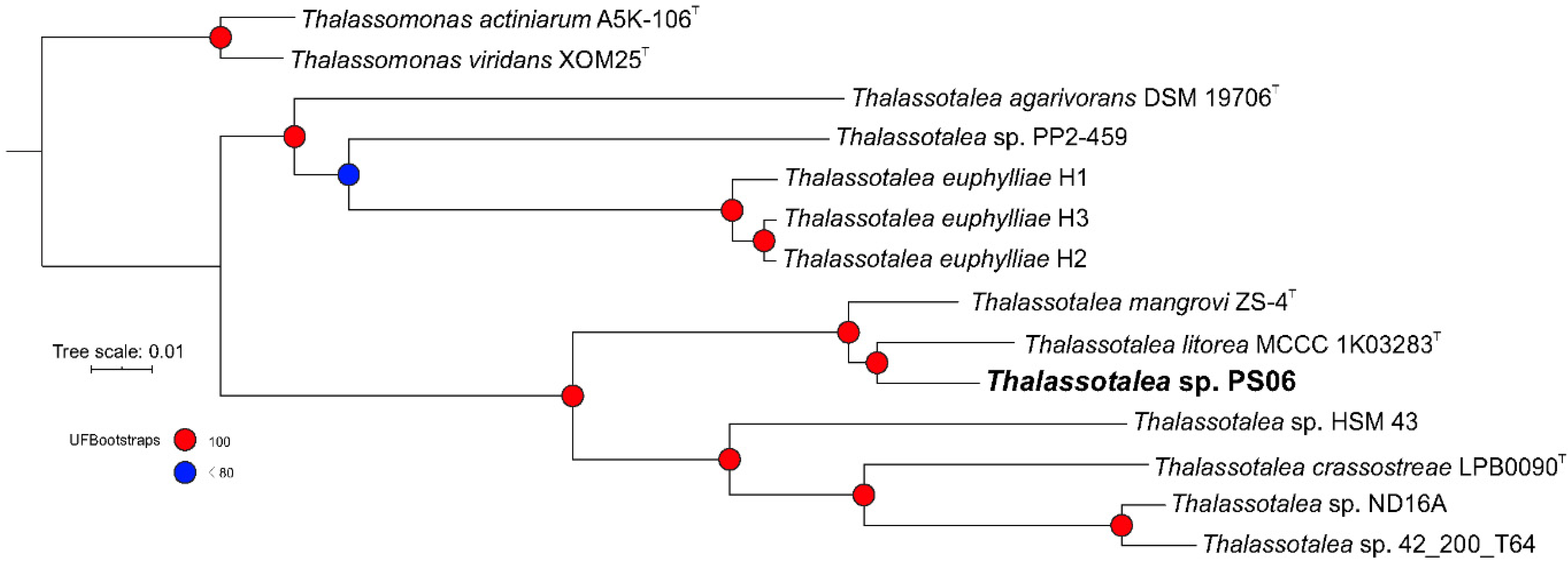
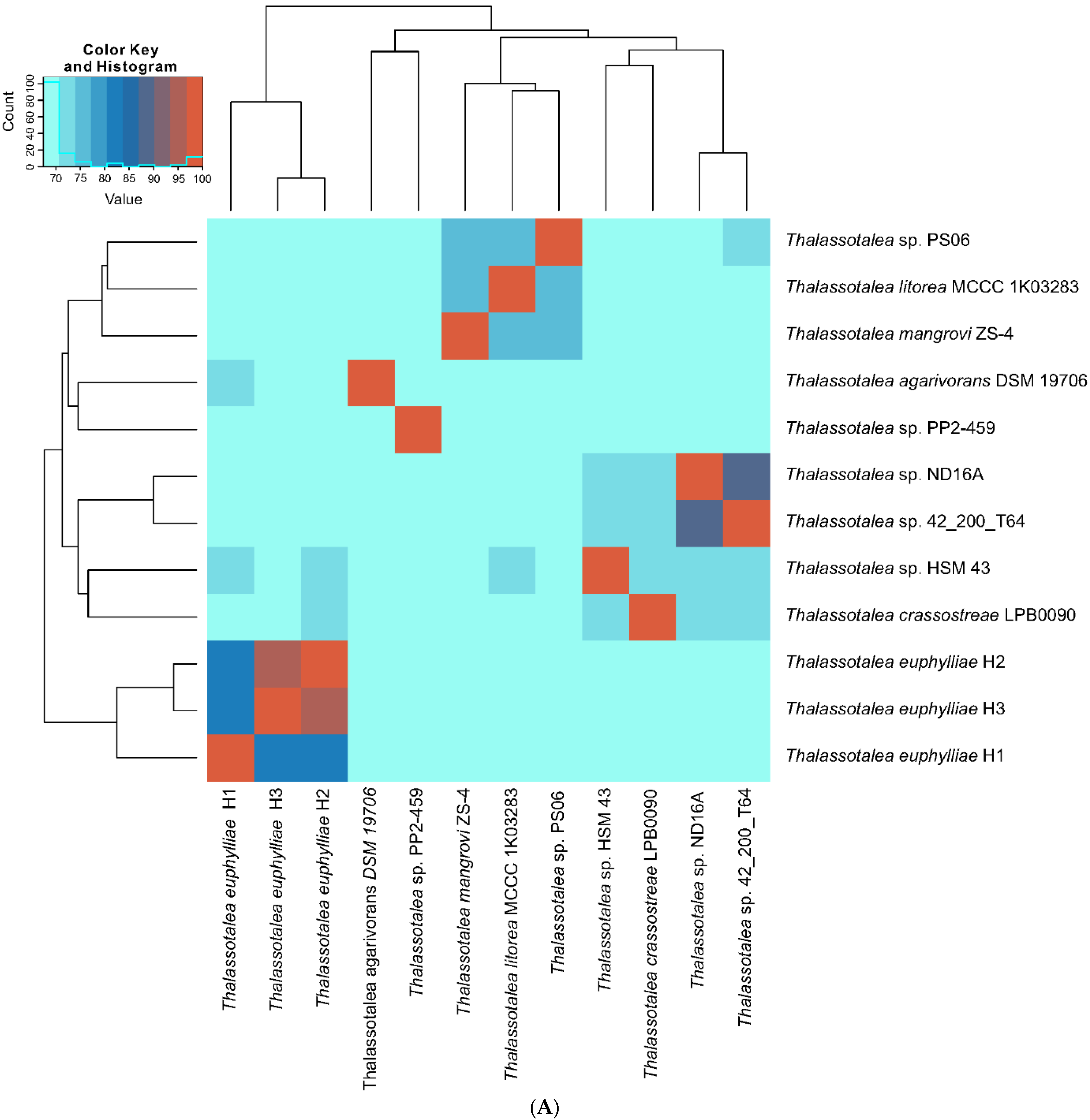
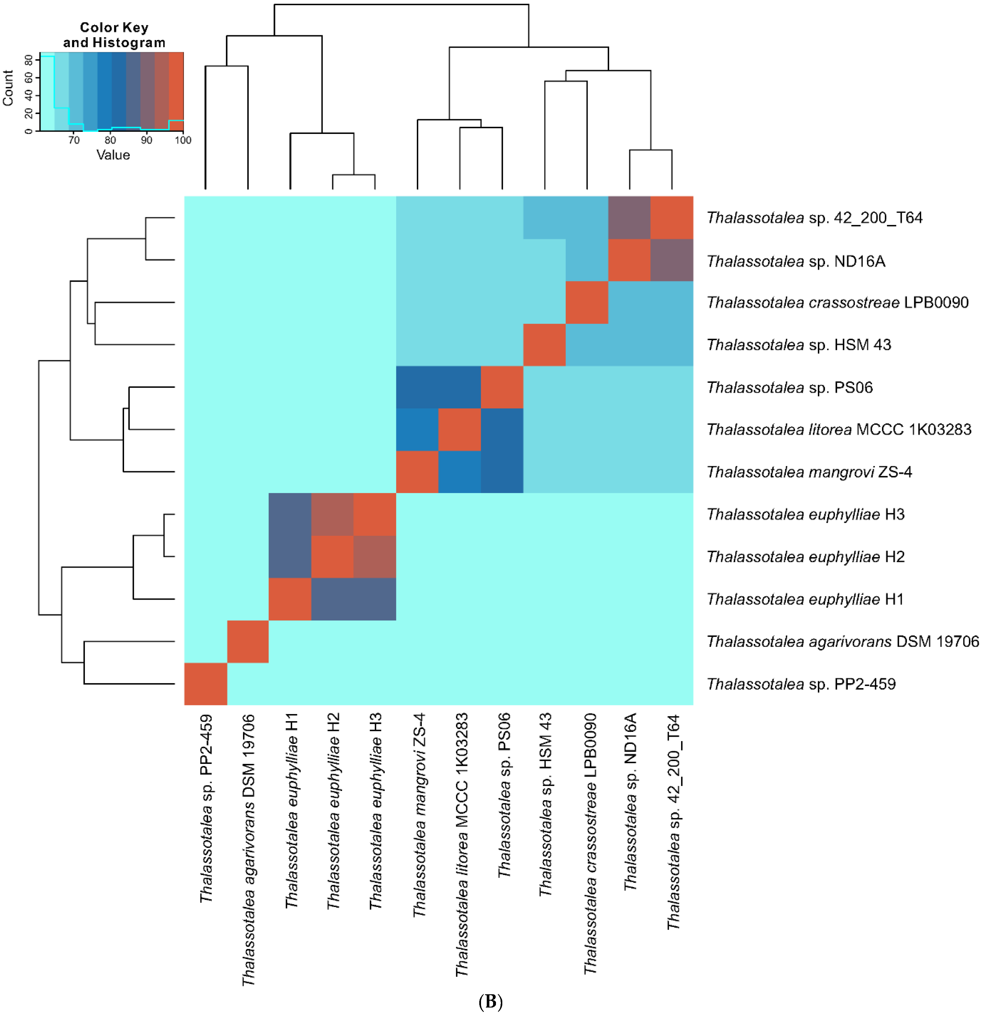
3.2. Phylogenetic and Phylogenomic Analyses

In a 16S rRNA gene sequence analysis, strain PS06 clustered with species belonging to the genus Thalassotalea (Figure S2). In a comparison of the 16S rRNA gene sequence extracted from the whole-genome sequence and that obtained by Sanger sequencing, no differences were apparent.
According to information available on the EzBioCloud server, strain PS06 was most closely related to Thalassotalea litorea HMF4135 (98.3% 16S rRNA gene sequence similarity), Thalassotalea ponticola GJSW-36 (95.9%), Thalassotalea agarivorans TMA1 (95.4%), and Thalassotalea fusca G-MB1 (94.9%). In addition, the phylogenetic tree based on 16S rRNA gene sequences showed that T. litorea, T. ponticola, T. crassostreae, and strain PS06 formed a cluster that was well separated from other strains in the genus Thalassotalea, and these strains were isolated from South Korea [3,12,49]. Based on the phylogenomic tree (Figure 1), strain PS06 clustered with T. litorea and Thalassotalea mangrovi [4], and three genomes (T. euphylliae H1, H2, and H3) isolated from corals in Hawaii, USA, formed a single cluster [10,11,14]. This result indicated that some species of the genus Thalassotalea might have more genomic relatedness than sequence similarity in their 16S rRNA genes and occupy the same ecological riches based on evolutionary relationships [50,51].
Genome-wide ANI and AAI comparisons further delineated strain PS06 into a single cluster (Figure 2), consistent with the phylogenomic tree. ANI and AAI values for the comparison between PS06 and the most closely related described species, T. litorea, were 76.7% and 82.1%, respectively. In accordance with the guidelines described by Konstantinidis et al. (30), these ANI and AAI values clearly support the designation of strain PS06 as a novel candidate species in the genus Thalassotalea.

3.3. Comparative Genome Analysis of Thalassotalea Strains

We analyzed 12 genomes of members of the genus Thalassotalea, including four validated type strains (Table S1). Seven genomes were obtained from public databases, and strain PS06 was newly sequenced in this study. Most strains were isolated from marine environments, including marine invertebrates (e.g., Crassostrea gigas, Ruditapes decussatus, or corals). One MAG, Thalassotalea sp. 42_200_T64, was obtained from a late-stage hydrocarbon degradation mimic-enriched culture in a deep-sea oil plume (Deepwater Horizon) [5]. These 12 genomes were nearly complete, with an average completeness of 97.8%, contamination of 1.04%, and strain heterogeneity of 0.6% (see Table S1). The expected genome sizes ranged from 3.37 Mb for T. agarivorans DSM 19706 to 4.75 Mb for T. euphylliae H1. Only two strains (T. crassostreae LPB0090 and Thalassotalea sp. PP2-459) isolated from calm areas had low GC contents (38.8% and 38.3%, respectively), whereas other genomes had average GC contents of 43.3% (41.6–45.9%). This high variation in genome size and GC content suggests that the members of the genus Thalassotalea experience different environmental conditions and have distinct physiological abilities [52,53], despite the similar functional profiles based on COG categories (Figure S1).
In a genome-wide comparison of orthologous genes across 12 genomes of the genus Thalassotalea, we detected 5877 clusters, 4560 orthologous clusters (containing at least two species), and 1317 single-copy gene clusters. Contrary to a previous hypothesis (i.e., different strategies for environmental adaptation), most orthologous genes were shared among all genomes in the analysis. Additionally, 44 unique CDSs in the strain PS06 genome were grouped into four clusters: response to oxidative stress, DNA replication, nucleotide binding, and transferase activity for glycosyl groups. These results indicated that each strain in the genus has a unique survival strategy, beyond the observed physiological characteristics.

3.4. Central Metabolism, Substrate Utilizations, and Respiration

The PS06 genome contained complete central metabolic pathways (Table S3): glycolysis (Embden–Meyerhof pathway), gluconeogenesis, pyruvate oxidation, tricarboxylic acid (TCA) cycle, pentose phosphate pathway, PRPP biosynthesis, and Entner–Doudoroff pathway. In addition, gene repertoires for glycogen biosynthesis and degradation in the genome were complete. Based on a KEGG analysis, we also deduced that strain PS06 has alternative pathways for carbon, despite some missing genes involved in the glyoxylate cycle, anabolic pathway for C2 compounds, malonate semialdehyde pathway (aldehyde oxidation) [54], and d-galactonate degradation (i.e., DeLey–Doudoroff pathway) [55]. Genes in the DeLey–Doudoroff pathway have been identified in algal polysaccharide-degrading marine microorganisms [56,57]. Although strain PS06 did not use d-galactose as a growth substrate, it might be able to use it for ascorbate biosynthesis [58,59].
To estimate substrate utilization, we screened transporters in the genome. Unexpectedly, although the strain has been recognized as a complete chemoheterotroph, the genome encoded few transporters of organic matter, such as lipoprotein (LolCED), phosphate (PstSCAB), capsular polysaccharide (KpsEMT), lipopolysaccharide (LptFGB), phospholipid (MlaCDEBF), and defensin (SapABCDF). The PS06 genome harbored transporters for inorganic matter, including molybdate (ModAB), iron (III) (AfuABC), sodium ion (NatBA), and heme [ccmDCBA (exporter)]. Based on the prediction for substrate utilization based on genomic traits, we evaluated physiological characteristics, as described previously [22]. Strain PS06 was able to utilize a limited number of substrates, including dextrin, d-trehalose, d-cellobiolose, and d-mannose (Table S4), which are related to some transport systems identified in the genome.
With respect to oxidative phosphorylation (i.e., ATP synthesis), the PS06 genome harbored genes necessary for aerobic respiration, including those for complex I (NADH:ubiquinone oxidoreductase), complex II (succinate dehydrogenase), and complex III (cytochrome bc1 complex). Genes encoding F-type ATPases were identified. Two types of terminal cytochrome oxidases were identified, complex IV: c type (CoxDCAB) and cbb3 type (CooNOQP). Additionally, the PS06 genome contained genes for other respiratory pathways. We found complete gene repertoires for assimilatory nitrate reduction (to ammonia, NapABCDEF and nirBD) and assimilatory sulfate reduction (to sulfide, cysNDCHJI) in the genome. The PS06 genome had a nitrate and nitrite transporter (NrtABCD). Also, only NasAB-encoding genes involved in assimilatory nitrate reduction were identified. However, genes coding for the dissimilatory nitrate reduction (denitrification and dissimilatory nitrate reduction to ammonia) were not identified in the PS06 genome, suggesting strain PS06 might be unable to be grown under anaerobic condition with inorganic electron acceptors (i.e., nitrate or nitrite) [60]. Moreover, anaerobic growth of the strain PS06 was not observed. In agreement with this, strain PS06 is likely capable of growth under high oxygen concentrations as well as microaerobic culture condition.

3.5. Metabolic Potentials

The PS06 genome contained few genes encoding transporters of cationic peptides, lipopolysaccharides, capsular polysaccharides, lipoproteins, vitamin B12, and phospholipids. Additionally, we found transporters for dissolved organic matter (DOM, cluster 3090 and 427) and siderophore/vitamins (cluster 410) in the PS06 genome based on the TBDT database [37], as well as in other genomes analyzed in this study (Table 1 and Data Set 2). These results suggest that members of the genus Thalassotalea play roles in marine nutrient cycling by the redistribution of DOM or inorganic compounds.
Moreover, strain PS06 can only grow on a complex medium supplied with yeast extract or peptone as a growth factor [61,62,63]. Consistent with this, the PS06 genome harbored few genes related to vitamin metabolism (i.e., biotin and tetrahydrofolate biosynthesis).
Two copies of the gene encoding poly-3-hydroxybutyrate (PHB) depolymerase were identified in the PS06 genome, despite the lack of genes involved in PHB biosynthesis. These results indicate that strain PS06 might contribute to the biodegradation of bio-polyester materials consisting of polyhydroxyalkanoic acids and utilize exogenous carbon or energy sources from extracellular PHB compounds [64,65].
As a branched polypeptide, cyanophycin (cyanophycin granule polypeptide, CGP) is an insoluble-reserve material of cyanobacteria [66] and non-cyanobacteria [67,68]. Interestingly, genes for type 1 glutamine amidotransferase (GATase1)-like domain and cyanophycinase (CphB) have been identified in the genome of the strain PS06 [69]. This result indicates that strain PS06 can use extracellular CGP as a valuable source of nitrogen, carbon, and energy [70,71], which would provide it with an ecological advantage over other bacteria in its habitat.

3.6. Repertoire of Carbohydrate-Active Enzymes (CAZymes) and Degrading Activities of the Genus Thalassotalea

The PS06 genome harbored various genes involved in carbohydrate metabolism, including genes encoding carbohydrate-active enzymes (CAZymes). The identified CAZymes provide insight into their lifestyles and potential applications in biomedicine or biotechnology [72,73]. CAZymes have been classified by following modules: glycoside hydrolases (GHs), glycosyltransferases (GTs), polysaccharide lyases (PLs), carbohydrate esterases (CEs), auxiliary activities (AAs), and carbohydrate-binding modules (CBMs). Also, bacterial CAZymes have been characterized for synthesis and degradation of polysaccharides as a rich source of bioactive compounds in algae including seaweeds and recognized as an eco-friendly enzyme for improving the yield and quality for derivatives of algal polysaccharides [73,74]. Although a number of marine heterotrophic bacteria are involved in the degradation of various biopolymers including, polysaccharide degradation, little is known about the gene repertories of hydrolytic systems in the genus Thalassotalea. The Thalassotalea genomes, including PS06, had genes encoding CAZymes in various functional classes (Table 2 and Data Set 2), as well as TonB-dependent transporters (TBDTs). In particular, GHs, GTs, and CEs were the most abundant CAZymes in all taxa analyzed in this study, suggesting that the members of the genus Thalassotalea have the ability to degrade the surface matrix of hosts (e.g., sponges and algae) [75]. Sixty-nine GH family genes (classified by DIAMOND with the removal of other families) were identified in all genomes. Most Thalassotalea genomes shared GH family genes (43 of 69 classes, n < 2 overlapping GH genes between genomes), indicating a lack of positive selection [76]. Nevertheless, the identification of unique GH families suggests specific adaptation for carbohydrate utilization and degradation according to habitats [77].
Genes encoding PLs were found in some genomes, including that of strain PS06, although only four PL family members were identified (i.e., PL1, 6, 7, or 17). PLs are a class of cleavage enzymes for uronic acid-containing polysaccharides (e.g., alginate) by a β-elimination mechanism, producing unsaturated products [78]. Therefore, the PLs of the Thalassotalea spp. could be used as highly active and specific enzymes for energy and chemical production via glycan saccharification [79]. Taken together, these enzymes for polysaccharide degradation of the Thalassotalea spp. might receive exceptionally attention to their biotechnological or medical potentials.
In addition, members of the genus Thalassotalea have the ability to hydrolyze polymer materials, such as agar, starch, casein, gelatin, DNA, Tweens, or chitin (Data Set 1). We confirmed that PS06 hydrolyzes gelatin, starch, and Tween 40 and 80, and shows positive activity for esterase (C4), esterase lipase (C8), lipase (C14), valine arylamidase, trypsin, acid phosphatase, Naphtol-AS-BI-phosphohydrolase, β-glucosidase, and N-acetyl-β-glucosaminidase. These results were supported by the identification of genes involved in carbohydrate assimilation and peptidase in the 12 genomes included in the study (Table 2 and Data Set 2). Therefore, these results provide further evidence that strain PS06 acts as a recycler for exogenous polymers, including detrital proteins, in marine environments. Additionally, genes involved in the twin-arginine translocation (Tat) system (tatABC) for protein translocation across the cytoplasmic membrane were clearly identified in the genome [80,81].

3.7. Environmental Stress and Adaptation

We determined the growth conditions according to previously described methods [22]. We found that strain PS06 has general mesophilic features, with growth temperatures ranging from 5 °C to 40 °C (optimal, 30 °C). Strain PS06 was an alkaliphile and moderate halophile based on growth at pH 7 to 10 (optimal, pH 9) and 1% to 10% w/v NaCl (optimal, 5%). As we mentioned above, the strain PS06 grew under aerobic and microaerobic culture conditions. Accordingly, we screened the genome for genes related to reactive oxygen species (ROS) stress, pH homeostasis, and osmoregulation.
The activity for catalase and oxidase was observed in strain PS06. In addition, enzymes involved in ROS stress (e.g., superoxide dismutase, glutathione peroxidase, putative iron-dependent peroxidase, and thiol peroxidase) were identified its genome. Generally, halophiles and/or alkaliphiles have genes encoding a vacuolar ATP synthase (V0V1-type, Ntp), multisubunit electrogenic sodium/proton antiporter (Mrp), compatible solutes (e.g., trehalose, ectoine, glycine betaine), osmoprotectant transport system (Opu), or cation/proton antiporter (CPA) superfamily (reviewed in [82]). Multisubunit Na+/H+ (Mnh), Ca2+/Na+, and Na+(K+)/H+ (Nha) antiporter genes were identified in the PS06 genome. Surprisingly, although this organism is a strict halophile, no genes involved in the synthesis of osmoprotection compounds (i.e., compatible solutes; trehalose, ectoine, and betaine) or osmoprotectant transporter genes (OpuCBDA) have been detected [83].
Additionally, the PS06 genome contained the complete set of genes for isoprenoid biosynthesis via the non-mevalonate pathway, even though full sets of genes in the pathway have only been reported in a few bacteria (reviewed in [84]). However, we did not find putative genes for squalene synthase (Hpn), which functions in the formation of squalene from its precursors [85]. These results indicated that strain PS06 might lack membrane properties for adaptation to haloalkaline environments, although the strain can grow in high-salt (up to 10%, w/v) or pH (up to 10) culture conditions. Nevertheless, the negatively charged cell membrane may be an alternative strategy for the survival of strain PS06 in haloalkaline environments [86,87].
Gene sets for flagellar biosynthesis and bacterial chemotaxis were lacking in the PS06 genome, whereas genes for Pil components essential for type IV pilus (Trp) assembly, tight adherence (Tad), and Flp (fimbrial low-molecular-weight protein) were found [88,89]. In addition, the genome contained genes for capsular polysaccharide biosynthesis (CPS), capsule polysaccharide export protein (kpsESC), and sugar phosphate isomerase (gutQ), involved in capsule formation and the type II secretion system (e.g., cell wall-degrading enzyme secretion), suggesting that PS06 has an advantage against competitors (e.g., prey) [90] or in harsh environments [91]. Additionally, using antiSMASH (version 5.1.0) [34] we attempted to identify putative antibiotic biosynthesis gene clusters. The PS06 genome had no genes related to secondary metabolite biosynthesis, whereas other genomes had a few secondary metabolite clusters, such as resorcinol, bacteriocin, hserlactone (homoserine lactone), ectoine, thiopeptide, or NRPS (non-ribosomal peptide synthetase cluster)-like (Table S5). Taken together, Thalassotalea spp. might have various strategies to cope with competitors in their habitats.
We further evaluated correlations between genomic traits and habitats or hosts. Generally, intergenic TRs are associated with various bacterial phenotypes (e.g., cell surface structure, motility, or restriction-modification systems) [92] related to survival in changing environments by genome flexibility. Most Thalassotalea genomes have hundreds of TRs (range 245 to 384, average 301), other than T. agarivorans (n = 80) and Thalassotalea sp. PP2-459 (n = 70). Although it is difficult to compare across genera or habitats, members of the genus Thalassotalea might have an advantage with respect to adaptation to environmental stresses. With respect to defense mechanisms against viral predation in the genomes, few clustered regularly interspaced short palindromic repeats (CRISPR) candidates and spacers were identified in the 11 genomes (Table S6). On the other hand, a number of CRISPR loci (n = 6) with spacers (n = 102) have been reported in the T. euphylliae H1 genome. The CRIPSR-Cas system has been identified in about half of all bacterial genomes as an adaptive immune system [93]. In addition, an array of repetitive and unique sequences (referred to as repeats and spacers, respectively) have evolved in their genetic relatedness. As mentioned above, except for the T. euphylliae H1 genome, most Thalassotalea genomes (n = 11) have a few CRISPR and spacers. This suggests that members of the genus Thalassotalea might be less subject to evolutionary (i.e., it is genomically stable) or phage infection stresses in their ecological niches [93,94,95].
To estimate the frequency of genomic rearrangements in Thalassotalea spp., we analyzed prophage regions [33]. Most genomes had incomplete or no prophages, whereas two questionable prophage regions were identified in the T. euphylliae H1 genome (Table S7). Taken together, the long-term evolutionary pressure on genomes of Thalassotalea spp. might be low, despite the lack of an efficient defense mechanism. This hypothesis should be evaluated using more isolates and genomes in the future.

4. Conclusions

Physiological and genomic analyses indicated that strain PS06 is capable of surviving under haloalkaline environments by alternative strategies in various marine environments. This inference is supported by the detection of TRs and genes encoding extracellular enzymes. In fact, the metabolic potential of PS06 suggests that the strain is an obligate chemoheterotroph (requiring complex organic compounds as an energy source, e.g., peptone or yeast extract), although it also harbors a limited set of genes for substrate transporters. In addition, Thalassotalea spp. including strain PS06, can degrade macroalgal-derived polysaccharides and contribute to nutrient cycling in marine environments. In addition, the ability for polysaccharide degradation of the genus Thalassotalea suggest an application to biotechnology or medicine. Finally, our results demonstrate that members of the genus Thalassotalea might have diverse metabolic pathways and ecological strategies in haloalkaline ecosystems.

Supplementary Materials

The following are available online at https://www.mdpi.com/2076-2607/8/9/1412/s1. Figure S1. Distribution of clusters of orthologous groups (COGs) for loci in PS06 and other genomes. The x-axis represents the percentage of genes in each COG category. Bars indicate COG functional classes. Figure S2. Phylogenetic tree inferred by the maximum likelihood method and Kimura 2-parameter model based on 16S rRNA gene sequences showing the phylogenetic relationships among strain PS06 and related strains, including members of the genus Thalassotalea and closely related taxa. The sequence of Halomonas alkaliantarctica CRSS (AJ564880) was used as the outgroup. The GenBank accession numbers are given in parentheses. The tree is drawn to scale, with branch lengths proportionate to the number of substitutions per site. This analysis involved 37 nucleotide sequences and 1473 total sites. Evolutionary analyses were conducted using MEGA X. Table S1. Summary statistics, including genomic completeness determined by CheckM, and detailed information about geographical locations and sites of strains included in this study. Table S2. TIGRfams markers used for the phylogenomic analysis. Table S3. Kyoto Encyclopedia of Genes and Genomes (KEGG) orthology annotation for PS06 and other genomes. Presence/absence information for listed KO numbers is shown for each individual MAG in columns F:Q (1 indicates present and 0 indicates absent). Table S4. Physiological characteristics (enzyme activity and substrate utilization) of strain PS06 determined by GEN III (Biolog) and API ZYM. Table S5. Predicted secondary metabolite clusters, as determined by antiSMASH5. All secondary metabolite clusters predicted for PS06 and other genomes. Table S6. Clustered regularly interspaced short palindromic repeat (CRISPR)-associated genes identified in PS06 and other genomes. Table S7. Questionable prophage regions identified in PS06 and other genomes.

Author Contributions

E.-Y.L. and S.-J.P. conceived and designed the experiments. M.K., I.-T.C., and S.-J.P. performed the experiments. K.-E.L. and E.-Y.L. contributed to sample preparation. M.K., I.-T.C., K.-E.L., and S.-J.P. contributed to the interpretation of the results. S.-J.P. took the lead in writing the manuscript. All authors have read and agreed to the published version of the manuscript.

Funding

This work was supported by grants from the National Research Foundation of Korea (2018R1C1B6006762 and 2020R1I1A3062110) and National Institute of Biological Resources funded by the Ministry of Environment (NIBR202002108).

Acknowledgments

In this section you can acknowledge any support given which is not covered by the author contribution or funding sections. This may include administrative and technical support, or donations in kind (e.g., materials used for experiments).

Conflicts of Interest

The authors declare no conflict of interest. The funders had no role in the design of the study; in the collection, analyses, or interpretation of data; in the writing of the manuscript; or in the decision to publish the results.

References

  1. Zhang, Y.; Tang, K.; Xiaochong, S.; Zhang, X.H. Description of Thalassotalea pisci.um Gen. Nov., sp. Nov., Isolated from Flounder (Paralichthys olivaceus), Reclassification of Four Species of the Genus Thalassomonas as Members of the Genus Thalassotalea Gen. Nov. and Emended Description of the Genus Thalassomonas. Int. J. Syst. Evol. Microbiol. 2014, 64, 1223–1228. [Google Scholar] [PubMed] [Green Version]
  2. Hou, T.T.; Liu, Y.; Zhong, Z.P.; Liu, H.C.; Liu, Z.P. Thalassotalea marina sp. Nov., Isolated from a Marine Recirculating Aquaculture System, Reclassification of Thalassomonas eurytherma as Thalassotalea eurytherma Comb. Nov. and Emended Description of the Genus Thalassotalea. Int. J. Syst. Evol. Microbiol. 2015, 65, 4710–4715. [Google Scholar] [CrossRef] [PubMed]
  3. Park, S.; Jung, Y.T.; Kang, C.H.; Park, J.M.; Yoon, J.H. Thalassotalea ponticola sp. Nov., Isolated from Seawater, Reclassification of Thalassomonas fusca as Thalassotalea Fusca Comb. Nov. And Emended Description of the Genus Thalassotalea. Int. J. Syst. Evol. Microbiol. 2014, 64, 3676–3682. [Google Scholar] [CrossRef] [PubMed]
  4. Zheng, S.; Zhang, D.; Gui, J.; Wang, J.; Zhu, X.; Lai, Q.; Wang, H.; Xu, H. Thalassotalea mangrovi sp. Nov., a Bacterium Isolated from Marine Mangrove Sediment. Int. J. Syst. Evol. Microbiol. 2019, 69, 3644–3649. [Google Scholar] [CrossRef] [PubMed]
  5. Hu, P.; Dubinsky, E.A.; Probst, A.J.; Wang, A.J.; Sieber, C.M.K.; Tom, L.M.; Gardinali, P.R.; Banfield, J.F.; Atlas, R.M.; Andersen, G.L. Simulation of Deepwater Horizon Oil Plume Reveals Substrate Specialization within a Complex Community of Hydrocarbon Degraders. Proc. Natl. Acad. Sci. USA 2017, 114, 7432–7437. [Google Scholar] [CrossRef] [Green Version]
  6. Stelling, S.C.; Techtmann, S.M.; Utturkar, S.M.; Alshibli, N.K.; Brown, S.D.; Hazen, T.C. Draft Genome Sequence of Thalassotalea sp. Strain Nd16a Isolated from Eastern Mediterranean Sea Water Collected from a Depth of 1055 Meters. Genome Announc. 2014, 2, 01231-14. [Google Scholar] [CrossRef] [Green Version]
  7. Dethlefsen, L.; Schmidt, T.M. Performance of the Translational Apparatus Varies with the Ecological Strategies of Bacteria. J. Bacteriol. 2007, 189, 3237–3245. [Google Scholar] [CrossRef] [Green Version]
  8. Espejo, R.T.; Plaza, N. Multiple Ribosomal Rna Operons in Bacteria; Their Concerted Evolution and Potential Consequences on the Rate of Evolution of Their 16s Rrna. Front. Microbiol. 2018, 9, 1232. [Google Scholar] [CrossRef]
  9. Acinas, S.G.; Marcelino, L.A.; Klepac-Ceraj, V.; Polz, M.F. Divergence and Redundancy of 16s Rrna Sequences in Genomes with Multiple Rrn Operons. J. Bacteriol. 2004, 186, 2629–2635. [Google Scholar] [CrossRef] [PubMed] [Green Version]
  10. Summers, S.; Freckelton, M.L.; Nedved, B.T.; Rice, S.A.; Hadfield, M.G. Full-Genome Sequence of Thalassotalea euphylliae H1, Isolated from a Montipora capitata Coral Located in Hawai’i. Microbiol. Resour. Announc. 2018, 7. [Google Scholar] [CrossRef] [Green Version]
  11. Summers, S.; Freckelton, M.L.; Nedved, B.T.; Rice, S.A.; Hadfield, M.G. Complete Genome Sequence of Thalassotalea euphylliae Strain H2. Microbiol. Resour. Announc. 2019, 8. [Google Scholar] [CrossRef] [PubMed] [Green Version]
  12. Choi, S.; Kim, E.; Shin, S.K.; Yi, H. Thalassotalea crassostreae sp. Nov., Isolated from Pacific Oyster. Int. J. Syst. Evol. Microbiol. 2017, 67, 2195–2198. [Google Scholar] [CrossRef] [PubMed]
  13. Sheu, D.S.; Sheu, S.Y.; Xie, P.B.; Tang, S.L.; Chen, W.M. Thalassotalea coralli sp. Nov., Isolated from the Torch Coral Euphyllia Glabrescens. Int. J. Syst. Evol. Microbiol. 2018, 68, 185–191. [Google Scholar] [CrossRef]
  14. Sheu, S.Y.; Liu, L.P.; Tang, S.L.; Chen, W.M. Thalassotalea euphylliae sp. Nov., Isolated from the Torch Coral Euphyllia glabrescens. Int. J. Syst. Evol. Microbiol. 2016, 66, 5039–5045. [Google Scholar] [CrossRef] [PubMed]
  15. Thompson, F.L.; Barash, Y.; Sawabe, T.; Sharon, G.; Swings, J.; Rosenberg, E. Thalassomonas loyana sp. Nov., a Causative Agent of the White Plague-Like Disease of Corals on the Eilat Coral Reef. Int. J. Syst. Evol. Microbiol. 2006, 56, 365–368. [Google Scholar] [CrossRef] [Green Version]
  16. Leal, M.C.; Sheridan, C.; Osinga, R.; Dionisio, G.; Rocha, R.J.; Silva, B.; Rosa, R.; Calado, R. Marine Microorganism-Invertebrate Assemblages: Perspectives to Solve the Supply Problem in the Initial Steps of Drug Discovery. Mar. Drugs 2014, 12, 3929–3952. [Google Scholar] [CrossRef] [Green Version]
  17. Parks, D.H.; Rinke, C.; Chuvochina, M.; Chaumeil, P.A.; Woodcroft, B.J.; Evans, P.M.; Hugenholtz, P.; Tyson, G.W. Recovery of Nearly 8,000 Metagenome-Assembled Genomes Substantially Expands the Tree of Life. Nat. Microbiol. 2017, 2, 1533–1542. [Google Scholar] [CrossRef]
  18. Parks, D.H.; Chuvochina, M.; Waite, D.W.; Rinke, C.; Skarshewski, A.; Chaumeil, P.A.; Hugenholtz, P. A Standardized Bacterial Taxonomy Based on Genome Phylogeny Substantially Revises the Tree of Life. Nat. Biotechnol. 2018, 36, 996–1004. [Google Scholar] [CrossRef]
  19. Parks, D.H.; Chuvochina, M.; Chaumeil, P.A.; Rinke, C.; Mussig, A.J.; Hugenholtz, P. A Complete Domain-to-Species Taxonomy for Bacteria Archaea. Nat. Biotechnol. 2020, 38, 1079–1086. [Google Scholar] [CrossRef]
  20. Reddy, C.A.; Beveridge, T.J.; Breznak, J.A.; Marzluf, G.A.; Schmidt, T.M.; Snyder, L.R. Methods for General and Molecular Microbiology, 3th ed.; American Society of Microbiology: Washington, DC, USA, 2007. [Google Scholar]
  21. Lane, D.J.; Pace, B.; Olsen, G.J.; Stahl, D.A.; Sogin, M.L.; Pace, L.R. Rapid Determination of 16s Ribosomal Rna Sequences for Phylogenetic Analyses. Proc. Natl. Acad. Sci. USA 1985, 82, 6955–6959. [Google Scholar] [CrossRef] [Green Version]
  22. Koh, H.W.; Rani, S.; Kim, S.J.; Moon, E.; Nam, S.Q.; Rhee, S.K.; Park, S.J. Halomonas Aestuarii Sp. Nov., a Moderately Halophilic Bacterium Isolated from a Tidal Flat. Int. J. Syst. Evol. Microbiol. 2017, 67, 4298–4303. [Google Scholar] [CrossRef] [PubMed]
  23. Hall, A.; Tom, A. Bioedit: A User-Friendly Biological Sequence Alignment Editor and Analysis Program for Windows 95/98/Nt. Nucleic Acids Symp. Ser. 1999, 41, 95–98. [Google Scholar]
  24. Thompson, J.D.; Gibson, T.J.; Plewniak, F.; Jeanmougin, F.; Higgins, D.G. The Clustal_X Windows Int.erface: Flexible Strategies for Multiple Sequence Alignment Aided by Quality Analysis Tools. Nucleic Acids Res. 1997, 25, 4876–4882. [Google Scholar] [CrossRef] [Green Version]
  25. Kimura, M. The Neutral Theory of Molecular Evolution and the World View of the Neutralists. Genome 1989, 31, 24–31. [Google Scholar] [CrossRef] [PubMed]
  26. Felsenstein, J. Evolutionary Trees from DNA Sequences: A Maximum Likelihood Approach. J. Mol. Evol. 1981, 17, 368–376. [Google Scholar] [CrossRef]
  27. Kumar, S.; Stecher, G.; Li, M.; Knyaz, C.; Tamura, K. Mega X: Molecular Evolutionary Genetics Analysis across Computing Platforms. Mol. Biol. Evol. 2018, 35, 1547–1549. [Google Scholar] [CrossRef]
  28. Felsenstein, J. Confidence Limits on Phylogenies: An Approach Using the Bootstrap. Evolution 1985, 39, 783–791. [Google Scholar] [CrossRef]
  29. Hyatt, D.; Chen, G.L.; Locascio, P.F.; Land, M.L.; Larimer, F.W.; Hauser, L.J. Prodigal: Prokaryotic Gene Recognition and Translation Initiation Site Identification. BMC Bioinform. 2010, 11, 119. [Google Scholar] [CrossRef] [Green Version]
  30. Lowe, T.M.; Eddy, S.R. Trnascan-Se: A Program for Improved Detection of Transfer Rna Genes in Genomic Sequence. Nucleic Acids Res. 1997, 25, 955–964. [Google Scholar] [CrossRef]
  31. Koh, H.W.; Hur, M.; Kang, M.S.; Ku, Y.B.; Ghai, R.; Park, S.J. Physiological and Genomic Insights into the Lifestyle of Arsenite-Oxidizing Herminiimonas Arsenitoxidans. Sci. Rep. 2017, 7, 15007. [Google Scholar] [CrossRef] [Green Version]
  32. Bulzu, P.A.; Andrei, A.S.; Salcher, M.M.; Mehrshad, M.; Inoue, K.; Kandori, H.; Beja, O.; Ghai, R.; Banciu, H.L. Casting Light on Asgardarchaeota Metabolism in a Sunlit Microoxic Niche. Nat. Microbiol. 2019, 4, 1129–1137. [Google Scholar] [CrossRef] [PubMed] [Green Version]
  33. Yu, W.J.; Lee, J.W.; Nguyen, N.L.; Rhee, S.K.; Park, S.L. The Characteristics and Comparative Analysis of Methanotrophs Reveal Genomic Insights into Methylomicrobium sp. Enriched from Marine Sediments. Syst. Appl. Microbiol. 2018, 41, 415–426. [Google Scholar] [CrossRef] [PubMed]
  34. Blin, K.; Shaw, S.; Steinke, K.; Villebro, R.; Ziemert, N.; Lee, S.Y.; Medema, M.H.; Weber, T. Antismash 5.0: Updates to the Secondary Metabolite Genome Mining Pipeline. Nucleic Acids Res. 2019, 47, W81–W87. [Google Scholar] [CrossRef] [Green Version]
  35. Yin, Y.; Mao, X.; Yang, J.; Chen, X.; Mao, F.; Xu, Y. Dbcan: A Web Resour.ce for Automated Carbohydrate-Active Enzyme Annotation. Nucleic Acids Res. 2012, 40, 445–451. [Google Scholar] [CrossRef]
  36. Parks, D.H.; Imelfort, M.; Skennerton, C.T.; Hugenholtz, P.; Tyson, G.W. Checkm: Assessing the Quality of Microbial Genomes Recovered from Isolates, Single Cells, and Metagenomes. Genome Res. 2015, 25, 1043–1055. [Google Scholar] [CrossRef] [PubMed] [Green Version]
  37. Tang, K.; Jiao, N.; Liu, K.; Zhang, Y.; Li, S. Distribution and Functions of Tonb-Dependent Transporters in Marine Bacteria and Environments: Implications for Dissolved Organic Matter Utilization. PLoS ONE 2012, 7, e41204. [Google Scholar] [CrossRef] [Green Version]
  38. Gelfand, Y.; Rodriguez, A.; Benson, G. Trdb-the Tandem Repeats Database. Nucleic Acids Res. 2007, 35, 80–87. [Google Scholar] [CrossRef]
  39. Xu, L.; Dong, Z.; Fang, L.; Luo, Y.; Wei, Z.; Guo, H.; Zhang, G.; Gu, Y.Q.; Coleman-Derr, D.; Xia, Q.; et al. Orthovenn2: A Web Server for Whole-Genome Comparison and Annotation of Orthologous Clusters across Multiple Species. Nucleic Acids Res. 2019, 47, W52–W58. [Google Scholar] [CrossRef] [Green Version]
  40. Arndt, D.; Grant, J.R.; Marcu, A.; Sajed, T.; Pon, A.; Liang, Y.; Wishart, D.S. Phaster: A Better, Faster Version of the Phast Phage Search Tool. Nucleic Acids Res. 2016, 44, 16–21. [Google Scholar] [CrossRef] [Green Version]
  41. Loytynoja, A. Phylogeny-Aware Alignment with Prank. Methods Mol. Biol 2014, 1079, 155–170. [Google Scholar]
  42. Criscuolo, A.; Gribaldo, S. Bmge (Block Mapping and Gathering with Entropy): A New Software for Selection of Phylogenetic Informative Regions from Multiple Sequence Alignments. BMC Evol. Biol 2010, 10, 210. [Google Scholar] [CrossRef] [PubMed] [Green Version]
  43. Susko, E.; Roger, A.J. On Reduced Amino Acid Alphabets for Phylogenetic Inference. Mol. Biol Evol. 2007, 24, 2139–2150. [Google Scholar] [CrossRef] [PubMed] [Green Version]
  44. Nguyen, L.T.; Schmidt, H.A.; Von Haeseler, A.; Minh, B.Q. Iq-Tree: A Fast and Effective Stochastic Algorithm for Estimating Maximum-Likelihood Phylogenies. Mol. Biol Evol. 2015, 32, 268–274. [Google Scholar] [CrossRef] [PubMed]
  45. Kalyaanamoorthy, S.; Minh, B.Q.; Wong, T.K.F.; Von Haeseler, A.; Jermiin, S.L. Modelfinder: Fast Model Selection for Accurate Phylogenetic Estimates. Nat. Methods 2017, 14, 587–589. [Google Scholar] [CrossRef] [Green Version]
  46. Hoang, D.T.; Chernomor, O.; Von Haeseler, A.; Minh, B.Q.; Vinh, S.L. Ufboot2: Improving the Ultrafast Bootstrap Approximation. Mol. Biol Evol. 2018, 35, 518–522. [Google Scholar] [CrossRef]
  47. Goris, J.; Konstantinidis, K.T.; Klappenbach, J.A.; Coenye, T.; Vandamme, P.; Tiedje, J.M. DNA-DNA Hybridization Values and Their Relationship to Whole-Genome Sequence Similarities. Int. J. Syst. Evol. Microbiol. 2007, 57, 81–91. [Google Scholar] [CrossRef] [Green Version]
  48. Konstantinidis, K.T.; Rossello-Mora, R.; Amann, R. Uncultivated Microbes in Need of Their Own Taxonomy. ISME J. 2017, 11, 2399–2406. [Google Scholar] [CrossRef]
  49. Kang, H.; Kim, H.; Nam, Y.I.; Joung, Y.; Jang, T.Y.; Joh, K. Thalassotalea litorea sp. Nov., Isolated from Seashore Sand. Int. J. Syst. Evol. Microbiol. 2017, 67, 2268–2273. [Google Scholar] [CrossRef]
  50. Kapli, P.; Yang, Z.; Telford, M.J. Phylogenetic Tree Building in the Genomic Age. Nat. Rev. Genet. 2020, 21, 428–444. [Google Scholar] [CrossRef]
  51. Zhu, Q.; Mai, U.; Pfeiffer, W.; Janssen, S.; Asnicar, F.; Sanders, J.G.; Belda-Ferre, P.; Al-Ghalith, G.A.; Kopylova, E.; McDonald, D.; et al. Phylogenomics of 10,575 Genomes Reveals Evolutionary Proximity between Domains Bacteria and Archaea. Nat. Commun. 2019, 10, 5477. [Google Scholar] [CrossRef] [Green Version]
  52. Almpanis, A.; Swain, M.; Gatherer, D.; McEwan, N. Correlation between Bacterial G+C Content, Genome Size and the G+C Content of Associated Plasmids and Bacteriophages. Microb. Genom. 2018, 4, e000168. [Google Scholar] [CrossRef] [PubMed]
  53. Zhang, S.; Song, W.; Yu, M.; Lin, X. Comparative Genomics Analysis of Five Psychrobacter Strains Isolated from World-Wide Habitats Reveal High Intra-Genus Variations. Extremophiles 2017, 21, 581–589. [Google Scholar] [CrossRef] [PubMed]
  54. Yamada, E.W.; Jakoby, W.B. Aldehyde Oxidation. V. Direct Conversion of Malonic Semialdehyde to Acetyl-Coenzyme, A. J. Biol Chem. 1960, 235, 589–594. [Google Scholar] [PubMed]
  55. De Ley, J.; Doudoroff, M. The Metabolism of D-Galactose in Pseudomonas saccharophila. J. Biol Chem. 1957, 227, 745–757. [Google Scholar] [PubMed]
  56. Gobet, A.; Barbeyron, T.; Matard-Mann, M.; Magdelenat, G.; Vallenet, D.; Duchaud, E.; Michel, G. Evolutionary Evidence of Algal Polysaccharide Degradation Acquisition by Pseudoalteromonas carrageenovora 9t to Adapt to Macroalgal Niches. Front. Microbiol. 2018, 9, 2740. [Google Scholar] [CrossRef] [Green Version]
  57. Christiansen, L.; Pathiraja, D.; Bech, P.K.; Schultz-Johansen, M.; Hennessy, R.; Teze, D.; Choi, I.C.; Stougaard, P. A Multifunctional Polysaccharide Utilization Gene Cluster in Colwellia echini Encodes Enzymes for the Complete Degradation of K-Carrageenan, I-Carrageenan, and Hybrid B/K-Carrageenan. MSphere 2020, 5. [Google Scholar] [CrossRef] [Green Version]
  58. Ishikawa, T.; Nishikawa, H.; Gao, Y.; Sawa, Y.; Shibata, H.; Yabuta, Y.; Maruta, T.; Shigeoka, S. The Pathway Via D-Galacturonate/L-Galactonate Is Significant for Ascorbate Biosynthesis in Euglena gracilis. J. Biol Chem. 2008, 283, 31133–31141. [Google Scholar] [CrossRef] [Green Version]
  59. Kuivanen, J.; Penttila, M.; Richard, P. Metabolic Engineering of the Fungal D-Galacturonate Pathway for L-Ascorbic Acid Production. Microb. Cell Fact. 2015, 14, 1–9. [Google Scholar] [CrossRef] [Green Version]
  60. Durand, A.; Bourbon, M.L.; Steunou, A.S.; Khalfaoui-Hassani, B.; Legrand, C.; Guitton, A.; Astier, C.; Ouchane, S. Biogenesis of the Bacterial Cbb3 Cytochrome C Oxidase: Active Subcomplexes Support a Sequential Assembly Model. J. Biol Chem. 2018, 293, 808–818. [Google Scholar] [CrossRef] [Green Version]
  61. Batista, K.A.; Bataus, L.A.M.; Ivan, T.N.C.; Fernandes, K.F. Development of Culture Medium Using Extruded Bean as a Nitrogen Source for Yeast Growth. J. Microbiol. Methods 2013, 92, 310–315. [Google Scholar] [CrossRef] [Green Version]
  62. Nancib, N.; Branlant, C.; Boudrant, J. Metabolic Roles of Peptone Yeast Extract for the Culture of a Recombinant Strain Ofescherichia Coli. J. Ind. Microbiol. 1991, 8, 165–169. [Google Scholar] [CrossRef] [PubMed]
  63. Plugge, C.M. Anoxic Media Design, Preparation, Considerations. In Methods in Enzymology; Academic Press: Cambridge, MA, USA, 2005; pp. 3–16. [Google Scholar]
  64. Mathuriya, A.S.; Yakhmi, J.V. Polyhydroxyalkanoates: Biodegradable Plastics Their Applications. In Handbook of Ecomaterials; Martínez, L.M.T., Kharissova, O.V., Kharisov, B.I., Eds.; Springer International Publishing: Cham, Switzerland, 2019; pp. 2873–2900. [Google Scholar]
  65. Jendrossek, D. Microbial Degradation of Polyesters. In Biopolyesters; Steinbüchel, W.B.A., Ed.; Springer: Berlin/Heidelberg, Germany, 2001; pp. 293–325. [Google Scholar]
  66. Simon, R.D.; Weathers, P. Determination of the Structure of the Novel Polypeptide Containing Aspartic Acid and Arginine Which Is Found in Cyanobacteria. Biochim. Biophys. Acta Protein Struct. 1976, 420, 165–176. [Google Scholar] [CrossRef]
  67. Krehenbrink, M.; Oppermann-Sanio, F.B.; Steinbuchel, A. Evaluation of Non-Cyanobacterial Genome Sequences for Occurrence of Genes Encoding Proteins Homologous to Cyanophycin Synthetase and Cloning of an Active Cyanophycin Synthetase from Acinetobacter Sp. Strain Dsm 587. Arch. Microbiol. 2002, 177, 371–380. [Google Scholar] [CrossRef]
  68. Ziegler, K.; Deutzmann, R.; Lockau, W. Cyanophycin Synthetase-Like Enzymes of Non-Cyanobacterial Eubacteria: Characterization of the Polymer Produced by a Recombinant Synthetase of Desulfitobacterium hafniense. Z. Nat. C 2002, 57, 522–529. [Google Scholar] [CrossRef]
  69. Obst, M.; Steinbuchel, A. Microbial Degradation of Poly(Amino Acid)S. Biomacromolecules 2004, 5, 1166–1176. [Google Scholar] [CrossRef]
  70. Obst, M.; Krug, A.; Luftmann, H.; Steinbuchel, A. Degradation of Cyanophycin by Sedimentibacter hongkongensis Strain Ki and Citrobacter amalonaticus Strain G Isolated from an Anaerobic Bacterial Consortium. Appl. Environ. Microbiol. 2005, 71, 3642–3652. [Google Scholar] [CrossRef] [PubMed] [Green Version]
  71. Obst, M.; Oppermann-Sanio, F.B.; Luftmann, H.; Steinbuchel, A. Isolation of Cyanophycin-Degrading Bacteria, Cloning Characterization of an Extracellular Cyanophycinase Gene (Cphe) from Pseudomonas anguilliseptica Strain Bi. J. Biol Chem. 2002, 277, 25096–25105. [Google Scholar] [CrossRef] [PubMed] [Green Version]
  72. Lapébie, P.; Lombard, V.; Drula, E.; Terrapon, N.; Henrissat, B. Bacteroidetes Use Thousands of Enzyme Combinations to Break Down Glycans. Nat. Commun. 2019, 10, 2043. [Google Scholar] [CrossRef]
  73. Chernysheva, N.; Bystritskaya, E.; Stenkova, A.; Golovkin, I.; Nedashkovskaya, O.; Isaeva, M. Comparative Genomics and Cazyme Genome Repertoires of Marine Zobellia amurskyensis Kmm 3526t and Zobellia laminariae Kmm 3676t. Mar. Drugs 2019, 17, 661. [Google Scholar] [CrossRef] [Green Version]
  74. Xu, S.Y.; Huang, X.; Cheong, K.L. Recent Advances in Marine Algae Polysaccharides: Isolation, Structure, and Activities. Mar. Drugs 2017, 15, 388. [Google Scholar] [CrossRef] [Green Version]
  75. Siegl, A.; Kamke, J.; Hochmuth, T.; Piel, J.; Richter, M.; Liang, C.L.; Dandekar, T.; Hentschel, U. Single-Cell Genomics Reveals the Lifestyle of Poribacteria, a Candidate Phylum Symbiotically Associated with Marine Sponges. ISME J. 2011, 5, 61–70. [Google Scholar] [CrossRef] [PubMed]
  76. Page, R.D.M.; Holmes, E.C. Molecular Evolution: A Phylogenetic Approach; Wiley: Hoboke, NJ, USA, 2009. [Google Scholar]
  77. Alex, A.; Antunes, A. Genus-Wide Comparison of Pseudovibrio Bacterial Genomes Reveal Diverse Adaptations to Different Marine Invertebrate Hosts. PLoS ONE 2018, 13, e0194368. [Google Scholar] [CrossRef] [PubMed] [Green Version]
  78. Garron, M.L.; Cygler, M. Structural and Mechanistic Classification of Uronic Acid-Containing Polysaccharide Lyases. Glycobiology 2010, 20, 1547–1573. [Google Scholar] [CrossRef] [PubMed] [Green Version]
  79. MacDonald, L.C.; Weiler, E.B.; Berger, W. Engineering Broad-Spectrum Digestion of Polyuronides from an Exolytic Polysaccharide Lyase. Biotechnol. Biofuels 2016, 9, 43. [Google Scholar] [CrossRef] [Green Version]
  80. Frobel, J.; Rose, P.; Muller, M. Twin-Arginine-Dependent Translocation of Folded Proteins. Philos Trans. R. Soc. Lond. B Biol. Sci. 2012, 367, 1029–1046. [Google Scholar] [CrossRef] [Green Version]
  81. Frain, K.M.; Robinson, C.; Van Dijl, M. Transport of Folded Proteins by the Tat System. Protein J. 2019, 38, 377–388. [Google Scholar] [CrossRef] [Green Version]
  82. Padan, E.; Bibi, E.; Ito, M.; Krulwich, T.A. Alkaline Ph Homeostasis in Bacteria: New Insights. Biochim. Biophys. Acta 2005, 1717, 67–88. [Google Scholar] [CrossRef] [Green Version]
  83. Bremer, E.; Kramer, R. Responses of Microorganisms to Osmotic Stress. Annu Rev. Microbiol. 2019, 73, 313–334. [Google Scholar] [CrossRef]
  84. Eisenreich, W.; Bacher, A.; Arigoni, D.; Rohdich, F. Biosynthesis of Isoprenoids Via the Non-Mevalonate Pathway. Cell Mol. Life Sci. 2004, 61, 1401–1426. [Google Scholar] [CrossRef]
  85. Pan, J.J.; Solbiati, J.O.; Ramamoorthy, G.; Hillerich, B.S.; Seidel, R.D.; Cronan, J.E.; Almo, C.S.; Poulter, C.D. Biosynthesis of Squalene from Farnesyl Diphosphate in Bacteria: Three Steps Catalyzed by Three Enzymes. ACS Cent. Sci. 2015, 1, 77–82. [Google Scholar] [CrossRef] [Green Version]
  86. Aono, R.; Masahiro, I.; Keith, N.J.; Horikoshi, K. A High Cell Wall Negative Charge Is Necessary for the Growth of the Alkaliphile Bacillus lentus C-125 at Elevated Ph. Microbiology 1995, 141, 2955–2964. [Google Scholar] [CrossRef] [Green Version]
  87. Melton, E.D.; Sorokin, D.Y.; Overmars, L.; Lapidus, A.L.; Pillay, M.; Ivanova, N.; Del Rio, T.G.; Kyrpides, N.; Woyke, T.; Muyzer, G. Draft Genome Sequence of Dethiobacter alkaliphilus Strain Aht1(T), a Gram-Positive Sulfidogenic Polyextremophile. Stand. Genom. Sci. 2017, 12, 57. [Google Scholar] [CrossRef] [PubMed] [Green Version]
  88. Sangermani, M.; Hug, I.; Sauter, N.; Pfohl, T.; Jenal, U. Tad Pili Play a Dynamic Role in Caulobacter crescentus Surface Colonization. mBio 2019, 10. [Google Scholar] [CrossRef] [PubMed] [Green Version]
  89. Goosens, V.J.; Busch, A.; Georgiadou, M.; Castagnini, M.; Forest, K.T.; Waksman, G.; Pelicic, V. Reconstitution of a Minimal Machinery Capable of Assembling Periplasmic Type Iv Pili. Proc. Natl. Acad. Sci. USA 2017, 114, E4978–E4986. [Google Scholar] [CrossRef] [Green Version]
  90. Shu, H.Y.; Fung, C.P.; Liu, Y.M.; Wu, K.M.; Chen, Y.T.; Li, L.H.; Liu, T.T.; Kirby, R.; Tsai, S.F. Genetic Diversity of Capsular Polysaccharide Biosynthesis in Klebsiella pneumoniae Clinical Isolates. Microbiology 2009, 155, 4170–4183. [Google Scholar] [CrossRef] [PubMed] [Green Version]
  91. Silhavy, T.J.; Kahne, D.; Walker, S. The Bacterial Cell Envelope. Cold Spring Harb. Perspect. Biol. 2010, 2, a000414. [Google Scholar] [CrossRef]
  92. Zhou, K.; Aertsen, A.; Michiels, C.W. The Role of Variable DNA Tandem Repeats in Bacterial Adaptation. FEMS Microbiol. Rev. 2014, 38, 119–141. [Google Scholar] [CrossRef] [Green Version]
  93. Grissa, I.; Vergnaud, G.; Pourcel, C. The Crisprdb Database and Tools to Display Crisprs and to Generae Dictionaries of Spacers and Repeats. BMC Bioinform. 2007, 8, 172. [Google Scholar] [CrossRef] [Green Version]
  94. Nasko, D.J.; Ferrell, B.D.; Moore, R.M.; Bhavsar, J.D.; Polson, S.W.; Wommack, K.E. Crispr Spacers Indicate Preferential Matching of Specific Virioplankton Genes. MBio 2019, 10. [Google Scholar] [CrossRef] [Green Version]
  95. Zheng, Z.; Zhang, Y.; Liu, Z.; Dong, Z.; Xie, C.; Bravo, A.; Soberon, M.; Mahillon, J.; Sun, M.; Peng, D. The Crispr-Cas Systems Were Selectively Inactivated During Evolution of Bacillus Cereus Group for Adaptation to Diverse Environments. ISME J. 2020, 14, 1479–1493. [Google Scholar] [CrossRef]
Figure 1. Phylogenomic tree based on draft or complete genomes of PS06, members of the genus Thalassotalea, and two Thalassomonas spp. Maximum likelihood phylogenies were inferred from 14 genomes. The PS06 genome is highlighted in bold. The strength of support for internal nodes was assessed by bootstrap resampling, and values are shown as colored circles (see legend). Midpoint rooting was used.
Microorganisms 08 01412 g001
Figure 2. Heatmap of genomic relatedness between PS06 and other members of the genus Thalassotalea. The average nucleotide identity (ANI; (A)) and average amino acid identity (AAI; (B)) between pairs of genomes were calculated.
Microorganisms 08 01412 g002aMicroorganisms 08 01412 g002b
Table 1. Summary of genes encoding TonB-dependent transporters (TBDTs) in Thalassotalea genomes. TBDTs are classified based on Tang et al. [37].
Function CategoriesLek Cluster NumberSubstratesThalassotalea sp. ND16AThalassotalea crassostreae LPB0090Thalassotalea sp. PP2-459Thalassotalea sp. 42_200_T64Thalassotalea euphylliae H1Thalassotalea euphylliae H3Thalassotalea euphylliae H2Thalassotalea sp. HSM 43Thalassotalea mangrovi ZS-4Thalassotalea litorea MCCC 1K03283Thalassotalea sp. PS06Thalassotalea agarivorans DSM 19706
Group I: Dissolved organic matter transportersCluster_3090Chito-oligosaccharides, phytate, maltodextrin, maltose, chitin, xylan, xylose, pectin131014774610913811
Cluster_720Digested proteins, starch/malto-oligo-saccharides, chondroitin sulfate/hyaluronic acid------------
Cluster_427Arabinose7475107769574
Cluster_952Sucrose--1---------
Group II: Siderophores/Vitamins transportersCluster_3303Ferric-citrate--1--11--1--
Cluster_410Aerobactin, alcaligin, anguibactin, catecholates, chrysobactin, coprogen, ferrioxamine B, rhodoturolic acid, desferrioxamine, ferric malleobactin, ferric ornibactin, ferrichrome, hexylsulfate, pseudobactin A, pseudobactin M114, pyochelin, pyoverdine, rhizobactin 1021, thiamin, vibriobactin, yersiniabactin7810417161491310118
Cluster_973Vitamin B12, catecholates, enterobactin, 2,3-dihydroxybenzoylserine(DHBS)236565421213
Cluster_325Vitamin B12---1------1-
Cluster_180Fibronectin, thiamin---1--------
Cluster_2835Thiamin----1-------
Group III: Heme/Hemophores/Iron(heme)-binding transportersCluster_1609Heme--2-11111111
Cluster_1856Heme1-2-433122--
Group IV: Metal transportersCluster_767Copper, Copper chelate--1-111-----
Cluster_987Nickel, Cobalt--1-------1-
Table 2. Number of genes involved in carbohydrate assimilation in the 12 genomes included in this study.
StrainTransporters by TCDB aCAZy bPeptidases by MEROPS cTBDT d
Thalassotalea sp. ND16A2151177530
Thalassotalea crassostreae LPB00901801385125
Thalassotalea sp. PP2-4592061188045
Thalassotalea sp. 42_200_T641801075823
Thalassotalea euphylliae H11991216347
Thalassotalea euphylliae H31961126538
Thalassotalea euphylliae H21931146537
Thalassotalea sp. HSM 432051457429
Thalassotalea mangrovi ZS-41891326235
Thalassotalea litorea MCCC 1K032831901476534
Thalassotalea sp. PS061761286730
Thalassotalea agarivorans DSM 197061711266527
a BLASTp search against the Transporter Classification Database (TCDB), cutoff: protein similarity ≥ 50%, coverage ≤ 70%. b BLASTp search against the Carbohydrate-active Enzymes Database (CAZy) annotated by the three methods: HMMER, diamond, and Hotpep by dbCAN. DIAMOND: E-Value < 1e-102, hits per query (-k) = 1, HMMER: E-Value < 1e-15, coverage > 0.35, Hotpep: Frequency > 2.6, Hits > 6. c BLASTp search against the peptidase database (MEROPS). d TonB-dependent transporters (TBDT).

