
Focused Cardiac Ultrasound Examination as a Tool for Diagnosis of Infective Endocarditis and Myocarditis in Dogs and Cats


 , , ,
, , , 
Abstract
:Simple Summary
Abstract
1. Introduction
2. Materials and Methods
2.1. The Persons Performing the Heart Ultrasound Examination
2.2. Ultrasound Device
2.3. Animals
2.3.1. Case No. 1
2.3.2. Case No. 2
2.3.3. Case No. 3
2.3.4. Case No. 4
2.3.5. Case No. 5
2.3.6. Case No. 6
2.3.7. Case No. 7
3. Results
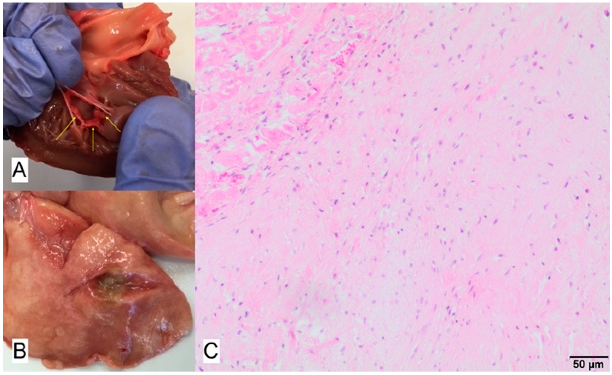
3.1. Case No. 1
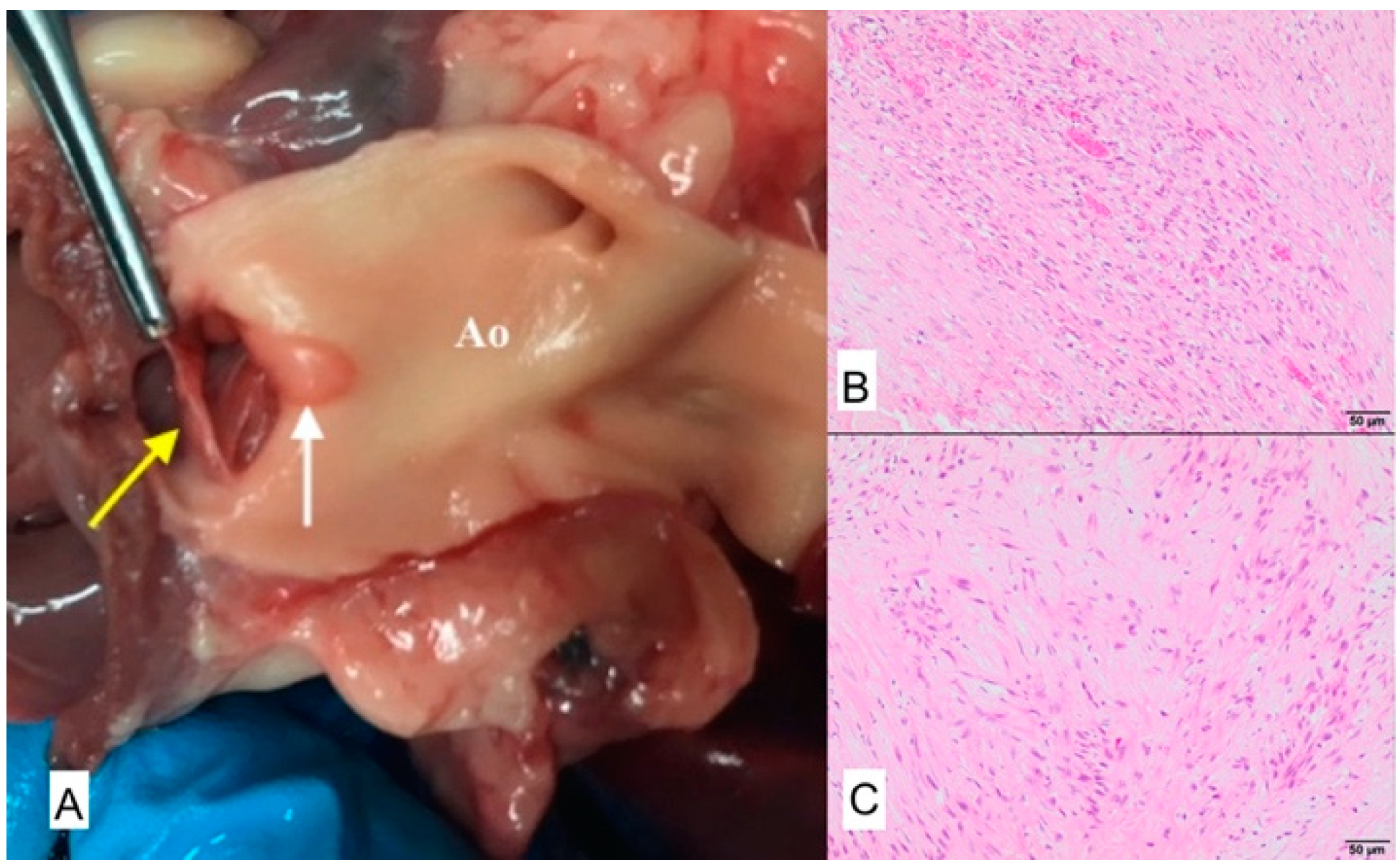
3.2. Case No. 2
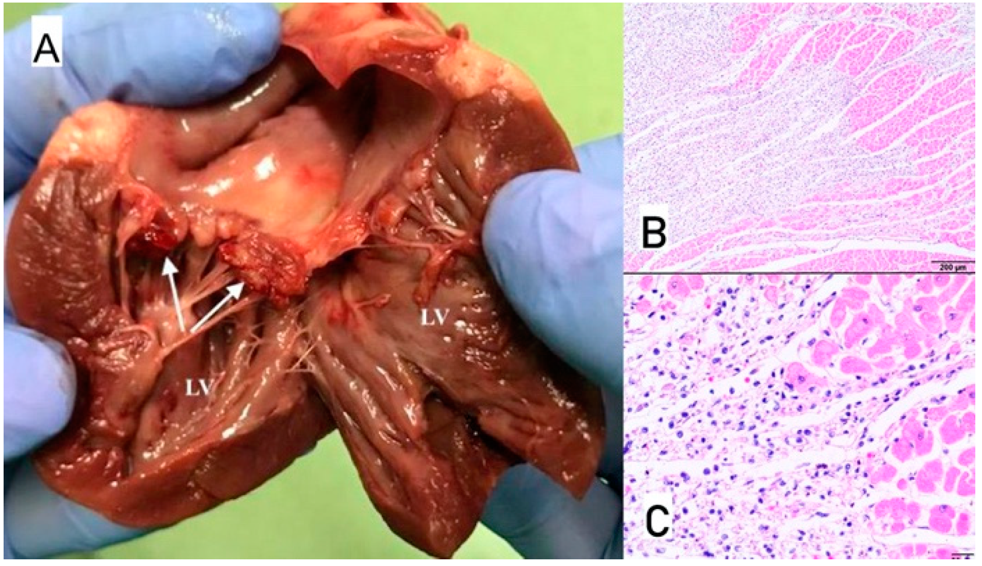
3.3. Case No. 3
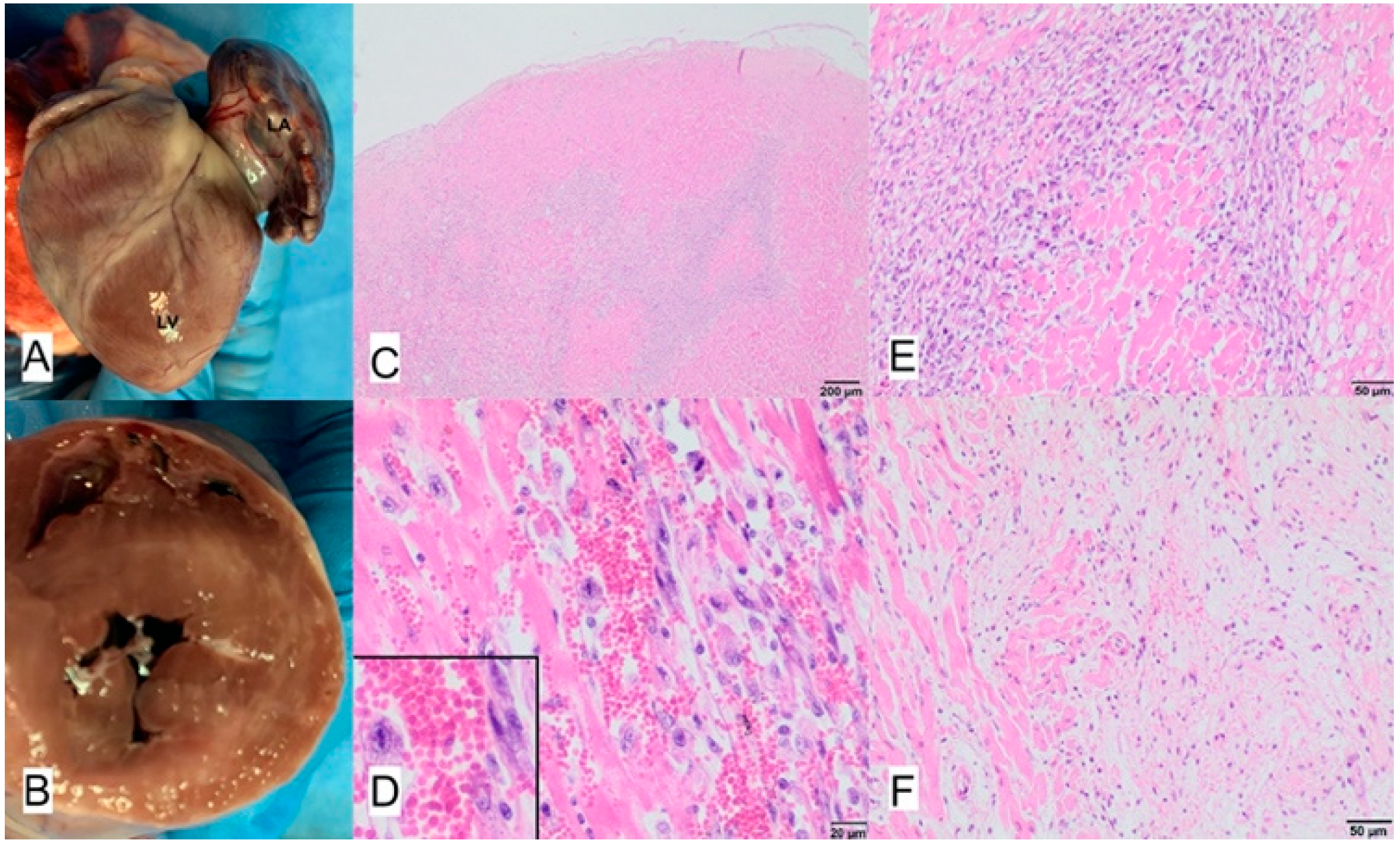
3.4. Case No. 4
3.5. Case No. 5
3.6. Case No. 6
3.7. Case No. 7
4. Discussion
5. Conclusions
Author Contributions
Funding
Institutional Review Board Statement
Informed Consent Statement
Data Availability Statement
Acknowledgments
Conflicts of Interest
References
- Zachary, J.F. Pathologic Basis of Veterinary Disease, 6th ed.; Elsevier: St. Louis, MO, USA, 2017; ISBN 978-0-323-35775-3. [Google Scholar]
- Balakrishnan, N.; Alexander, K.; Keene, B.; Kolluru, S.; Fauls, M.L.; Rawdon, I.; Breitschwerdt, E.B. Successful Treatment of Mitral Valve Endocarditis in a Dog Associated with ‘Actinomyces Canis -like’ Infection. J. Vet. Cardiol. 2016, 18, 271–277. [Google Scholar] [CrossRef] [PubMed]
- Kittleson, M.D.; Kienle, R.D. Small Animal Cardiovascular Medicine; Mosby: St. Louis, MO, USA, 1998; ISBN 978-0-8151-5140-1. [Google Scholar]
- Aoki, T.; Sunahara, H.; Sugimoto, K.; Ito, T.; Kanai, E.; Fujii, Y. Infective Endocarditis of the Aortic Valve in a Border Collie Dog with Patent Ductus Arteriosus. J. Vet. Med. Sci. 2015, 77, 331–336. [Google Scholar] [CrossRef] [Green Version]
- Felker, G.M.; Boehmer, J.P.; Hruban, R.H.; Hutchins, G.M.; Kasper, E.K.; Baughman, K.L.; Hare, J.M. Echocardiographic Findings in Fulminant and Acute Myocarditis. J. Am. Coll. Cardiol. 2000, 36, 227–232. [Google Scholar] [CrossRef] [Green Version]
- Coffin, S.T.; Benton, S.M.; Lenihan, D.J.; Naftilan, A.J.; Mendes, L.A.; Lenihan, D.J. Eosinophilic Myocarditis—An Unusual Cause of Left Ventricular Hypertrophy. Am. J. Med. Sci. 2015, 349, 358–362. [Google Scholar] [CrossRef] [PubMed]
- Sagrera, M.R.; Sala, M.F.; Barcena, J.P.; Garcia, J.B.; Canellas, C.F.; Juve, J.I. Acute Myocarditis and Left Ventricular “Hypertrophy”. Echocardiography 2000, 17, 567–570. [Google Scholar] [CrossRef]
- Lai, W.W.; Mertens, L.; Cohen, M.; Geva, T. (Eds.) Echocardiography in Pediatric and Congenital Heart Disease: From Fetus to Adult, 2nd ed.; John Wiley & Sons Ltd.: Chichester, West Sussex, UK; Hoboken, NJ, USA, 2015; ISBN 978-1-118-74221-1. [Google Scholar]
- Janus, I.; Noszczyk-Nowak, A.; Nowak, M.; Cepiel, A.; Ciaputa, R.; Pasławska, U.; Dzięgiel, P.; Jabłońska, K. Myocarditis in Dogs: Etiology, Clinical and Histopathological Features (11 Cases: 2007–2013). Ir. Vet. J. 2014, 67, 28. [Google Scholar] [CrossRef] [PubMed] [Green Version]
- Sykes, J.E.; Kittleson, M.D.; Chomel, B.B.; MacDonald, K.A.; Pesavento, P.A. Clinicopathologic Findings and Outcome in Dogs with Infective Endocarditis: 71 Cases (1992–2005). J. Am. Vet. Med. Assoc. 2006, 228, 1735–1747. [Google Scholar] [CrossRef]
- Szatmári, V. Incidence of Postoperative Implant-Related Bacterial Endocarditis in Dogs That Underwent Trans-Catheter Embolization of a Patent Ductus Arteriosus without Intra- and Post-Procedural Prophylactic Antibiotics. Vet. Microbiol. 2017, 207, 25–28. [Google Scholar] [CrossRef] [PubMed] [Green Version]
- Tabar, M.-D.; Altet, L.; Maggi, R.G.; Altimira, J.; Roura, X. First Description of Bartonella Koehlerae Infection in a Spanish Dog with Infective Endocarditis. Parasites Vectors 2017, 10, 247. [Google Scholar] [CrossRef] [Green Version]
- Roura, X.; Santamarina, G.; Tabar, M.-D.; Francino, O.; Altet, L. Polymerase Chain Reaction Detection of Bartonella Spp. in Dogs from Spain with Blood Culture-Negative Infectious Endocarditis. J. Vet. Cardiol. 2018, 20, 267–275. [Google Scholar] [CrossRef] [PubMed]
- Vicent, L.; Saldivar, H.G.; Bouza, E.; Muñoz, P.; Cuerpo, G.; de Alarcón, A.; Vidal, B.; Cobo, M.; Goenaga, M.Á.; Carrasco-Chinchilla, F.; et al. Prognostic Implications of a Negative Echocardiography in Patients with Infective Endocarditis. Eur. J. Intern. Med. 2018, 52, 40–48. [Google Scholar] [CrossRef]
- Brandão, T.J.D.; Januario-da-Silva, C.A.; Correia, M.G.; Zappa, M.; Abrantes, J.A.; Dantas, A.M.R.; Golebiovski, W.; Barbosa, G.I.F.; Weksler, C.; Lamas, C.C. Histopathology of Valves in Infective Endocarditis, Diagnostic Criteria and Treatment Considerations. Infection 2017, 45, 199–207. [Google Scholar] [CrossRef]
- MacDonald, K.A.; Chomel, B.B.; Kittleson, M.D.; Kasten, R.W.; Thomas, W.P.; Pesavento, P. A Prospective Study of Canine Infective Endocarditis in Northern California (1999–2001): Emergence of Bartonella as a Prevalent Etiologic Agent. J. Vet. Intern. Med. 2004, 18, 56–64. [Google Scholar] [CrossRef] [PubMed]
- O’Connor, M.J. Imaging the Itis: Endocarditis, Myocarditis, and Pericarditis. Curr. Opin. Cardiol. 2019, 34, 57–64. [Google Scholar] [CrossRef]
- Von Reyn, C.F. Infective Endocarditis: An Analysis Based on Strict Case Definitions. Ann. Intern. Med. 1981, 94, 505. [Google Scholar] [CrossRef] [PubMed]
- Seif, D.; Meeks, A.; Mailhot, T.; Perera, P. Emergency Department Diagnosis of Infective Endocarditis Using Bedside Emergency Ultrasound. Cr. Ultrasound J. 2013, 5, 1. [Google Scholar] [CrossRef] [Green Version]
- Pathak, N.J.; Ng, L.; Saul, T.; Lewiss, R.E. Focused Cardiac Ultrasound Diagnosis of Right-Sided Endocarditis. Am. J. Emerg. Med. 2013, 31, 998.e3–998.e4. [Google Scholar] [CrossRef] [PubMed]
- Zhang, L.; Wang, B.; Zhou, J.; Kirkpatrick, J.; Xie, M.; Johri, A.M. Bedside Focused Cardiac Ultrasound in COVID-19 from the Wuhan Epicenter: The Role of Cardiac Point-of-Care Ultrasound, Limited Transthoracic Echocardiography, and Critical Care Echocardiography. J. Am. Soc. Echocardiogr. 2020, 33, 676–682. [Google Scholar] [CrossRef] [PubMed]
- Blaivas, M. Unexpected Finding of Myocardial Depression in 2 Healthy Young Patients with COVID-19 Pneumonia: Possible Support for COVID-19-related Myocarditis. J. Am. Coll. Emerg. Physicians Open 2020, 1, 375–378. [Google Scholar] [CrossRef] [PubMed]
- Murchan, S.; Kaufmann, M.E.; Deplano, A.; de Ryck, R.; Struelens, M.; Zinn, C.E.; Fussing, V.; Salmenlinna, S.; Vuopio-Varkila, J.; El Solh, N.; et al. Harmonization of Pulsed-Field Gel Electrophoresis Protocols for Epidemiological Typing of Strains of Methicillin-Resistant Staphylococcus Aureus: A Single Approach Developed by Consensus in 10 European Laboratories and Its Application for Tracing the Spread of Related Strains. J. Clin. Microbiol. 2003, 41, 1574–1585. [Google Scholar] [CrossRef] [Green Version]
- Fischer, B.G.; Baduashvili, A. Cardiac Point-of-Care Ultrasound for the Diagnosis of Infective Endocarditis in a Patient with Non-Specific Rheumatologic Symptoms and Glomerulonephritis. Am. J. Case Rep. 2019, 20, 542–547. [Google Scholar] [CrossRef]
- Muñoz, P.; Bouza, E.; Marín, M.; Alcalá, L.; Rodríguez Créixems, M.; Valerio, M.; Pinto, A.; on behalf of the Group for the Management of Infective Endocarditis of the Gregorio Marañón Hospital. Heart Valves Should Not Be Routinely Cultured. J. Clin. Microbiol. 2008, 46, 2897–2901. [Google Scholar] [CrossRef] [Green Version]
- Greub, G.; Lepidi, H.; Rovery, C.; Casalta, J.-P.; Habib, G.; Collard, F.; Fournier, P.-E.; Raoult, D. Diagnosis of Infectious Endocarditis in Patients Undergoing Valve Surgery. Am. J. Med. 2005, 118, 230–238. [Google Scholar] [CrossRef]
- Morris, A.J.; Drinković, D.; Pottumarthy, S.; MacCulloch, D.; Kerr, A.R.; West, T. Bacteriological Outcome after Valve Surgery for Active Infective Endocarditis: Implications for Duration of Treatment after Surgery. Clin. Infect. Dis. 2005, 41, 187–194. [Google Scholar] [CrossRef] [Green Version]
- MacDonald, K. Infective Endocarditis in Dogs: Diagnosis and Therapy. Vet. Clin. N. Am. Small Anim. Pract. 2010, 40, 665–684. [Google Scholar] [CrossRef] [PubMed]
- Spina, G.S.; Sampaio, R.O.; Branco, C.E.; Miranda, G.B.; Rosa, V.E.E.; Tarasoutchi, F. Incidental Histological Diagnosis of Acute Rheumatic Myocarditis: Case Report and Review of the Literature. Front. Pediatr. 2014, 2, 126. [Google Scholar] [CrossRef] [Green Version]
- Collins, J.A.; Zhang, Y.; Burke, A.P. Pathologic Findings in Native Infective Endocarditis. Pathol. Res. Pract. 2014, 210, 997–1004. [Google Scholar] [CrossRef] [PubMed]


Publisher’s Note: MDPI stays neutral with regard to jurisdictional claims in published maps and institutional affiliations. |
© 2021 by the authors. Licensee MDPI, Basel, Switzerland. This article is an open access article distributed under the terms and conditions of the Creative Commons Attribution (CC BY) license (https://creativecommons.org/licenses/by/4.0/).
Share and Cite
Szaluś-Jordanow, O.; Stabińska-Smolarz, M.; Czopowicz, M.; Moroz, A.; Mickiewicz, M.; Łobaczewski, A.; Chrobak-Chmiel, D.; Kizerwetter-Świda, M.; Rzewuska, M.; Sapierzyński, R.; et al. Focused Cardiac Ultrasound Examination as a Tool for Diagnosis of Infective Endocarditis and Myocarditis in Dogs and Cats. Animals 2021, 11, 3162. https://doi.org/10.3390/ani11113162
Szaluś-Jordanow O, Stabińska-Smolarz M, Czopowicz M, Moroz A, Mickiewicz M, Łobaczewski A, Chrobak-Chmiel D, Kizerwetter-Świda M, Rzewuska M, Sapierzyński R, et al. Focused Cardiac Ultrasound Examination as a Tool for Diagnosis of Infective Endocarditis and Myocarditis in Dogs and Cats. Animals. 2021; 11(11):3162. https://doi.org/10.3390/ani11113162
Chicago/Turabian StyleSzaluś-Jordanow, Olga, Marta Stabińska-Smolarz, Michał Czopowicz, Agata Moroz, Marcin Mickiewicz, Andrzej Łobaczewski, Dorota Chrobak-Chmiel, Magdalena Kizerwetter-Świda, Magdalena Rzewuska, Rafał Sapierzyński, and et al. 2021. "Focused Cardiac Ultrasound Examination as a Tool for Diagnosis of Infective Endocarditis and Myocarditis in Dogs and Cats" Animals 11, no. 11: 3162. https://doi.org/10.3390/ani11113162