Article

Effects of Cardiac Contractility Modulation Therapy on Right Ventricular Function: An Echocardiographic Study

1
Heart Failure Unit, Department of Cardiology, AORN dei Colli Monaldi Hospital, 80131 Naples, Italy
2
Electrophysiology Unit, Department of Cardiology, AORN dei Colli Monaldi Hospital, 80131 Naples, Italy
3
Cardiology Unit, Department of Medical Translational Sciences, University of Campania “Luigi Vanvitelli”, 80131 Naples, Italy
*
Author to whom correspondence should be addressed.
Appl. Sci. 2022, 12(15), 7917; https://doi.org/10.3390/app12157917
Submission received: 23 June 2022 / Revised: 4 August 2022 / Accepted: 5 August 2022 / Published: 7 August 2022
(This article belongs to the Special Issue Biomedical Imaging Technologies for Cardiovascular Disease)

Abstract

:
Background. Cardiac contractility modulation (CCM) is a novel device-based therapy for patients with heart failure with reduced and mild reduced ejection fraction (HFrEF/HFmrEF). CCM increases cardiac performance and produces reverse left ventricular remodeling, with improved symptoms, functional capacity, quality of life, and reduced HF-related hospitalizations. However, to date, little evidence is available on the effects of CCM on right ventricle (RV) function. Therefore, we analyzed the effects of CCM on RV systolic function and RV–pulmonary artery (PA) coupling. Methods. Twenty-one (65 ± 12.5 years) patients with NYHA class III, ejection fraction < 40% and QRS < 120 ms were assessed at baseline. During follow up, two patients had died, and so nineteen patients were evaluated six months after CCM therapy. Using echocardiography, tricuspid annular systolic excursion (TAPSE), myocardial systolic excursion velocity (RVs), and RV free-wall strain was measured. PA systolic pressure (PASP) was estimated from tricuspid regurgitation, adding the right atrial pressure estimation. The RV-PA coupling was calculated as TAPSE/PASP ratio. Results. After six months, patients who underwent CCM therapy showed a reduction in RV diameters and improved RV systolic function, as evidenced by the increase in both TAPSE (16.6 ± 4.2 mm vs. 18.5 ± 3.6 mm; p < 0.05), RVs (10.1 ± 1.8 cm/s vs. 11.3 ± 11.4 cm/s; p < 0.05), and RV strain (−13.7 ± 1.8% vs. −15.6 ± 2.3%; p < 0.05). CCM also determined a reduction in PASP (34.2 ± 9.8 mmHg vs. 28 ± 6.2 mmHg; p < 0.05) and an increase in the TAPSE/PASP ratio (0.52 ± 0.14 mm/mmHg vs. 0.66 ± 0.23 mm/mmHg; p < 0.05). Conclusions. At six months, CCM increases RV reverse remodeling and performance, reducing RV size and improving RV systolic function, PASP, and RV-PA coupling.

1. Introduction

Heart failure (HF) is the most common cardiovascular disease worldwide, with more than 25 million people being affected in industrialized countries [1].
Despite advances in pharmacologic therapies for treating patients with HF with reduced ejection fraction (HFrEF), the prognosis of such patients remains poor [2,3]; therefore, device-based therapy has become increasingly important in recent years for the treatment of HFrEF.
The most widely used device-based therapy for the treatment of HFrEF is cardiac resynchronization therapy, which can lead to improved cardiac performance and prognosis in patients with HFrEF and wide QRS (duration > 150 ms) [4].
Unfortunately, only 30% of HFrEF have a QRS duration > 150 ms [5]. For patients with persistent symptoms or frequent HF-related hospitalizations but with narrow QRS, a new device (Optimizer Smart®) capable of delivering cardiac contractility modulation (CCM) therapy has been available for several years [6].
Optimizer Smart delivers non-excitatory electrical impulses on the interventricular septum during the absolute refractory period of cardiomyocytes; therefore, it does not alter the cellular action potential and does not affect heart rhythm [7].
CCM affects Ca2+ handling in cardiomyocytes, the expression of genes encoding factors that play a crucial role in Ca2+ metabolism, and molecular pathways involved in interstitial fibrosis [8,9].
These actions result in reverse left ventricular remodeling and increased cardiac performance, improving symptoms, functional capacity, quality of life, and reduced HF-related hospitalizations [10,11].
However, to date, there is little evidence available on the effect of CCM on right ventricular performance; thus, in this study, we analyzed the effects of CCM on right ventricular systolic function and right ventricle–pulmonary artery (RV-PA) coupling in patients with HFrEF.

2. Materials and Methods

2.1. Study Population

We prospectively and consecutively enrolled all patients diagnosed with HFrEF undergoing Optimizer Smart® implant between November 2019 and November 2021.
The following inclusion criteria were used:
-
HFrEF with a left ventricular ejection fraction < 40%;
-
NYHA class II–III;
-
QRS duration < 120 ms.
-
The following exclusion criteria were used:
-
Acute coronary syndrome in the previous three month;
-
Non-optimal medical therapy with disease modifier drugs for HFrEF.
Demographic, clinical, and laboratory data were acquired from stable patients 24 h before device implantation.
The research was conducted according to the Declaration of Helsinki and approved by the ethics committee of the AORN dei Colli-Ospedale Monaldi (resolution No. 903/2020).
Signed informed consent was obtained for all patients.

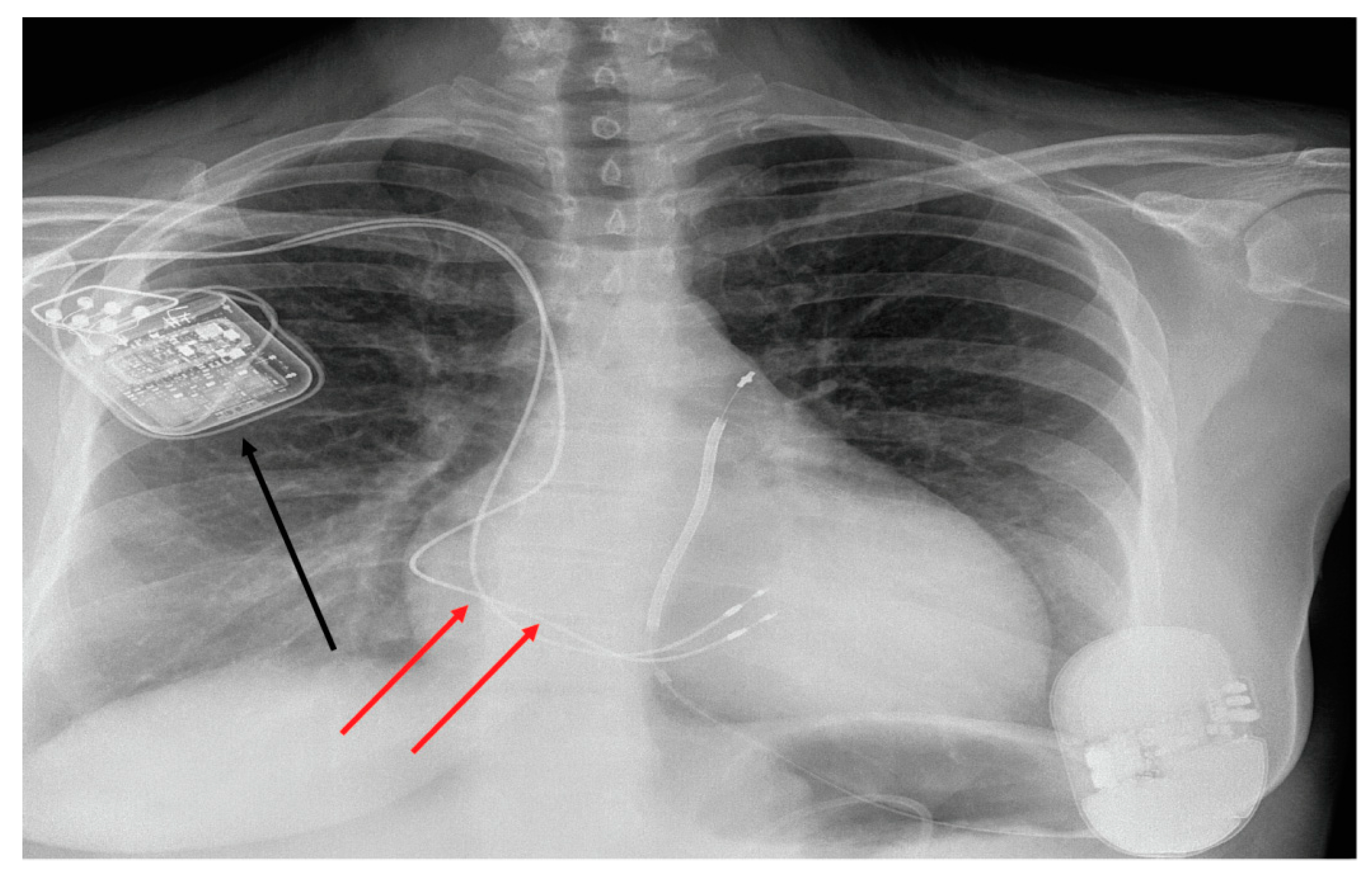
2.2. Optimizer Smart Implant

Implantation of the Optimizer Smart® (Impulse Dynamics Inc., Marlton, NJ, USA) was performed under local anaesthesia.
Two electrodes, required to detect ventricular activity and subsequent delivery of CCM signals, were placed on the right interventricular septum via the subclavian vein.
The ends of both ventricular electrodes were actively attached to the right side of the interventricular septum at least 3 cm away from the implantable defibrillator lead.
Finally, both leads were connected to the Smart Optimizer®, and the device was implanted in a subcutaneous pocket (Figure 1)

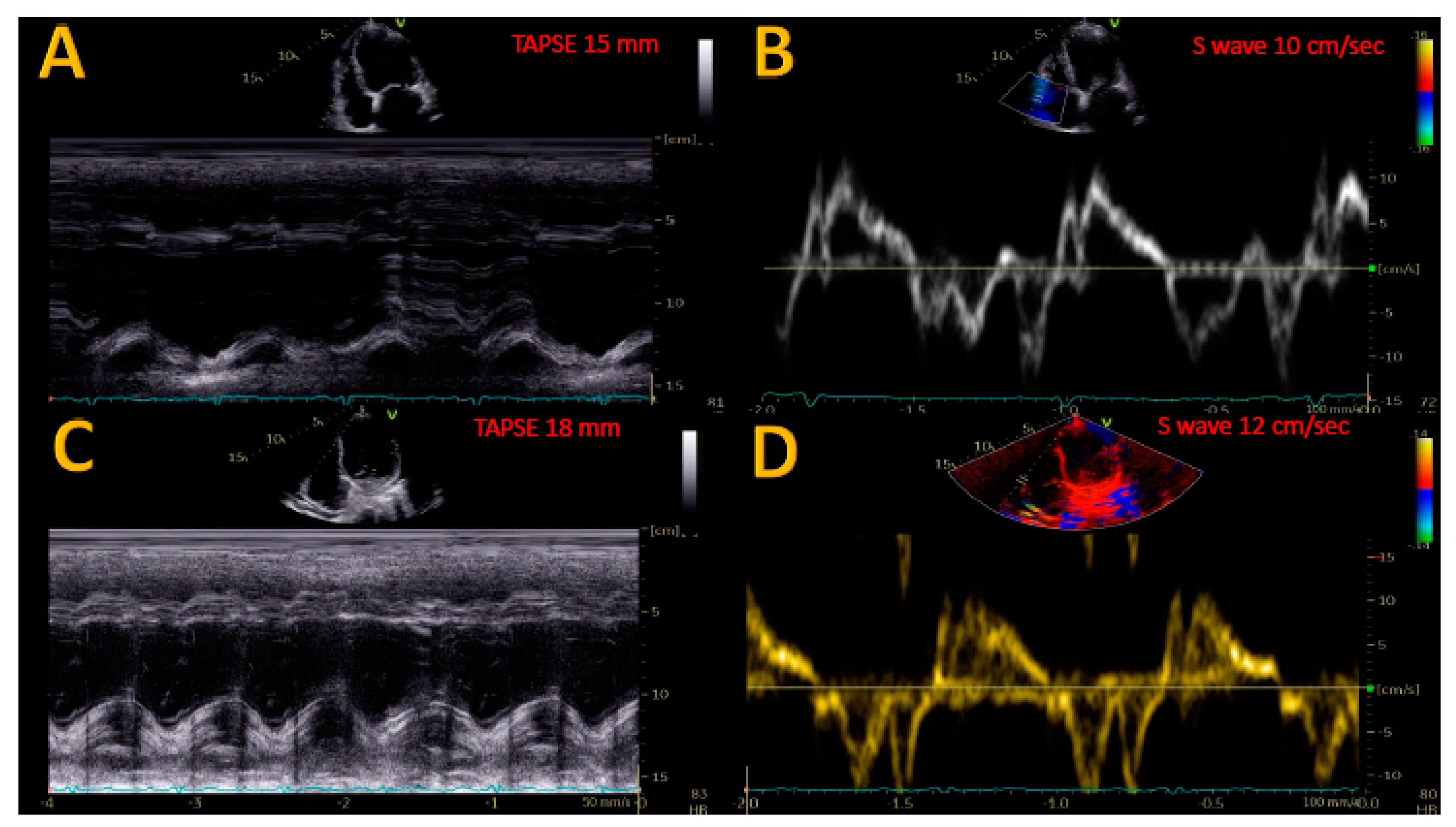
2.3. Echocardiographic Evaluation

Standard transthoracic echocardiography and Doppler assessment were performed with available market equipment (Vivid E9—GE Healthcare, Chicago, IL, USA) as recommended elsewhere [12,13].
Two expert echocardiographers analysed all echocardiographic studies, and the value of specific measurements was obtained from the average of 3–5 cardiac cycles.
Systolic excursion in the tricuspid annular plane (TAPSE) was measured in the optimized 4-chamber apical view to obtain the correct M-mode orientation.
Myocardial systolic excursion velocity (S’) was measured at the lateral tricuspid annulus with pulsed tissue Doppler imaging (TDI).
The pulmonary artery systolic pressure (PASP) was inferred from the maximum velocity of the tricuspid regurgitant jet (in accordance with the modified Bernoulli equation). Adding to the obtained value, the right atrial pressure was calculated according to the size and collapsibility of the inferior vena cava following international recommendations. LV and LA volumes were measured using the modified Simpson’s rule with biplane planimetry [13].
Right ventricular myocardial strain parameters were measured using the Q-Analysis software package (EchoPAC BT2.02; GE Vingmed, Horten, Norway). After manually identifying the end-systolic endocardial boundary of the right ventricle by locating three points (two on the tricuspid annulus) and one on the apex of the right ventricle, a region of interest (ROI) was automatically generated.
Next, the width of the ROI was manually adjusted to include the entire myocardial wall. Finally, according to international recommendations, we calculated the RV free-wall longitudinal strain (RVFWLS) value by averaging the values obtained from the three free-wall segments.
Right ventricular arterial coupling was estimated as the ratio of the TAPSE and PASP value.
Echocardiographic evaluations were performed for all patients 24 h before and 6 months after Optimizer Smart® implantation.

2.4. Statistical Analysis

Statistical analyses were performed using Prism 9 (GraphPad Software, San Diego, CA, USA).
Demographic and clinical variables were expressed as medians and standard deviations. Qualitative variables were expressed as both numbers and percentages. Wilcoxon-rank test was used to compare the differences between values at baseline and treatment values in case of non-normal distribution; a t-test was used for variables with normal distribution. All p-values were two-sided; p < 0.05 indicated statistical significance.
ROC (Receiver Operating Feature) curve analysis was performed to select the cut-off of echocardiographic measures. The reproducibility of measurements was determined in all patients, considering inter-observer and intra-observer variability by using intraclass correlation (ICC).

3. Results

During the study period, we enrolled 21 patients with HFrEF who underwent Optimizer Smart implant®. Demographic and clinical characteristics are reported in Table 1.
Of the overall population enrolled, 9 patients (42%) had an ischemic form of HFrEF, (left ventricular ejection 29 ± 6%) and 4 patients (19%) had a low TAPSE (<16 mm) and S wave (<10 cm/s) values.
At six months, 2 patients (10%) died, while the follow-up echocardiographic data were collected for 19 patients (90%). No significant changes in disease modifiers drugs doses occurred during follow up (Table 2). The r coefficient value for intra-observed variability was 0.82 and 0.78 for inter-observed variability.

3.1. Effects of CCM on RV Reverse Remodeling and Contractility Index

The echocardiographic index of RV contractility improved in patients enrolled in the study during follow up.
There was a significant reduction in RV end-diastolic transversal diameters (p < 0.05) (Table 3), while the severity of tricuspid regurgitation remained stable (Table 3).
As shown in Figure 2 and Figure 3, a statistically significant increase in both TAPSE (16.6 ± 4.2 mm vs. 18.5 ± 3.6 mm; p < 0.05) and S wave (10.1 ± 1.8 cm/s vs. 11.3 ± 11.4 cm/s; p < 0.05) occurred at six months.
Concurrently, a significant increase in more specific and reproducible index of RV function, the RVFWLS occurred (−13.7 ± 1.8% vs. −15.6 ± 2.3%; p < 0.05; Figure 3).
Notably, the increase in all the parameters of RV function occurred in patients with a standard RV function rather than in patients with reduced ventricular function at baseline (p = 0.12).

3.2. Effects of CCM on PASP

Positive effects of CCM on right ventricular performance determine hemodynamic benefits, too.
As shown in Figure 4, CCM determines, at six months, a significant reduction in PASP (34.2 ± 9.8 mmHg vs. 28 ± 6.2 mmHg; p < 0.05); also, in this case, these effects are present both in patients with normal values at baseline and in patients with a high PASP value at enrollment (p = 0.78).

3.3. Effects of CCM on RV-PA Coupling

The positive effects that CCM therapy brings about both in terms of increasing right ventricular contractility indices and reducing pressures in the pulmonary circulation result in improved coupling between RV and PA. In fact, at six months of follow up, the TAPSE/PAPS ratio improves significantly (0.52 ± 0.14 mm/mmHg vs. 0.66 ± 0.23 mm/mmHg; p < 0.05; Figure 5).

4. Discussion

For the first time, our data show that in patients with HFrEF, CCM therapy can improve after six months of follow-up RV systolic function, as evidenced by an increase in both tapes, RVS wave value and RVFWLS, which resulted in RV reverse remodeling. All three indices of RV function increased in both normal and reduced RV function patients. At six months follow up, the degree of tricuspid regurgitation remained stable.
At six months, CCM also determined a significant reduction in PASP in patients with normal values at baseline and those with high PASP values at enrollment.
Furthermore, the positive effects of CCM in terms of increasing RV contractility and reducing pulmonary pressures resulted in improved coupling between RV and PA, as evidenced by an increase in the TAPSE/PAPS ratio.

4.1. Improvement of RV Reverse Remodeling Systolic Function after CCM

Although several studies reported improvements in symptoms, exercise capacity, and LV global and regional LV contractility, whether CCM would induce favorable changes in RV systolic function was largely unknown.
As guidelines [13] recommended, RV size was measured from a four-chamber view in the context of the left ventricle. Both basal and mid-cavity diameters were reduced, suggesting a favorable RV remodeling; however, the severity of tricuspid regurgitation remained stable, maybe because of the presence of leads passing from the right atrium to the RV through the valve.
In this study, we found that all three RV systolic function indices (TAPSE, S wave, RVFWLS) improved after 6 months of CCM therapy.
TAPSE is a measure of longitudinal contraction of the RV. It requires no geometric assumptions, it has the advantage of being obtained even with poor images, and it can be measured on all ultrasound machines [14].
TAPSE has prognostic value in a variety of conditions that may affect the RV, such as HFrEF [15] and pulmonary hypertension [16,17].
TDI-derived lateral tricuspid annulus S-wave velocity is easy to measure, reliable, and reproducible. Like TAPSE, it is a unidimensional measure and evaluates RV longitudinal function [16].
Unlike wall motion displacement (TAPSE) or velocity (S wave), RV free-wall myocardial strain (myocardial deformation) is unaffected by the motion of the entire heart and allows distinction between active and passive myocardial tissue movement. RVFWLS is an angle-independent speckle-tracking echocardiography (STE)-derived parameter that is very useful for the evaluation of RV contractile function. It has prognostic value for cardiovascular morbidity and mortality [18,19].
Therefore, as previously demonstrated for LV global and regional systolic function [20], in our study the CCM also seems to improve RV systolic function, increasing global RV contractility, not just limited to the septal wall where CCM signals were delivered.
Similar to the findings in our study, in an animal model of chronic right ventricular dysfunction, CCM has been shown to improve global cardiac function significantly inboth ventricles [21].
CCM seems to be useful in HFrEF patients to improve both LV and RV systolic function, probably given its proven effects on regional and global myocardial contractility [20] and given the key role played by the interventricular septum in the contractility of both ventricles via ventricular interdependence [15,22,23]. Furthermore, Yucel G et al. showed better positive biventricular echocardiographic results in patients with lower LVEF, probably because the intensity of LVEF reduction appears to be correlated partly with impaired neuro-humoral activation associated with HF, assuming that the underlying mechanism could be “the worse the LVEF at baseline, the more can be repaired” [24].

4.2. Improvement of PASP after CCM

Similar to a recent case report, which shows the positive effect of CCM on pulmonary pressure measured by Cardio MEMS [25], in this study we found that CCM determined a significant reduction in PASP at six months. The favorable effect of CCM on pulmonary pressure would be due to the improvement in both ventricles’ contractility.

4.3. Improvement of RV-PA Coupling after CCM

We found an improvement RV-PA coupling after 6 months of CCM, evidenced by the increase TAPSE/PASP ratio, which incorporates both RV longitudinal displacement and load. This index assesses RV contraction by plotting fiber longitudinal shortening versus the force generated to overcome the imposed load [26,27].
Unlike TAPSE, S wave and FWRVLS depend on RV loading conditions. TAPSE/PASP ratio allows estimation of RV performance, and it is also a non-invasive index of RV to pulmonary circulation coupling based on the correlation with invasively evaluated RV systolic elastance/arterial elastance [27]. This echocardiographic index is a predictor of mortality in patients with HFrEF and with severe PAH [28,29].

5. Study Limitations

The relatively small sample size, single-centre study design, and observational nature of the study may affect our results. STE can be affected by RV loading conditions.
We do not assess RV with 3D echocardiography.

6. Conclusions

At six months follow up, CCM therapy increased RV performance, improving RV systolic function, PASP, and coupling between RV and PA. A better forward ejection of blood could be useful for RV reverse remodeling.
Additional larger studies are needed to provide a greater understanding of the long-term impact of CCM on RV.

Author Contributions

Conceptualization, C.C. and S.D.V.; and methodology, C.C., S.D.V., M.L.M., A.D., E.A. and G.N.; data curation, C.C., M.L.M., V.E. and D.M.; writing—original draft preparation, C.C., M.L.M. and D.M.; writing—review and editing, C.C., S.D.V., A.D., E.A., G.N. and G.P.; visualization, C.C.; supervision, G.P. and D.M. All authors have read and agreed to the published version of the manuscript.

Funding

This research received no external funding.

Institutional Review Board Statement

The study was conducted in accordance with the Declaration of Helsinki, andapproved byethics committee of the AORN dei Colli-Ospedale Monaldi (resolution No. 903/2020).

Informed Consent Statement

Informed consent was obtained from all subjects involved in the study.

Data Availability Statement

The data presented in this study are available on request from the corresponding author.

Conflicts of Interest

The authors declare no conflict of interest.

References

  1. Savarese, G.; Lund, L.H. Global Public Health Burden of Heart Failure. Card. Fail. Rev. 2017, 3, 7–11. [Google Scholar] [CrossRef]
  2. Benjamin, E.J.; Virani, S.S.; Callaway, C.W.; Chamberlain, A.M.; Chang, A.R.; Cheng, S.; Chiuve, S.E.; Cushman, M.; Delling, F.N.; Deo, R.; et al. Heart Disease and Stroke Statistics-2018 Update: A Report from the American Heart Association. Circulation 2018, 137, e67–e492, Erratum in Circulation 2018, 137, e493. [Google Scholar] [CrossRef]
  3. Komajda, M.; Böhm, M.; Borer, J.S.; Ford, I.; Tavazzi, L.; Pannaux, M.; Swedberg, K. Incremental benefit of drug therapies for chronic heart failure with reduced ejection fraction: A network meta-analysis. Eur. J. Heart Fail. 2018, 20, 1315–1322. [Google Scholar] [CrossRef] [PubMed] [Green Version]
  4. Moskovitch, J.; Voskoboinik, A. Cardiac resynchronization therapy: A comprehensive review. Minerva Medica 2019, 110, 121–138. [Google Scholar] [CrossRef] [PubMed]
  5. Abraham, W.T. Cardiac Resynchronization Therapy and Cardiac Contractility Modulation in Patients with Advanced Heart Failure. Heart Fail. Clin. 2021, 17, 599–606. [Google Scholar] [CrossRef]
  6. Campbell, C.M.; Kahwash, R.; Abraham, W.T. Optimizer Smart in the treatment of moderate-to-severe chronic heart failure. Future Cardiol. 2020, 16, 13–25. [Google Scholar] [CrossRef]
  7. Lawo, T.; Borggrefe, M.; Butter, C.; Hindricks, G.; Schmidinger, H.; Mika, Y.; Burkhoff, D.; Pappone, C.; Sabbah, H.N. Electrical Signals Applied during the Absolute Refractory Period: An Investigational Treatment for Advanced Heart Failure in Patients with Normal QRS Duration. J. Am. Coll. Cardiol. 2005, 46, 2229–2236. [Google Scholar] [CrossRef] [Green Version]
  8. Butter, C.; Rastogi, S.; Minden, H.-H.; Meyhöfer, J.; Burkhoff, D.; Sabbah, H.N. Cardiac Contractility Modulation Electrical Signals Improve Myocardial Gene Expression in Patients with Heart Failure. J. Am. Coll. Cardiol. 2008, 51, 1784–1789. [Google Scholar] [CrossRef] [Green Version]
  9. Zhang, F.; Dang, Y.; Li, Y.; Hao, Q.; Li, R.; Qi, X. Cardiac Contractility Modulation Attenuate Myocardial Fibrosis by Inhibiting TGF-β1/Smad3 Signaling Pathway in a Rabbit Model of Chronic Heart Failure. Cell. Physiol. Biochem. 2016, 39, 294–302. [Google Scholar] [CrossRef]
  10. Lyon, A.R.; Samara, M.A.; Feldman, D.S. Cardiac contractility modulation therapy in advanced systolic heart failure. Nat. Rev. Cardiol. 2013, 10, 584–598, Erratum in Nat. Rev. Cardiol. 2014, 11, 188. [Google Scholar] [CrossRef]
  11. Cappannoli, L.; Scacciavillani, R.; Rocco, E.; Perna, F.; Narducci, M.L.; Vaccarella, M.; D’Amario, D.; Pelargonio, G.; Massetti, M.; Crea, F.; et al. Cardiac contractility modulation for patient with refractory heart failure: An updated evidence-based review. Heart Fail. Rev. 2021, 26, 227–235. [Google Scholar] [CrossRef] [PubMed]
  12. Nagueh, S.F.; Smiseth, O.A.; Appleton, C.P.; Byrd, B.F., 3rd; Dokainish, H.; Edvardsen, T.; Flachskampf, F.A.; Gillebert, T.C.; Klein, A.L.; Lancellotti, P.; et al. Recommendations for the Evaluation of Left Ventricular Diastolic Function by Echocardiography: An Update from the American Society of Echocardiography and the European Association of Cardiovascular Imaging. Eur. Heart J. Cardiovasc. Imaging 2016, 17, 1321–1360. [Google Scholar] [CrossRef] [PubMed]
  13. Lang, R.M.; Badano, L.P.; Mor-Avi, V.; Afilalo, J.; Armstrong, A.; Ernande, L.; Flachskampf, F.A.; Foster, E.; Goldstein, S.A.; Kuznetsova, T.; et al. Recommendations for Cardiac Chamber Quantification by Echocardiography in Adults: An Update from the American Society of Echocardiography and the European Association of Cardiovascular Imaging. Eur. Heart J. Cardiovasc. Imaging 2015, 16, 233–271, Erratum in Eur. Heart J. Cardiovasc. Imaging 2016, 17, 412. [Google Scholar] [CrossRef] [PubMed]
  14. Jones, N.; Burns, A.T.; Prior, D.L. Echocardiographic Assessment of the Right Ventricle–State of the Art. Heart Lung Circ. 2019, 28, 1339–1350. [Google Scholar] [CrossRef] [Green Version]
  15. Santamore, W.P.; Dell’Italia, L.J. Ventricular interdependence: Significant left ventricular contributions to right ventricular systolic function. Prog. Cardiovasc. Dis. 1998, 40, 289–308. [Google Scholar] [CrossRef]
  16. Rudski, L.G.; Lai, W.W.; Afilalo, J.; Hua, L.; Handschumacher, M.D.; Chandrasekaran, K.; Solomon, S.D.; Louie, E.K.; Schiller, N.B. Guidelines for the Echocardiographic Assessment of the Right Heart in Adults: A Report from the American Society of Echocardiography: Endorsed by the European Association of Echocardiography, a registered branch of the European Society of Cardiology, and the Canadian Society of Echocardiography. J. Am. Soc. Echocardiogr. 2010, 23, 685–713. [Google Scholar] [CrossRef]
  17. Kind, T.; Mauritz, G.-J.; Marcus, J.T.; van de Veerdonk, M.; Westerhof, N.; Vonk-Noordegraaf, A. Right ventricular ejection fraction is better reflected by transverse rather than longitudinal wall motion in pulmonary hypertension. J. Cardiovasc. Magn. Reson. 2010, 12, 35. [Google Scholar] [CrossRef] [Green Version]
  18. Dandel, M.; Hetzer, R. Echocardiographic strain and strain rate imaging–Clinical applications. Int. J. Cardiol. 2009, 132, 11–24. [Google Scholar] [CrossRef]
  19. Badano, L.P.; Kolias, T.J.; Muraru, D.; Abraham, T.P.; Aurigemma, G.; Edvardsen, T.; D’Hooge, J.; Donal, E.; Fraser, A.G.; Marwick, T.; et al. Standardization of left atrial, right ventricular, and right atrial deformation imaging using two-dimensional speckle tracking echocardiography: A consensus document of the EACVI/ASE/Industry Task Force to standardize deformation imaging. Eur. Heart J. Cardiovasc. Imaging 2018, 19, 591–600, Erratum in Eur. Heart J. Cardiovasc. Imaging 2018, 19, 830–833. [Google Scholar] [CrossRef]
  20. Yu, C.-M.; Chan, J.Y.-S.; Zhang, Q.; Yip, G.W.; Lam, Y.-Y.; Chan, A.; Burkhoff, D.; Lee, A.; Fung, J.W.-H. Impact of Cardiac Contractility Modulation on Left Ventricular Global and Regional Function and Remodeling. JACC Cardiovasc. Imaging 2009, 2, 1341–1349. [Google Scholar] [CrossRef] [Green Version]
  21. Roubertie, F.; Eschalier, R.; Zemmoura, A.; Thambo, J.-B.; Rooryck, C.; Labrousse, L.; Ploux, S.; Ritter, P.; Haïssaguerre, M.; Dos Santos, P.; et al. Cardiac Contractility Modulation in a Model of Repaired Tetralogy of Fallot: A Sheep Model. Pediatr. Cardiol. 2016, 37, 826–833. [Google Scholar] [CrossRef] [PubMed]
  22. Sanz, J.; Sánchez-Quintana, D.; Bossone, E.; Bogaard, H.J.; Naeije, R. Anatomy, Function, and Dysfunction of the Right Ventricle. J. Am. Coll. Cardiol. 2019, 73, 1463–1482. [Google Scholar] [CrossRef] [PubMed]
  23. Haddad, F.; Hunt, S.A.; Rosenthal, D.; Murphy, D.J. Right Ventricular Function in Cardiovascular Disease, Part I. Circulation 2008, 117, 1436–1448. [Google Scholar] [CrossRef] [PubMed]
  24. Yücel, G.; Fastner, C.; Hetjens, S.; Toepel, M.; Schmiel, G.; Yazdani, B.; Husain-Syed, F.; Liebe, V.; Rudic, B.; Akin, I.; et al. Impact of baseline left ventricular ejection fraction on long-term outcomes in cardiac contractility modulation therapy. Pacing Clin. Electrophysiol. 2022, 45, 639–648. [Google Scholar] [CrossRef] [PubMed]
  25. Visco, V.; Esposito, C.; Manzo, M.; Fiorentino, A.; Galasso, G.; Vecchione, C.; Ciccarelli, M. A Multistep Approach to Deal with Advanced Heart Failure: A Case Report on the Positive Effect of Cardiac Contractility Modulation Therapy on Pulmonary Pressure Measured by CardioMEMS. Front. Cardiovasc. Med. 2022, 9, 874433. [Google Scholar] [CrossRef]
  26. Guazzi, M.; Bandera, F.; Pelissero, G.; Castelvecchio, S.; Menicanti, L.; Ghio, S.; Temporelli, P.L.; Arena, R. Tricuspid annular plane systolic excursion and pulmonary arterial systolic pressure relationship in heart failure: An index of right ventricular contractile function and prognosis. Am. J. Physiol. Circ. Physiol. 2013, 305, H1373–H1381. [Google Scholar] [CrossRef]
  27. Guazzi, M.; Dixon, D.; Labate, V.; Beussink-Nelson, L.; Bandera, F.; Cuttica, M.J.; Shah, S.J. RV Contractile Function and its Coupling to Pulmonary Circulation in Heart Failure with Preserved Ejection Fraction. JACC Cardiovasc. Imaging 2017, 10, 1211–1221. [Google Scholar] [CrossRef]
  28. Amsallem, M.; Boulate, D.; Aymami, M.; Guihaire, J.; Selej, M.; Huo, J.; Denault, A.Y.; McConnell, M.V.; Schnittger, I.; Fadel, E.; et al. Load Adaptability in Patients with Pulmonary Arterial Hypertension. Am. J. Cardiol. 2017, 120, 874–882. [Google Scholar] [CrossRef]
  29. Topilsky, Y.; Oh, J.K.; Shah, D.K.; Boilson, B.A.; Schirger, J.A.; Kushwaha, S.S.; Pereira, N.L.; Park, S.J. Echocardiographic Predictors of Adverse Outcomes After Continuous Left Ventricular Assist Device Implantation. JACC Cardiovasc. Imaging 2011, 4, 211–222. [Google Scholar] [CrossRef] [Green Version]
Figure 1. An example of the Optimizer Smart® implant in a female patient with a subcutaneous ICD. Note the presence of the impulse pocket generator (black arrow) and of two electrodes for the delivery of CCM therapy (red arrows).
Applsci 12 07917 g001
Figure 2. Effects of CCM therapy of right ventricular contractility indexes. * = p < 0.05.
Applsci 12 07917 g002
Figure 3. Example of the effects of CCM on TAPSE (panel (A,C)) and S wave (panel (B,D)) at six months follow-up.
Applsci 12 07917 g003
Figure 4. Effects of CCM on pulmonary artery systolic pressure. * = p < 0.05.
Applsci 12 07917 g004
Figure 5. Effects of CCM on right ventricular arterial coupling. * = p < 0.05.
Applsci 12 07917 g005
Table 1. Baseline demographic, clinical, and echocardiographic patients’ characteristics.
VariableOverall Population (21)
Age (mean ± SD)65 ± 12.5 years
Female sex (n, %)3 (14.2%)
Ischemic (n, %)12 (57%)
Hypertension (n, %)10 (47%)
Diabetes (n, %)7 (33%)
COPD (n, %)6 (28 %)
NYHA class III (n, %)13 (62%)
NYHA class IV (n, %)8 (38%)
ICD-DR (n, %)16 (71%)
S-ICD2 (9%)
CRT-D3 (14%)
SBP (mean ± SD)108 ± 18 mmHg
DBP (mean ± SD)65 ± 9 mmHg
NT-pro BNP (mean ± SD)2665 ± 1298 pg/mL
Atrial fibrillation7 (33%)
LVEDV (mean ± SD)224.2 ± 69.8 mL
LVESV(mean ± SD)154.8 ± 53.6 mL
LVEF (mean ± SD)30.2 ± 6.1%
LAVi47.2 ± 7.9 mL/m2
Loop diuretic (n, %)14 (66%)
Beta-Blockers (n, %)21 (100%)
ARNI (n %)21 (100%)
MRA (n, %)16 (76%)
Table 2. Comparison of disease modifiers drugs dosage before and after CCM implants.
DrugsBaseline Dose
(Mean ± SD)
Follow-Up Dose
(Mean ± SD)
p-Value
Bisoprolol7.8 ± 1.9 mg7.3 ± 2.1 mg0.163
Carvedilol40.8 ± 9.8 mg43.3 ± 11.4 mg0.098
Sacubitril/valsartan95.4 ± 58.7 mg103.8 ± 62.5 mg0.087
Eplerenone22.8 ± 12.6 mg20.5 ± 10.20.34
Table 3. Effects of CCM on LV and RV echocardiographic variables at six months follow up.
VariableBaseline6 Months Follow Upp-Value
LVEDV (mL)224.2 ± 69.8198.3 ± 45.7<0.05
LVESV (mL)154.8 ± 53.6122.6 ± 66.3<0.05
LVEF (%) 30.2 ± 6.135.4 ± 7.3<0.05
TAPSE (mm)16.6 ± 4.2 18.5 ± 3.6 <0.05
S wave (cm/s)10.1 ± 1.8 11.3 ± 1.4 <0.05
PASP (mmHg)34.2 ± 9.6 28.1 ± 6.9<0.05
RVFWLS (%)−13.7 ± 2.5−15.1 ± 2.8<0.05
TAPSE/PASP (mm/mmHg)0.52 ± 0.220.66 ± 0.21<0.05
RVOT PLAX (mm)28.2 ± 3.127.1 ± 4.20.062
RVD 1 26.8 ± 5.325.7 ± 4.1<0.05
RVD 228.1 ± 4.326.2 ± 3.2<0.05
TI mild (n, %)16 (76%)18 (85%)NA
TI moderate (n, %)3 (14%)2 (9%)NA
TI severe (n, %)2 (10%)1 (4%)NA
Publisher’s Note: MDPI stays neutral with regard to jurisdictional claims in published maps and institutional affiliations.

