
Oncholaimus tripapillatus sp. nov., a New Free-Living Marine Nematode of the Genus Oncholaimus Dujardin, 1845 (Nematoda: Enoplida: Oncholaimidae) from the Subtidal Sediment of Dokdo Island, East Sea, Korea, with a New Record of O. qingdaoensis Zhang & Platt, 1983


Abstract
:1. Introduction
2. Materials and Methods
2.1. Sampling of Taxa
2.2. Sample Processing and Preparation of the Specimens
2.3. Terminology and Abbreviations
3. Results
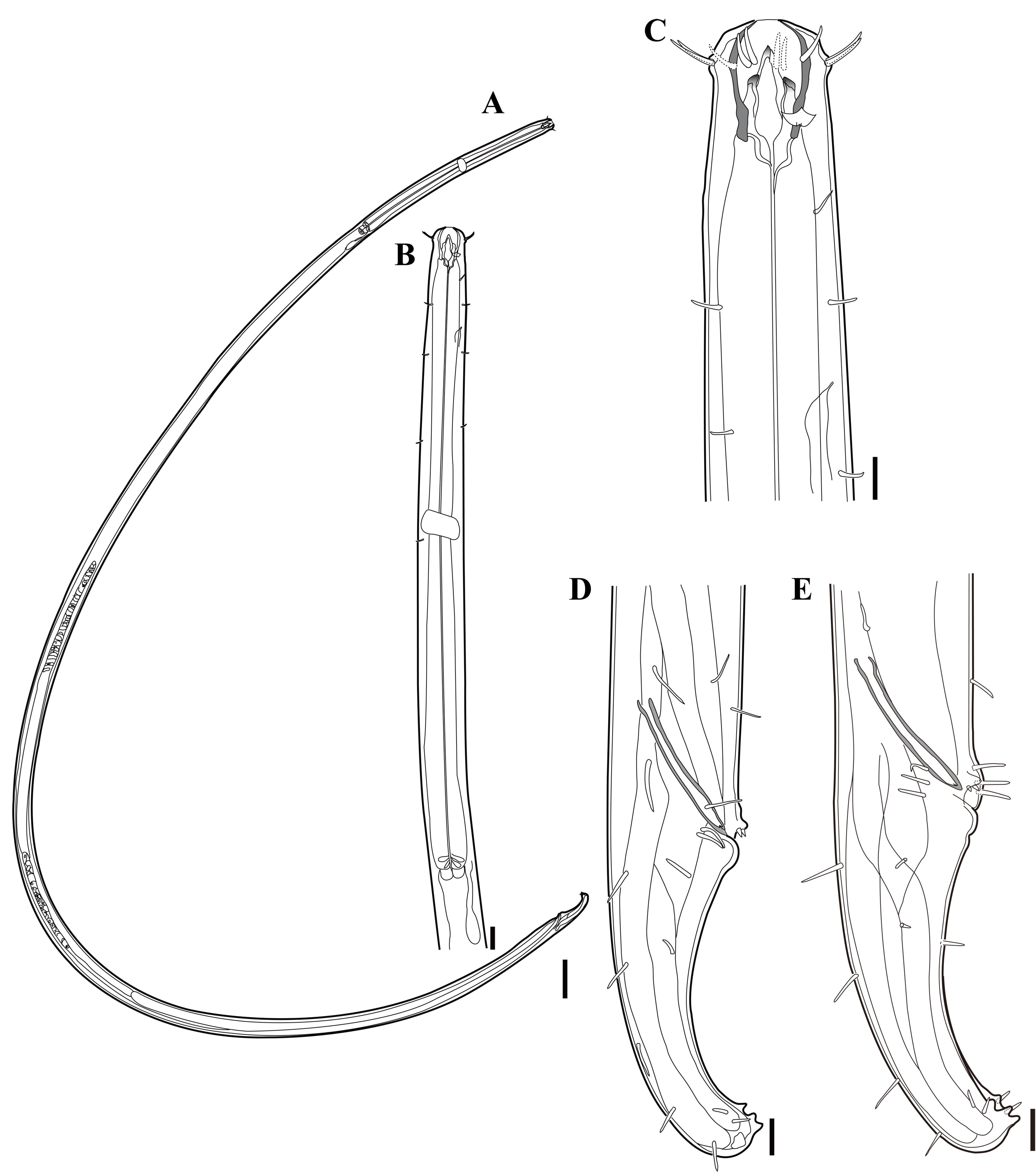
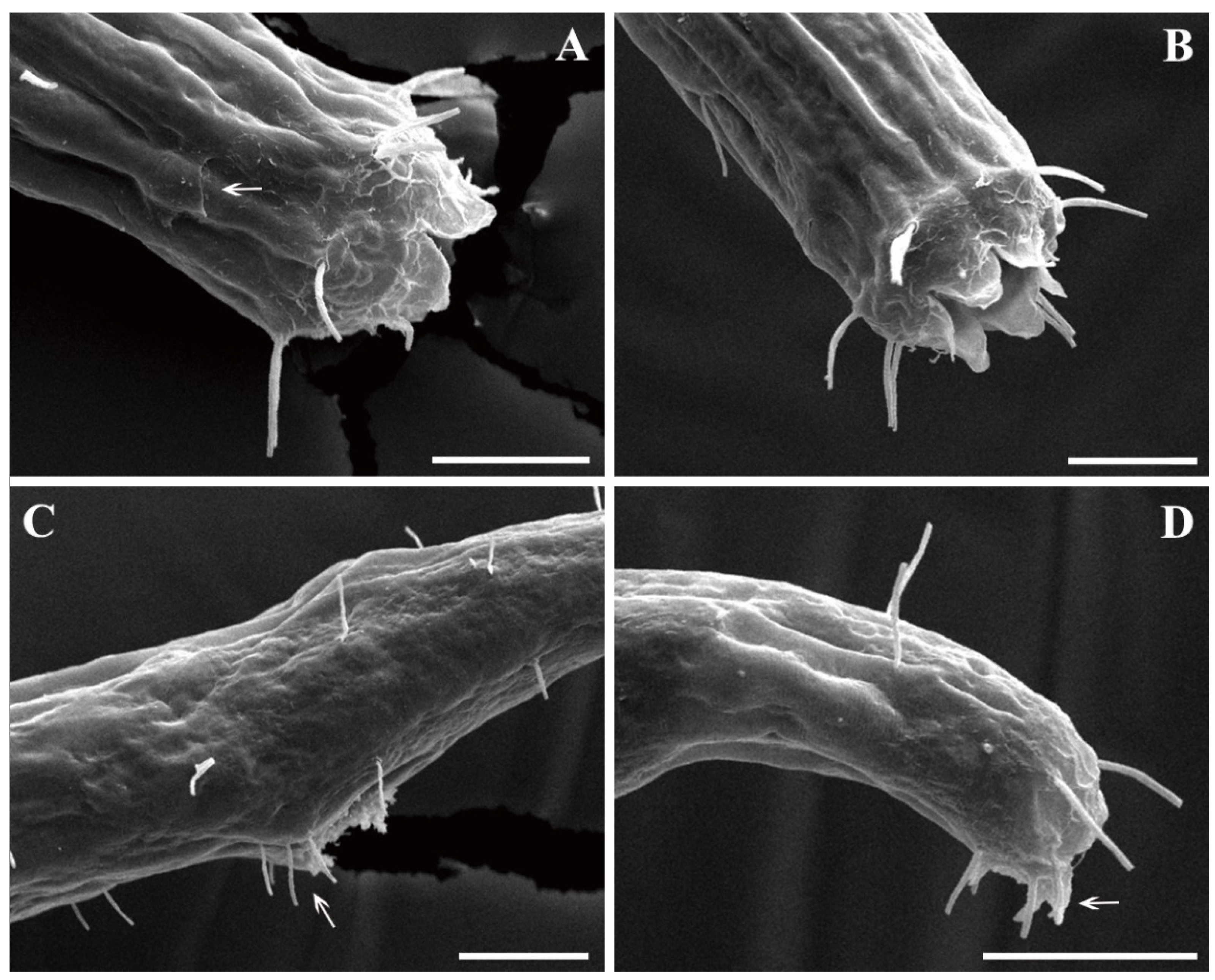
3.1. Description of Oncholaimus tripapillatus sp. nov.
3.1.1. Type Material
3.1.2. Type Locality and Habitat
3.1.3. Etymology
3.1.4. Measurements
3.1.5. Diagnosis
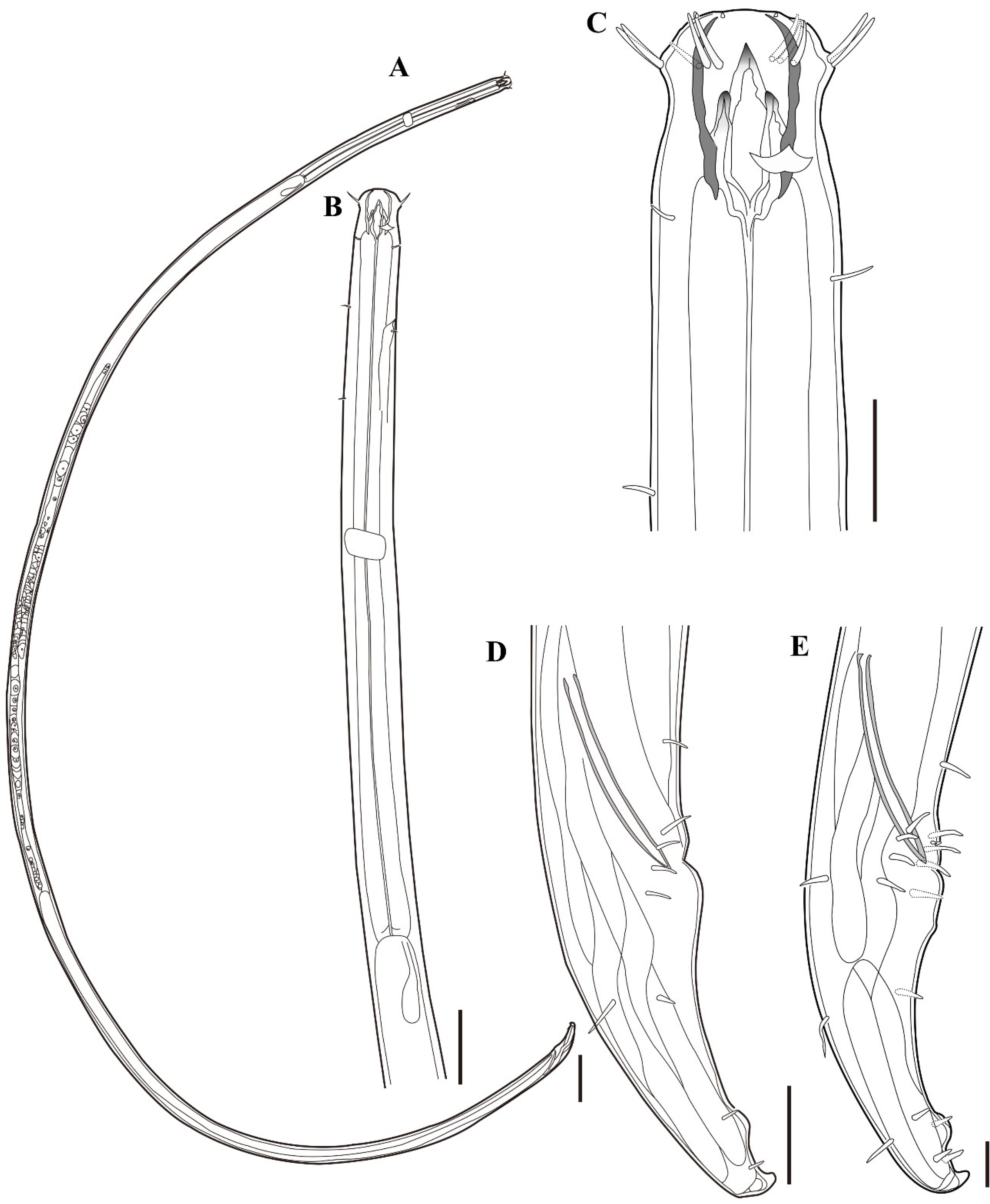
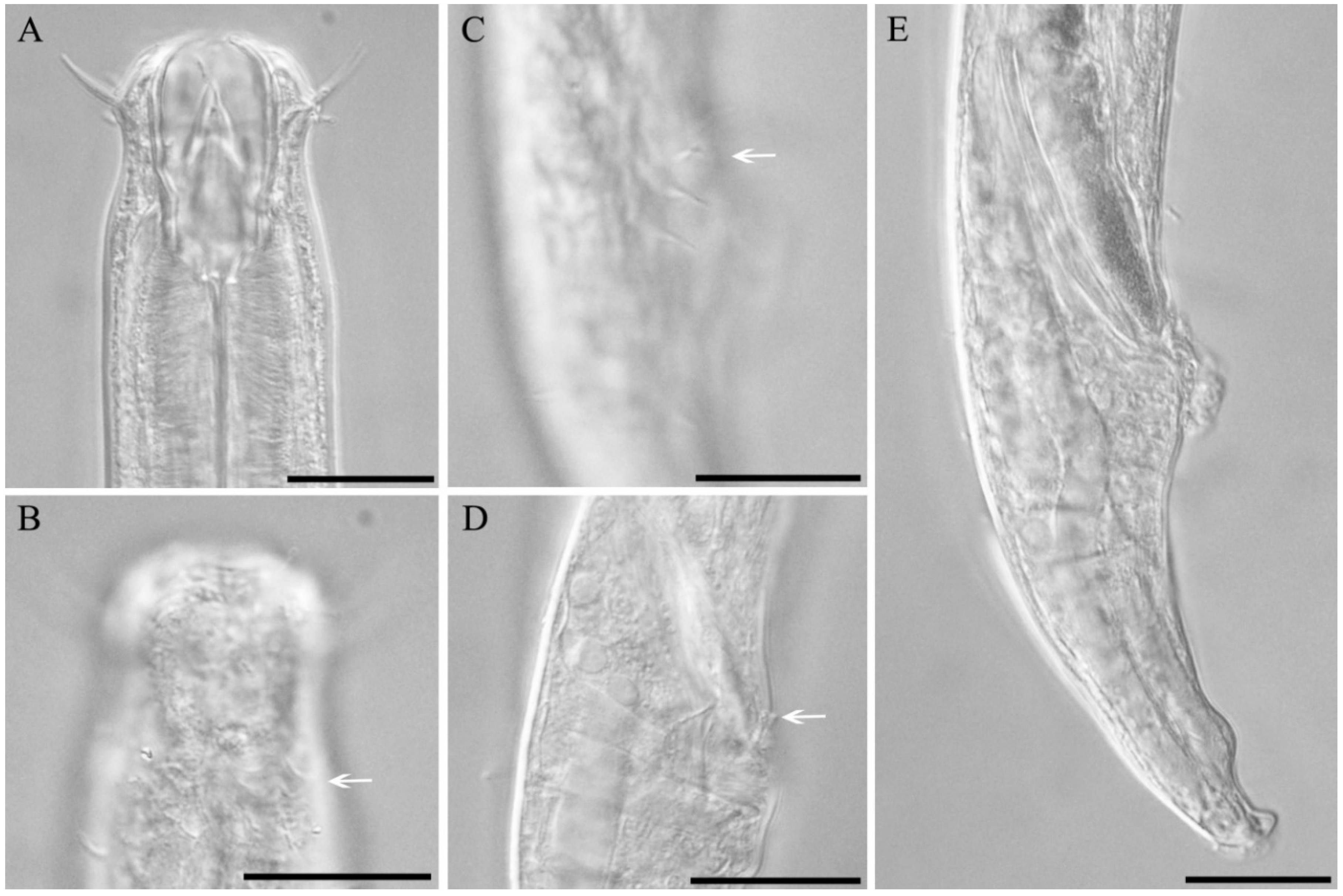
3.1.6. Description
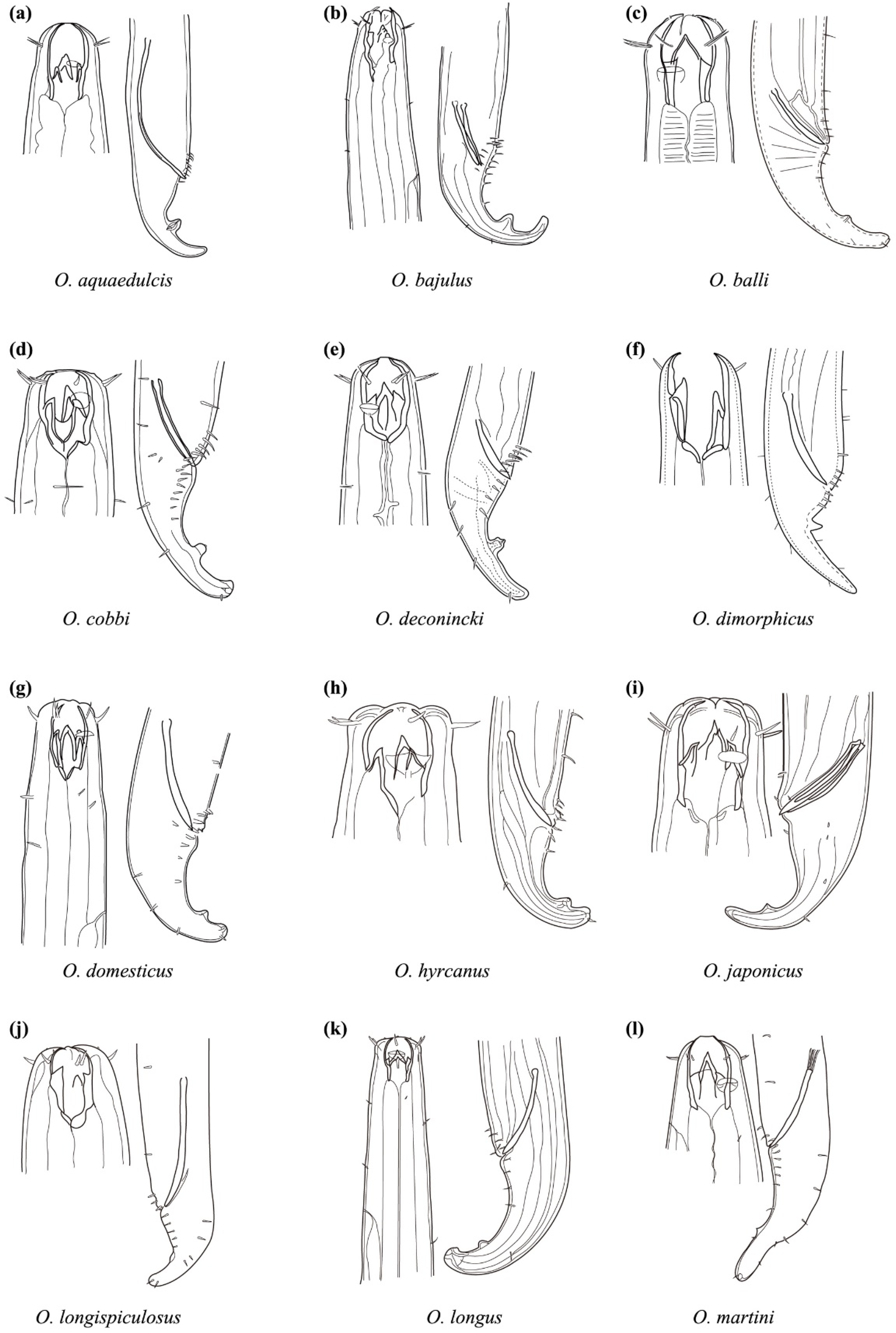
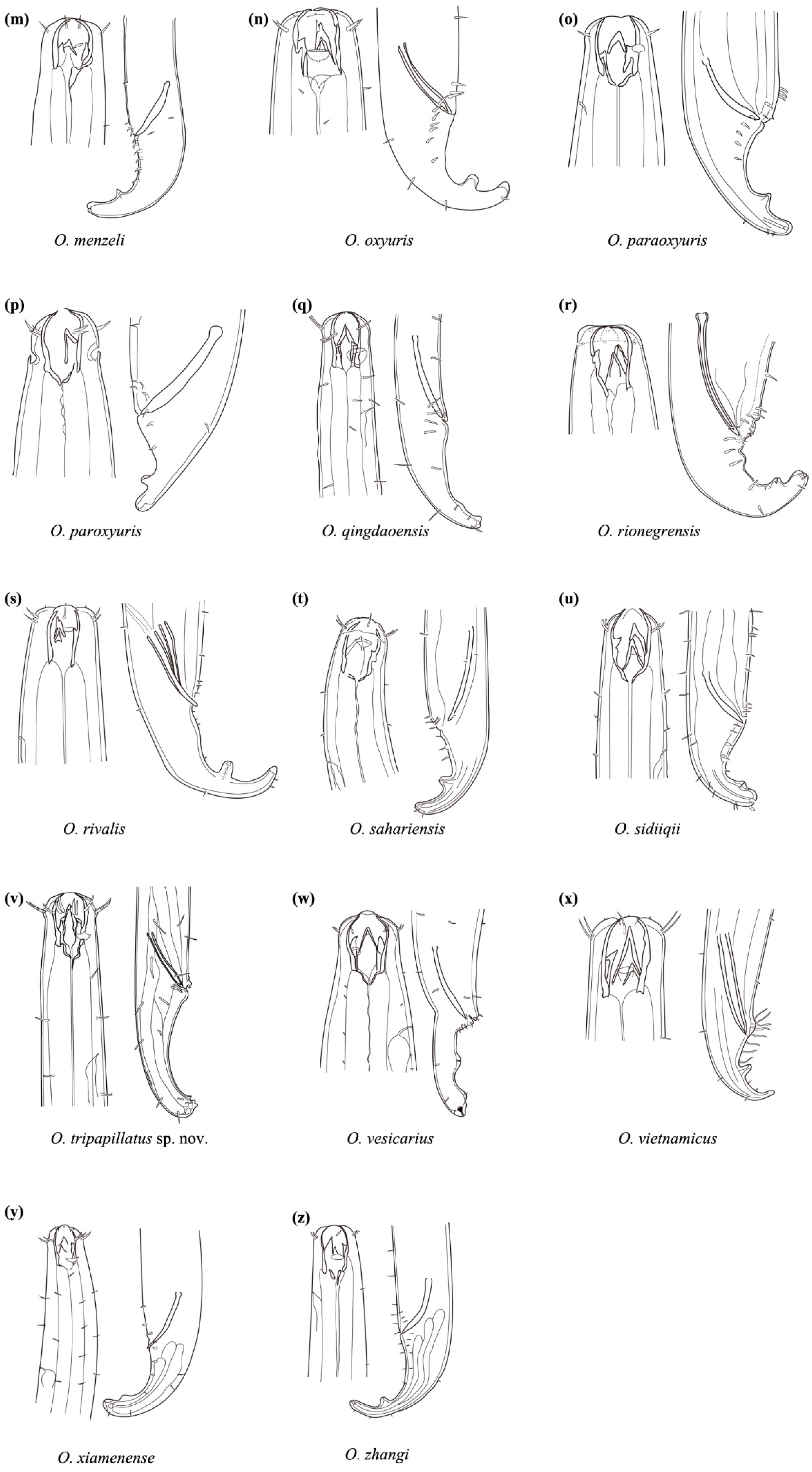
3.1.7. Differential Diagnosis and Relationships
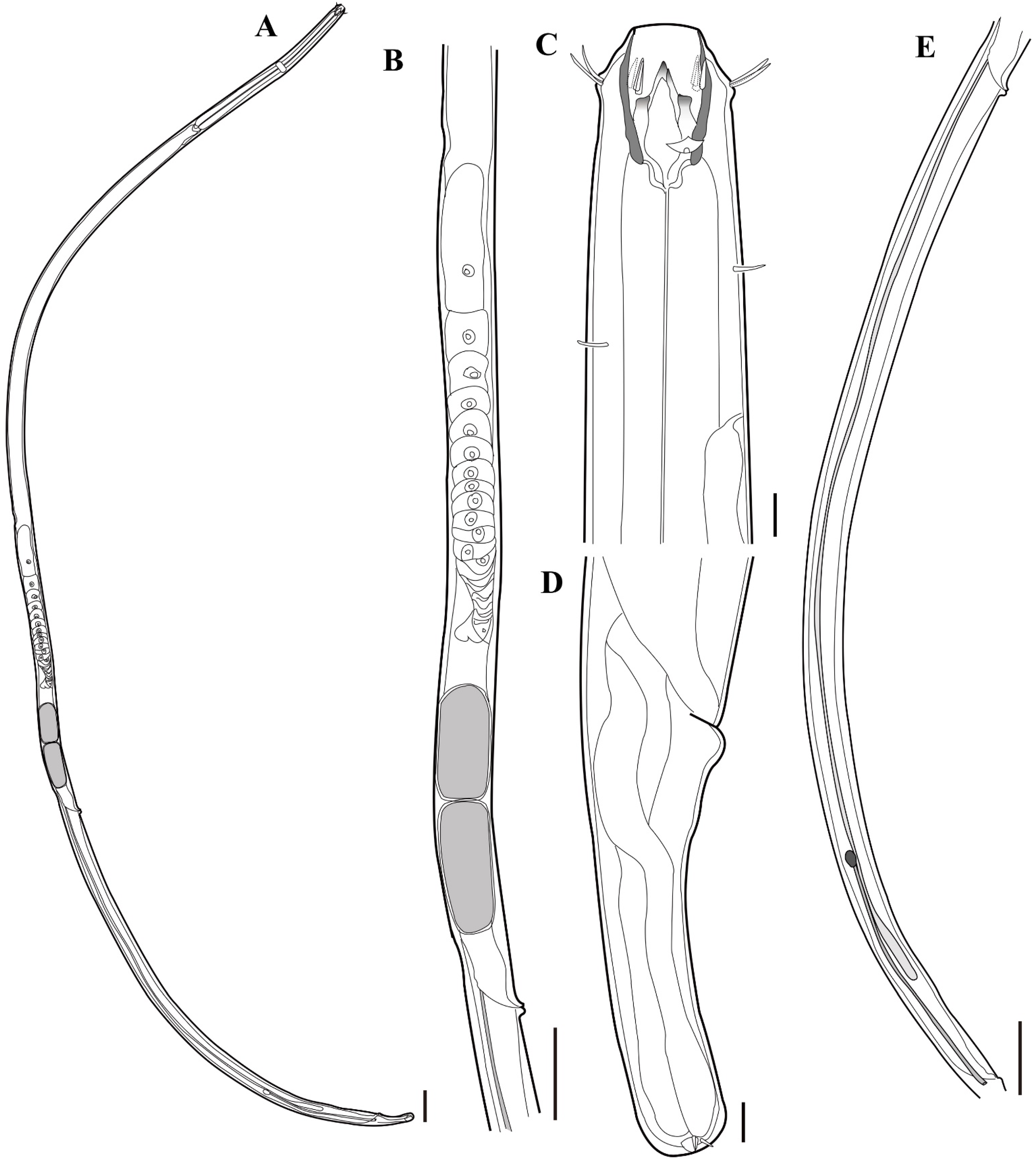
3.2. Description of Oncholaimus Qingdaoensis Zhang & Platt, 1983
3.2.1. Material Examined
3.2.2. Locality and Habitats
3.2.3. Measurements
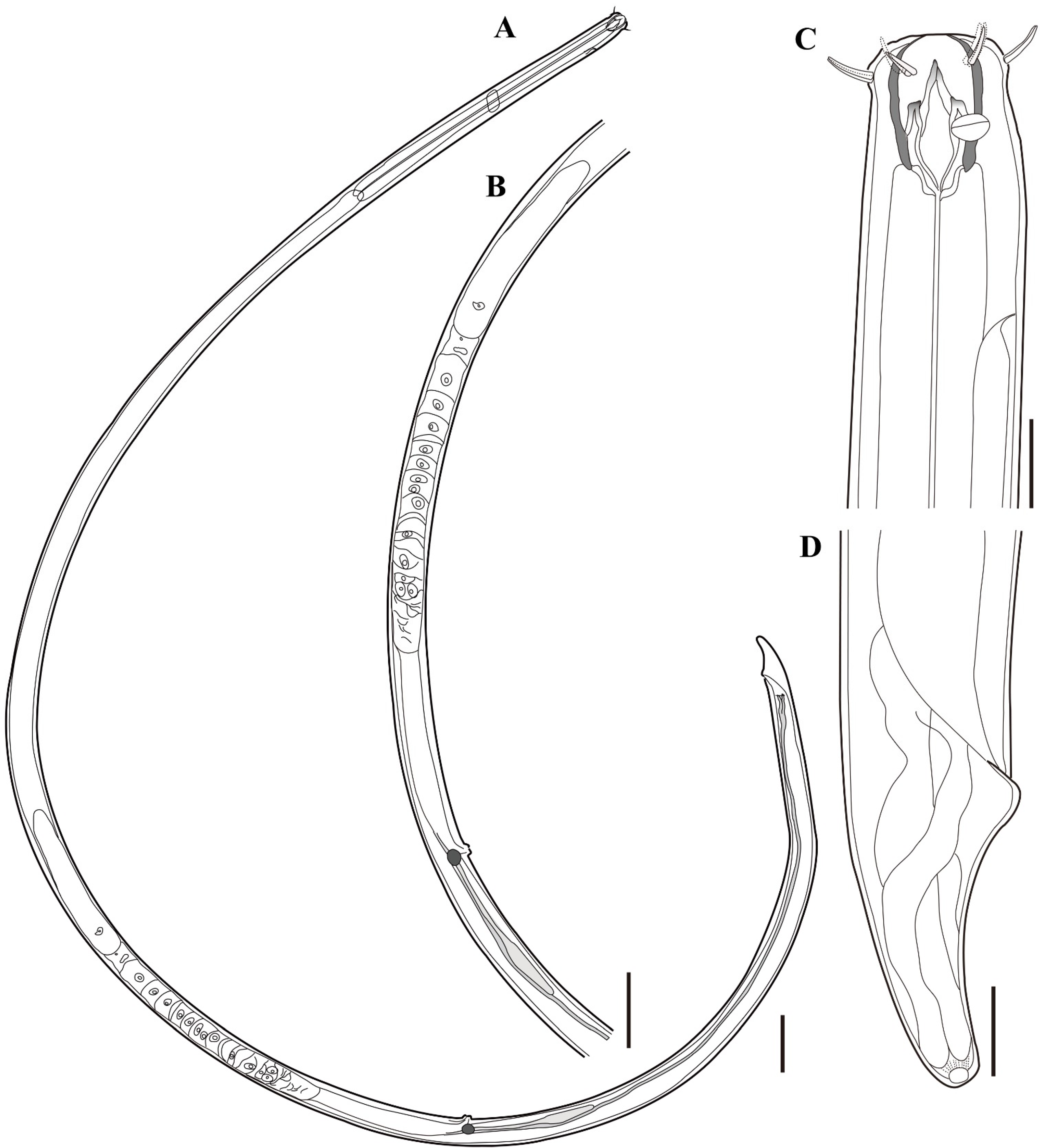
3.2.4. Description
3.2.5. Distribution
3.2.6. Differential Diagnosis and Relationships
Author Contributions
Funding
Institutional Review Board Statement
Informed Consent Statement
Data Availability Statement
Conflicts of Interest
References
- Smol, N.; Muthumbi, A.; Sharma, J. Handbook of Zoology. Gastrotricha, Cycloneuralia and Gnathifera; Nematoda. 7.3 Order Enoplida; Walter De Gruyter: Berlin, Germany, 2013; Volume 2. [Google Scholar]
- Gerlach, S.; Riemann, F. The Bremerhaven Checklist of Aquatic Nematodes. A Catalogue of Nematoda Adenophorea excluding the Dorylaimida. Veröffentlichungen Inst. Meeresforsch. Bremerhav. Suppl. 1974, 4, 405–736. [Google Scholar]
- Zeppilli, D.; Bellec, L.; Cambon-Bonavita, M.-A.; Decraemer, W.; Fontaneto, D.; Fuchs, S.; Gayet, N.; Mandon, P.; Michel, L.N.; Portail, M.; et al. Ecology and trophic role of Oncholaimus dyvae sp. nov.(Nematoda: Oncholaimidae) from the lucky strike hydrothermal vent field (Mid-Atlantic Ridge). BMC Zool. 2019, 4, 1–15. [Google Scholar] [CrossRef]
- Shimada, D.; Suzuki, A.C.; Tsujimoto, M.; Imura, S.; Kakui, K. Oncholaimus langhovdensis sp. nov.(Nematoda: Enoplea: Oncholaimida), a new species of free-living marine nematode from Langhovde, Dronning Maud Land, East Antarctica. Species Divers. 2017, 22, 151–159. [Google Scholar] [CrossRef] [Green Version]
- Belogurov, O.; Belogurova, L.; Pavlyuk, O. Morphology and systematic position of two new species of marine nematodes (Nematoda: Oncholaimidae) from Far-Eastern seas: Oncholaimium olium sp. n. and Pseudoncholaimus vesicarius (Wieser, 1959) comb. nov. Biol. Morya 1975, 2, 25–30. [Google Scholar]
- Belogurov, O.; Belogurova, L. Morphology of Oncholaimus paropisthonchus sp. n., Oncholaimus zinus sp. n., Oncholimium unicum sp. n. and key for genera of the subfamily Oncholaiminae(Nematoda). Zool. Zhurnal 1978, 57, 977–986. [Google Scholar]
- Belogurov, O.; Belogurova, L.; Pavlyuk, O. Three new species of free-living nematodes from the family Oncholaimidae from the Far-Eastern seas of the USSR, and a differential key for Pseudoncholaimus species. In Coastal Plankton and Benthos of the Northern Parts of the Sea of Japan; Akademiya Nauk SSSR, Dal’nevostochnyĭ Nauchnyĭ Tsentr; Institut Biologii Morya: Vladivostok, Russia, 1980; pp. 57–64. [Google Scholar]
- Smolyanko, O.; Belogyrov, O. On the study of the morphology and taxonomy on two nematode species of the genus Oncholaimium (Nematoda, Oncholaimidae) and comments on the independence of this genus and on synonymy of O. olium and O. sinensis. Zool. Zhurnal 1991, 70, 17–24. [Google Scholar]
- Wieser, W. A collection of marine nematodes from Japan. Publ. Seto Mar. Biol. Lab. 1955, 4, 159–181. [Google Scholar] [CrossRef] [Green Version]
- Shimada, D.; Kajihara, H.; Mawatari, S.F. Three new species of free-living marine nematodes (Nematoda: Enoplida) from northern Japan. Species Divers. 2009, 14, 137–150. [Google Scholar] [CrossRef] [Green Version]
- Zhang, Z.; Platt, H.M. New species of marine nematodes from Qingdao, China. Bull. Br. Mus. Nat. Hist. Zool. 1983, 45, 253–561. [Google Scholar] [CrossRef]
- Chen, Y.; Guo, Y. Three new species of free-living marine nematodes from East China Sea. Zootaxa 2014, 3841, 117–126. [Google Scholar] [CrossRef] [PubMed] [Green Version]
- Gao, Q.; Huang, Y. Oncholaimus zhangi sp. nov.(Oncholaimidae, Nematoda) from the intertidal zone of the East China Sea. Chin. J. Oceanol. Limnol. 2017, 35, 1212–1217. [Google Scholar] [CrossRef]
- Lee, H.J.; Rho, H.S.; Jung, J. New Record of the Genus Oncholaimus Nematode Species (Nematoda: Oncholaimidae) from the East Sea of Korea. Korean J. Environ. Biol. 2015, 33, 170–176. [Google Scholar] [CrossRef]
- Kristensen, R.M.; Higgins, R.P. Marine Tardigrada from the southeastern United States coastal waters I. Paradoxipus orzeliscoides n. gen., n. sp.(Arthrotardigrada: Halechiniscidae). Trans. Am. Microsc. Soc. 1989, 262–282. [Google Scholar] [CrossRef]
- Shirayama, Y.; Kaku, T.; Higgins, R.P. Double-sided microscopic observation of meiofauna using an HS-slide. Benthos Res. 1993, 44, 41–44. [Google Scholar] [CrossRef]


| Characters | Holotype | Paratypes | ||||
|---|---|---|---|---|---|---|
| 1♂ | 1♂ | 2♂ | 1♀ | 2♀ | 3♀ | |
| Total body length | 4281 | 4435 | 4071 | 4514 | 4661 | 4520 |
| a | 82 | 86 | 83 | 70 | 64 | 72 |
| b | 8 | 8 | 8 | 7 | 8 | 8 |
| c | 46 | 48 | 42 | 41 | 41 | 41 |
| Head diameter at the level of cephalic setae | 30 | 30 | 30 | 31 | 32 | 30 |
| Body diameter at the level of cardia | 42 | 42 | 43 | 52 | 54 | 46 |
| Maximum body diameter | 52 | 51 | 49 | 65 | 73 | 63 |
| Cephalic setae length | 10 | 11 | 12 | 11 | 11 | 11 |
| Amphideal fovea diameter | 9 | 8 | 11 | 10 | 9 | 10 |
| Amphideal fovea length | 5 | 6 | 7 | 6 | 6 | 5 |
| Buccal cavity diameter | 19 | 20 | 20 | 20 | 21 | 22 |
| Buccal cavity length | 29 | 31 | 31 | 31 | 34 | 33 |
| Anterior end to the largest tooth tip | 7 | 8 | 7 | 9 | 8 | 9 |
| Anterior end to the other teeth tips | 14 | 16 | 14 | 18 | 15 | 17 |
| Anterior end to amphids | 22 | 23 | 25 | 22 | 22 | 23 |
| Anterior end to excretory pores | 88 | 80 | 85 | 95 | 87 | 91 |
| Anterior end to the nerve ring | 251 | 254 | 247 | 274 | 271 | 264 |
| Pharynx length | 563 | 548 | 545 | 606 | 602 | 589 |
| Spicule length | 44 | 42 | 41 | - | - | - |
| Cloacal setae length | 8 | 7 | 7 | - | - | - |
| Anterior end to the vulva | - | - | - | 2987 | 3107 | 2968 |
| Body diameter at level of the vulva | - | - | - | 61 | 70 | 61 |
| V (%) | - | - | - | 66 | 67 | 66 |
| Anal body diameter | 35 | 36 | 37 | 37 | 36 | 35 |
| Tail length | 94 | 92 | 96 | 110 | 114 | 111 |
| Tail length/anal body diameter | 2.7 | 2.5 | 2.6 | 3 | 3.2 | 3.1 |
| Species | Characters | ||||||
|---|---|---|---|---|---|---|---|
| Body Length (µm) | a | b | c | Buccal Cavity Height (µm) | Spicule Length (µm) | Number of Tail Papillae | |
| O. aquaedulcis Schneider, 1937 | 2600–2900 | 60–71 | 7–9 | 28–42 | 39 | 100 | 1 |
| O. bajulus Paramonov, 1937 | 3144–3676 | 54–61 | 7–8 | 48–50 | 35 | 40 | 1 |
| O. balli Nicholas & Stewart, 1984 | 1834–2314 | 52–69 | 7–8 | 33–51 | 26–28 | 26–34 | 1 |
| O. cobbi Gerlach, 1954 | 3452 | 40 | 7 | 47 | 42 | 57 | 1 |
| O. deconincki Heyns & Coomans, 1977 | 3200–3770 | 66–80 | 7–9 | 36–47 | 32–41 | 35–41 | 1 |
| O. dimorphicus Tahseen et. al., 2016 | 1925–2076 | 69–77 | 6 | 48–57 | - | 36–38 | 1 |
| O. domesticus Chitwood, 1960 | 2700–3350 | 54–78 | 7 | 39–47 | 36–38 | 51–54 | 1 |
| O. hyrcanus Tchesunov, 1976 | 2810 | 57 | 7 | 36 | 29 | 55 | 1 |
| O. japonicas Belogurov & Belogurova, 1981 | 3250–3420 | 60–89 | 9–10 | 52–62 | 26–31 | 37–41 | 1 |
| O. longispiculosus Gerlach, 1955 | 3090 | 31 | 7 | 46 | 40 | 108 | 1 |
| O. longus Wieser, 1953 | 5000–6740 | 130–150 | 13 | 104–108 | 26 | 42 | 1 |
| O. martini Wieser, 1959 | 3890 | 65 | 8 | 43 | 36 | 51 | 1 |
| O. menzeli Schneider, 1937 | 1885–2084 | 75–83 | 6 | 34–45 | 22 | 30 | 1 |
| O. oxyuris Gerlach, 1957 | 2970 | 62 | 7 | 42 | 32 | 45 | 1 |
| O. paraoxyuris Salma et. al, 2017 | 4100 | 83 | 11 | 64 | 38 | 50 | 1 |
| O. paroxyuris Schuurmans Stekhoven, 1942 | 1512 | 42 | 6 | 47 | - | - | 1 |
| O. qingdaoensis Zhang & Platt, 1983 | 2380–2640 | 92–98 | 6–7 | 48–53 | 25–27 | 34–36 | 1 |
| O. rionegrensis Pastor de Ward et. al., 2013 | 2440–3030 | 37–51 | 6–7 | 30–45 | 26–33 | 47–57 | 1 |
| O. rivalis Gagarin & Gusakov, 2012 | 3730 | 57 | 8 | 47 | 35 | 53 | 1 |
| O. sahariensis Coomans & Heyns, 1983 | 3070 | 63 | 7 | 46 | 27 | 48 | 1 |
| O. sidiiqii Tauheed & Ahmad, 2016 | 2420–2820 | 59–66 | 6 | 39–50 | 34–38 | 35–40 | 1 |
| O. tripapillatus sp. nov. | 4071–4435 | 82–86 | 7–8 | 42–48 | 29–31 | 41–44 | 3 |
| O. vesicarius Wieser, 1959 | 3650 | 52 | 8 | 72 | 32 | 38 | 2 |
| O. vietnamicus Gagarin & Thanh, 2011 | 1758–2226 | 70–77 | 5–7 | 55–59 | 26–29 | 52–56 | 1 |
| O. xiamenense Chen & Guo, 2014 | 2480–3020 | 57–81 | 6–7 | 37–42 | 27–32 | 44–52 | 1 |
| O. zhangi Gao & Huang, 2016 | 3718–3934 | 55–62 | 7 | 34–37 | 33–35 | 52–56 | 1 |
| Characters | 1♂ | 2♂ | 3♂ | 1♀ | 2♀ |
|---|---|---|---|---|---|
| Body length | 3379 | 3505 | 3715 | 4061 | 3550 |
| a | 82 | 86 | 85 | 75 | 67 |
| b | 7 | 7 | 7 | 7 | 7 |
| c | 46 | 45 | 44 | 56 | 43 |
| Head diameter at the level of cephalic setae | 28 | 29 | 29 | 32 | 31 |
| Body diameter at the level of the cardia | 36 | 40 | 38 | 44 | 47 |
| Maximum body diameter | 41 | 41 | 44 | 54 | 53 |
| Cephalic setae length | 10 | 12 | 11 | 11 | 11 |
| Amphideal fovea diameter | 10 | 12 | 11 | 10 | 12 |
| Amphideal fovea length | 5 | 6 | 7 | 5 | 7 |
| Buccal cavity diameter | 17 | 19 | 18 | 17 | 21 |
| Buccal cavity length | 32 | 31 | 33 | 35 | 34 |
| Anterior end to the largest tooth tip | 5 | 5 | 7 | 6 | 8 |
| Anterior end to the other teeth tips | 13 | 12 | 13 | 12 | 14 |
| Anterior end to amphids | 22 | 20 | 23 | 22 | 22 |
| Anterior end to excretory pores | 84 | 79 | 77 | 68 | 81 |
| Anterior end to the nerve ring | 229 | 241 | 243 | 265 | 247 |
| Pharynx length | 492 | 533 | 555 | 560 | 514 |
| Spicule length | 47 | 50 | 52 | - | - |
| Cloacal setae length | 8 | 8 | 8 | - | - |
| Anterior end to the vulva | - | - | - | 2812 | 2990 |
| Body diameter at level of the vulva | - | - | - | 54 | 56 |
| V (%) | - | - | - | 69 | 84 |
| Anal body diameter | 30 | 29 | 32 | 38 | 40 |
| Tail length | 74 | 79 | 85 | 72 | 83 |
| Tail length/anal body diameter | 2.5 | 2.7 | 2.7 | 1.9 | 2.1 |
Publisher’s Note: MDPI stays neutral with regard to jurisdictional claims in published maps and institutional affiliations. |
© 2021 by the authors. Licensee MDPI, Basel, Switzerland. This article is an open access article distributed under the terms and conditions of the Creative Commons Attribution (CC BY) license (https://creativecommons.org/licenses/by/4.0/).
Share and Cite
Lee, H.; Lee, H.; Rho, H. Oncholaimus tripapillatus sp. nov., a New Free-Living Marine Nematode of the Genus Oncholaimus Dujardin, 1845 (Nematoda: Enoplida: Oncholaimidae) from the Subtidal Sediment of Dokdo Island, East Sea, Korea, with a New Record of O. qingdaoensis Zhang & Platt, 1983. J. Mar. Sci. Eng. 2021, 9, 1334. https://doi.org/10.3390/jmse9121334
Lee H, Lee H, Rho H. Oncholaimus tripapillatus sp. nov., a New Free-Living Marine Nematode of the Genus Oncholaimus Dujardin, 1845 (Nematoda: Enoplida: Oncholaimidae) from the Subtidal Sediment of Dokdo Island, East Sea, Korea, with a New Record of O. qingdaoensis Zhang & Platt, 1983. Journal of Marine Science and Engineering. 2021; 9(12):1334. https://doi.org/10.3390/jmse9121334
Chicago/Turabian StyleLee, Hyojin, Heegab Lee, and Hyunsoo Rho. 2021. "Oncholaimus tripapillatus sp. nov., a New Free-Living Marine Nematode of the Genus Oncholaimus Dujardin, 1845 (Nematoda: Enoplida: Oncholaimidae) from the Subtidal Sediment of Dokdo Island, East Sea, Korea, with a New Record of O. qingdaoensis Zhang & Platt, 1983" Journal of Marine Science and Engineering 9, no. 12: 1334. https://doi.org/10.3390/jmse9121334