
The Pandemic and Your Skin—Direct and Indirect Impact of COVID-19
 , , , ,
  , and
, and 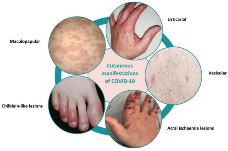
Abstract
:1. Introduction
2. Skin Manifestations Related to COVID-19
2.1. Cutaneous Manifestations Associated with COVID-19
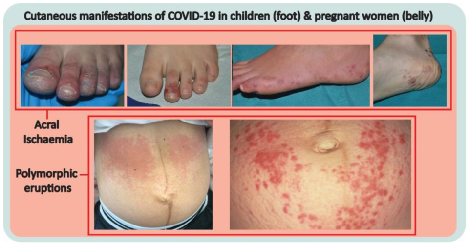
2.2. Cutaneous Manifestations in Children and Pregnant Women
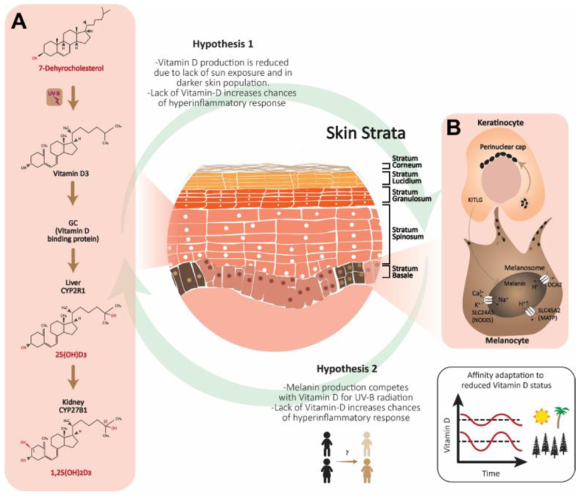
3. Role of Vitamin D Deficiency and Skin Demographics with COVID-19
3.1. Understanding the Molecular Mechanisms behind Vitamin D Deficiency
3.2. Role of Vitamin D: Skin Pigmentation and Skin Color Demographics
4. The Health of the Skin
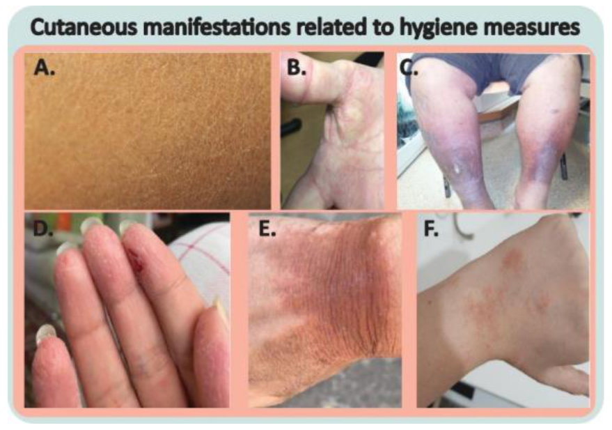
4.1. The Consequences of Excess Hygiene Measures

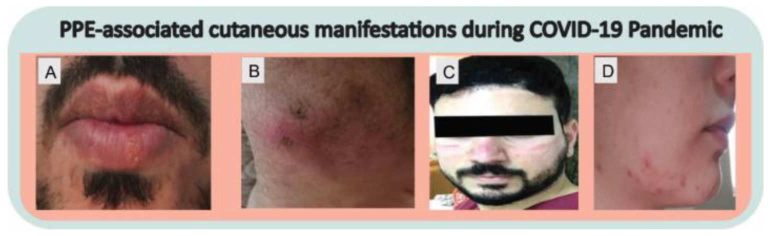
4.2. PPE-Associated Cutaneous Manifestations
5. Protection of Skin from Harmful UV Radiation
5.1. Sunscreens
5.2. Other Methods to Protect the Skin from Harmful UV Radiation
- Slop a broad-spectrum and water-resistant sunscreen labeled SPF30 or higher 20 min before going outdoors and reapplying every two hours;
- Slap on a hat that provides shade to the head, ears, face, and neck (high protection factor);
- Seek shade coverage.
- Slide on direct, scattered, and reflected solar UVR glasses tested to Australian standards and labeled as “sunglasses or as special purposes sunglasses.”
6. Conclusions
Funding
Institutional Review Board Statement
Informed Consent Statement
Data Availability Statement
Acknowledgments
Conflicts of Interest
References
- Li, H.; Liu, Z.; Ge, J. Scientific research progress of COVID-19/SARS-CoV-2 in the first five months. J. Cell. Mol. Med. 2020, 24, 6558–6570. [Google Scholar] [CrossRef]
- Criado, P.R.; Ianhez, M.; Silva de Castro, C.C.; Talhari, C.; Ramos, P.M.; Miot, H.A. COVID-19 and skin diseases: Results from a survey of 843 patients with atopic dermatitis, psoriasis, vitiligo and chronic urticaria. J. Eur. Acad. Dermatol. Venereol. 2022, 36, e1–e3. [Google Scholar] [CrossRef] [PubMed]
- Dellino, M.; Cascardi, E.; Vinciguerra, M.; Lamanna, B.; Malvasi, A.; Scacco, S.; Acquaviva, S.; Pinto, V.; Di Vagno, G.; Cormio, G.; et al. Nutrition as Personalized Medicine against SARS-CoV-2 Infections: Clinical and Oncological Options with a Specific Female Groups Overview. Int. J. Mol. Sci. 2022, 23, 9136. [Google Scholar] [CrossRef] [PubMed]
- Novak, N.; Peng, W.; Naegeli, M.C.; Galvan, C.; Kolm-Djamei, I.; Brüggen, C.; Cabanillas, B.; Schmid-Grendelmeier, P.; Catala, A. SARS-CoV-2, COVID-19, skin and immunology—What do we know so far? Allergy 2021, 76, 698–713. [Google Scholar] [CrossRef] [PubMed]
- Swarnkar, B.; Bhari, N. Skin amidst COVID-19 pandemic. Dermatol. Ther. 2020, 33, e13789. [Google Scholar] [CrossRef]
- Xu, Q.; Zhang, L.; Chen, L.; Zhao, X.; Wang, X.; Hu, M.; Le, Y.; Xue, F.; Li, X.; Zheng, J. SARS-CoV-2 might transmit through the skin while the skin barrier function could be the mediator. Med. Hypotheses 2022, 159, 110752. [Google Scholar] [CrossRef]
- Imran, M.; Iqubal, M.K.; Imtiyaz, K.; Saleem, S.; Mittal, S.; Rizvi, M.M.A.; Ali, J.; Baboota, S. Topical nanostructured lipid carrier gel of quercetin and resveratrol: Formulation, optimization, in vitro and ex vivo study for the treatment of skin cancer. Int. J. Pharm. 2020, 587, 119705. [Google Scholar] [CrossRef]
- Asai, Y.; Nguyen, P.; Hanna, T.P. Impact of the COVID-19 pandemic on skin cancer diagnosis: A population-based study. PLoS ONE 2021, 16, e0248492. [Google Scholar] [CrossRef]
- Seretis, K.; Boptsi, E.; Boptsi, A.; Lykoudis, E.G. The impact of treatment delay on skin cancer in COVID-19 era: A case-control study. World J. Surg. Oncol. 2021, 19, 350. [Google Scholar] [CrossRef]
- Gül, Ü. COVID-19 and dermatology. Turk. J. Med. Sci. 2020, 50, 1751–1759. [Google Scholar] [CrossRef]
- Genovese, G.; Moltrasio, C.; Berti, E.; Marzano, A.V. Skin Manifestations Associated with COVID-19: Current Knowledge and Future Perspectives. Dermatology 2021, 237, 1–12. [Google Scholar] [CrossRef]
- Shinkai, K.; Bruckner, A.L. Dermatology and COVID-19. JAMA 2020, 324, 1133–1134. [Google Scholar] [CrossRef]
- Jia, J.L.; Kamceva, M.; Rao, S.A.; Linos, E. Cutaneous manifestations of COVID-19: A preliminary review. J. Am. Acad. Dermatol. 2020, 83, 687–690. [Google Scholar] [CrossRef]
- Athwani, V.; Gothwal, S. Dermatological Manifestations of COVID-19 in Children. J. Ski. Stem Cell 2020, 7, e106890. [Google Scholar] [CrossRef]
- Mariyath, O.K.R.; Samad, K.A.; Devi, K.; Surya, V.S.; Effeena, M.D.; Ajina, M.; Diseases, S.T. Atypical maculopapular rash as the initial sign of COVID-19: A case report from a COVID hospital. J. Ski. Sex. Transm. Dis. 2021, 3, 87–90. [Google Scholar] [CrossRef]
- Polly, S.; Fernandez, A.P. Common skin signs of COVID-19 in adults: An update. Cleve. Clin. J. Med. 2022, 89, 161–167. [Google Scholar] [CrossRef]
- Galván Casas, C.; Català, A.; Carretero Hernández, G.; Rodríguez-Jiménez, P.; Fernández-Nieto, D.; Rodríguez-Villa Lario, A.; Navarro Fernández, I.; Ruiz-Villaverde, R.; Falkenhain-López, D.; Llamas Velasco, M.; et al. Classification of the cutaneous manifestations of COVID-19: A rapid prospective nationwide consensus study in Spain with 375 cases. Br. J. Dermatol. 2020, 183, 71–77. [Google Scholar] [CrossRef]
- Calvão, J.; Relvas, M.; Pinho, A.; Brinca, A.; Cardoso, J.C. Acro-ischaemia and COVID-19 infection: Clinical and histopathological features. J. Eur. Acad. Dermatol. Venereol. 2020, 34, e653–e754. [Google Scholar] [CrossRef]
- Henry, D.; Ackerman, M.; Sancelme, E.; Finon, A.; Esteve, E. Urticarial eruption in COVID-19 infection. J. Eur. Acad. Dermatol. Venereol. 2020, 34, e244–e245. [Google Scholar] [CrossRef] [Green Version]
- Palaniappan, V.; Subramaniam, K.; Karthikeyan, K. Papular-Vesicular Rash in COVID-19. Am. J. Trop. Med. Hyg. 2021, 105, 551–552. [Google Scholar] [CrossRef]
- Cazzato, G.; Cascardi, E.; Colagrande, A.; Foti, C.; Stellacci, A.; Marrone, M.; Ingravallo, G.; Arezzo, F.; Loizzi, V.; Solimando, A.G.; et al. SARS-CoV-2 and Skin: New Insights and Perspectives. Biomolecules 2022, 12, 1212. [Google Scholar] [CrossRef] [PubMed]
- Lavery, M.J.; Bouvier, C.A.; Thompson, B. Cutaneous manifestations of COVID-19 in children (and adults): A virus that does not discriminate. Clin. Dermatol. 2021, 39, 323–328. [Google Scholar] [CrossRef] [PubMed]
- Andina, D.; Belloni-Fortina, A.; Bodemer, C.; Bonifazi, E.; Chiriac, A.; Colmenero, I.; Diociaiuti, A.; El-Hachem, M.; Fertitta, L.; van Gysel, D.; et al. Skin manifestations of COVID-19 in children: Part 1. Clin. Exp. Dermatol. 2021, 46, 444–450. [Google Scholar] [CrossRef] [PubMed]
- Visconti, A.; Murray, B.; Rossi, N.; Wolf, J.; Ourselin, S.; Spector, T.D.; Freeman, E.E.; Bataille, V.; Falchi, M. Cutaneous Manifestations of SARS-CoV-2 infection during the Delta and Omicron waves in 348,691 UK users of the UK ZOE COVID Study App. Br. J. Dermatol. 2022, 187, 900–908. [Google Scholar] [CrossRef]
- Zulfiqar, A.-A.; Lorenzo Villalba, N.; Severac, F.; Andrès, E. Infection liée à la COVID-19 chez une série de sujets âgés: Résultats d’une étude préliminaire COVID-19 infection in a cohort of elderly patients in France: Results of a preliminary study. La Presse Médicale Form. 2020, 1, 460–463. [Google Scholar] [CrossRef]
- Proietti, I.; Bernardini, N.; Tolino, E.; Mambrin, A.; Balduzzi, V.; Marchesiello, A.; Michelini, S.; Skroza, N.; Potenza, C. Polymorphic eruption of pregnancy as a possible COVID-19 manifestation. Dermatol. Ther. 2020, 33, e14117. [Google Scholar] [CrossRef]
- Lim, R.K.; Kalagara, S.; Chen, K.K.; Mylonakis, E.; Kroumpouzos, G. Dermatology in a multidisciplinary approach with infectious disease and obstetric medicine against COVID-19. Int. J. Womens Dermatol. 2021, 7, 640–646. [Google Scholar] [CrossRef]
- Jan, Y.; Malik, M.; Yaseen, M.; Ahmad, S.; Imran, M.; Rasool, S.; Haq, A. Vitamin D fortification of foods in India: Present and past scenario. J. Steroid Biochem. Mol. Biol. 2019, 193, 105417. [Google Scholar] [CrossRef]
- Mohan, M.; Cherian, J.J.; Sharma, A. Exploring links between vitamin D deficiency and COVID-19. PLoS Pathog. 2020, 16, e1008874. [Google Scholar] [CrossRef]
- Demir, M.; Demir, F.; Aygun, H. Vitamin D deficiency is associated with COVID-19 positivity and severity of the disease. J. Med. Virol. 2021, 93, 2992–2999. [Google Scholar] [CrossRef]
- Lippi, G.; Ferrari, A.; Targher, G. Is COVID-19 lockdown associated with vitamin D deficiency? Eur. J. Public Health 2021, 31, 278–279. [Google Scholar] [CrossRef]
- Patel, P.; Hiam, L.; Sowemimo, A.; Devakumar, D.; McKee, M. Ethnicity and COVID-19. BMJ 2020, 369, 1–2. [Google Scholar] [CrossRef]
- Pareek, M.; Bangash, M.N.; Pareek, N.; Pan, D.; Sze, S.; Minhas, J.S.; Hanif, W.; Khunti, K. Ethnicity and COVID-19: An urgent public health research priority. Lancet 2020, 395, 1421–1422. [Google Scholar] [CrossRef]
- Aldridge, R.W.; Lewer, D.; Katikireddi, S.V.; Mathur, R.; Pathak, N.; Burns, R.; Fragaszy, E.B.; Johnson, A.M.; Devakumar, D.; Abubakar, I. Black, Asian and Minority Ethnic groups in England are at increased risk of death from COVID-19: Indirect standardisation of NHS mortality data. Wellcome Open Res. 2020, 5, 88. [Google Scholar] [CrossRef]
- Khunti, K.; Singh, A.K.; Pareek, M.; Hanif, W. Is ethnicity linked to incidence or outcomes of COVID-19? BMJ 2020, 369, 1–2. [Google Scholar] [CrossRef] [Green Version]
- Pan, D.; Sze, S.; Minhas, J.S.; Bangash, M.N.; Pareek, N.; Divall, P.; Williams, C.M.; Oggioni, M.R.; Squire, I.B.; Nellums, L.B. The impact of ethnicity on clinical outcomes in COVID-19: A systematic review. EClinicalMedicine 2020, 23, 100404. [Google Scholar] [CrossRef]
- Sze, S.; Pan, D.; Nevill, C.R.; Gray, L.J.; Martin, C.A.; Nazareth, J.; Minhas, J.S.; Divall, P.; Khunti, K.; Abrams, K.R. Ethnicity and clinical outcomes in COVID-19: A systematic review and meta-analysis. EClinicalMedicine 2020, 29, 100630. [Google Scholar] [CrossRef]
- Darling, A.L.; Ahmadi, K.R.; Ward, K.A.; Harvey, N.C.; Alves, A.C.; Dunn-Walters, D.K.; Lanham-New, S.A.; Cooper, C.; Blackbourn, D.J. Vitamin D status, body mass index, ethnicity and COVID-19: Initial analysis of the first-reported UK Biobank COVID-19 positive cases (n 580) compared with negative controls (n 723). MedRxiv 2020, 4. [Google Scholar] [CrossRef]
- Herrling, T.; Jung, K.; Fuchs, J. The role of melanin as protector against free radicals in skin and its role as free radical indicator in hair. Spectrochim. Acta Part A Mol. Biomol. Spectrosc. 2008, 69, 1429–1435. [Google Scholar] [CrossRef]
- Islam, M.T.; Salehi, B.; Karampelas, O.; Sharifi-Rad, J.; Docea, A.O.; Martorell, M.; Calina, D. High skin melanin content, vitamin D deficiency and immunity: Potential interference for severity of COVID-19. Farmacia 2020, 68, 970–983. [Google Scholar] [CrossRef]
- Chen, T.C.; Chimeh, F.; Lu, Z.; Mathieu, J.; Person, K.S.; Zhang, A.; Kohn, N.; Martinello, S.; Berkowitz, R.; Holick, M.F. Factors that influence the cutaneous synthesis and dietary sources of vitamin D. Arch. Biochem. Biophys. 2007, 460, 213–217. [Google Scholar] [CrossRef] [PubMed] [Green Version]
- Jagannath, V.A.; Filippini, G.; Di Pietrantonj, C.; Asokan, G.; Robak, E.W.; Whamond, L.; Robinson, S.A. Vitamin D for the management of multiple sclerosis. Cochrane Database Syst. Rev. 2018, 24, 9. [Google Scholar] [CrossRef] [PubMed]
- Aglipay, M.; Birken, C.S.; Parkin, P.C.; Loeb, M.B.; Thorpe, K.; Chen, Y.; Laupacis, A.; Mamdani, M.; Macarthur, C.; Hoch, J.S. Effect of high-dose vs standard-dose wintertime vitamin D supplementation on viral upper respiratory tract infections in young healthy children. JAMA 2017, 318, 245–254. [Google Scholar] [CrossRef] [PubMed]
- Esposito, S.; Lelii, M. Vitamin D and respiratory tract infections in childhood. BMC Infect. Dis. 2015, 15, 1–10. [Google Scholar] [CrossRef] [PubMed] [Green Version]
- Lester, J.; Jia, J.; Zhang, L.; Okoye, G.; Linos, E. Absence of images of skin of colour in publications of COVID-19 skin manifestations. Br. J. Dermatol. 2020, 183, 593–595. [Google Scholar] [CrossRef]
- Sangha, A.M. Dermatological Conditions in SKIN OF COLOR—: Managing Atopic Dermatitis. J. Clin. Aesthetic Dermatol. 2021, 14, S20. [Google Scholar]
- Kaufman, B.P.; Guttman-Yassky, E.; Alexis, A.F. Atopic dermatitis in diverse racial and ethnic groups—Variations in epidemiology, genetics, clinical presentation and treatment. Exp. Dermatol. 2018, 27, 340–357. [Google Scholar] [CrossRef] [Green Version]
- Akuffo-Addo, E.; Nicholas, M.N.; Joseph, M. COVID-19 Skin Manifestations in Skin of Colour. J. Cutan. Med. Surg. 2022, 26, 189–197. [Google Scholar] [CrossRef]
- Batarseh, E.; Kersten, B.P.; Pinelo, A.C.; Nadler, J.N.; Schwartz, S.A. Angioedema in African American patients hospitalized for COVID-19. Am. J. Respir. Crit. Care Med. 2020, 202, 1581–1584. [Google Scholar] [CrossRef]
- Freeman, E.E.; McMahon, D.E.; Lipoff, J.B.; Rosenbach, M.; Kovarik, C.; Takeshita, J.; French, L.E.; Thiers, B.H.; Hruza, G.J.; Fox, L.P. Pernio-like skin lesions associated with COVID-19: A case series of 318 patients from 8 countries. J. Am. Acad. Dermatol. 2020, 83, 486–492. [Google Scholar] [CrossRef]
- Miot, H.; Ianhez, M.; Ramos, P.M. Self-reported cutaneous manifestations in 1429 Brazilian COVID-19 patients. J. Eur. Acad. Dermatol. Venereol. 2021, 35, e172–e173. [Google Scholar] [CrossRef]
- Daneshjou, R.; Rana, J.; Dickman, M.; Yost, J.M.; Chiou, A.; Ko, J. Pernio-like eruption associated with COVID-19 in skin of color. JAAD Case Rep. 2020, 6, 892–897. [Google Scholar] [CrossRef]
- Strom, M.A.; Trager, M.H.; Timerman, D.; Coromilas, A.J.; Burris, K.; Belsito, D.V.; Eber, A.; Greenberg, S.; Husain, S.; Lewin, J.M. Cutaneous findings in hospitalized and critically ill patients with COVID-19: A case series of 15 patients. J. Am. Acad. Dermatol. 2021, 84, 510–511. [Google Scholar] [CrossRef]
- Prasad, S.; Bassett, I.V.; Freeman, E.E. Dermatology on the global stage: The role of dermatologists in international health advocacy and COVID-19 research. Int. J. Womens Dermatol. 2021, 7, 653–659. [Google Scholar] [CrossRef]
- Hasan, N.; Imran, M.; Nadeem, M.; Jain, D.; Haider, K.; Moshahid Alam Rizvi, M.; Sheikh, A.; Kesharwani, P.; Kumar Jain, G.; Jalees Ahmad, F. Formulation and development of novel lipid-based combinatorial advanced nanoformulation for effective treatment of non-melanoma skin cancer. Int. J. Pharm. 2023, 632, 122580. [Google Scholar] [CrossRef]
- Schwartz, R.A.; Lambert, W.C. COVID-19-specific skin changes related to SARS-CoV-2: Visualizing a monumental public health challenge. Clin. Dermatol. 2021, 39, 374–379. [Google Scholar] [CrossRef]
- Golpanian, R.S.; Yosipovitch, G. Geriatric Skin Care in the Era of COVID-19. J. Am. Geriatr. Soc. 2020, 68, 1680–1682. [Google Scholar] [CrossRef]
- Hadaway, A. Handwashing: Clean hands save lives. J. Consum. Health Internet 2020, 24, 43–49. [Google Scholar] [CrossRef]
- Lin, P.; Zhu, S.; Huang, Y.; Li, L.; Tao, J.; Lei, T.; Song, J.; Liu, D.; Chen, L.; Shi, Y. Adverse skin reactions among healthcare workers during the coronavirus disease 2019 outbreak: A survey in Wuhan and its surrounding regions. Br. J. Dermatol. 2020, 183, 190–192. [Google Scholar] [CrossRef] [Green Version]
- Ale, I.S.; Maibach, H.I. Irritant contact dermatitis. Rev. Environ. Health 2014, 29, 195–206. [Google Scholar] [CrossRef]
- Wilhelm, K.-P. Prevention of surfactant-induced irritant contact dermatitis. Prev. Contact Dermat. 1996, 25, 78–85. [Google Scholar]
- Jing, J.L.J.; Pei Yi, T.; Bose, R.J.; McCarthy, J.R.; Tharmalingam, N.; Madheswaran, T. Hand sanitizers: A review on formulation aspects, adverse effects, and regulations. Int. J. Environ. Res. Public Health 2020, 17, 3326. [Google Scholar] [CrossRef] [PubMed]
- Dindarloo, K.; Aghamolaei, T.; Ghanbarnejad, A.; Turki, H.; Hoseinvandtabar, S.; Pasalari, H.; Ghaffari, H.R. Pattern of disinfectants use and their adverse effects on the consumers after COVID-19 outbreak. J. Environ. Health Sci. Eng. 2020, 18, 1301–1310. [Google Scholar] [CrossRef] [PubMed]
- Mushtaq, S.; Terzi, E.; Recalcati, S.; Salas-Alanis, J.C.; Amin, S.; Faizi, N. Cutaneous adverse effects due to personal protective measures during COVID-19 pandemic: A study of 101 patients. Int. J. Dermatol. 2021, 60, 327–331. [Google Scholar] [CrossRef]
- Erasmus, V.; Daha, T.J.; Brug, H.; Richardus, J.H.; Behrendt, M.D.; Vos, M.C.; van Beeck, E.F. Systematic review of studies on compliance with hand hygiene guidelines in hospital care. Infect. Control. Hosp. Epidemiol. 2010, 31, 283–294. [Google Scholar] [CrossRef] [Green Version]
- Guin, J.D.; Goodman, J. Contact urticaria from benzyl alcohol presenting as intolerance to saline soaks. Contact Dermat. 2001, 45, 182–183. [Google Scholar] [CrossRef]
- de Groot, A.C. Contact allergy to cosmetics: Causative ingredients. Contact Dermat. 1987, 17, 26–34. [Google Scholar] [CrossRef]
- Podda, M.; Zollner, T.; Grundmann-Kollmann, M.; Kaufmann, R.; Boehncke, W.-H. Allergic contact dermatitis from benzyl alcohol during topical antimycotic treatment. Contact Dermat. 1999, 41, 302–303. [Google Scholar] [CrossRef]
- Cimiotti, J.P.; Marmur, E.S.; Nesin, M.; Hamlin-Cook, P.; Larson, E.L. Adverse reactions associated with an alcohol-based hand antiseptic among nurses in a neonatal intensive care unit. Am. J. Infect. Control 2003, 31, 43–48. [Google Scholar] [CrossRef] [Green Version]
- Ophaswongse, S.; Maibach, H.I. Alcohol dermatitis: Allergic contact dermatitis and contact urticaria syndrome: A review. Contact Dermat. 1994, 30, 1–6. [Google Scholar] [CrossRef]
- Rosenberg, A.; Alatary, S.; Peterson, A. Safety and efficacy of the antiseptic chlorhexidine gluconate. Surg. Gynecol. Obstet. 1976, 143, 789–792. [Google Scholar]
- Finlay, B.B.; Amato, K.R.; Azad, M.; Blaser, M.J.; Bosch, T.C.G.; Chu, H.; Dominguez-Bello, M.G.; Ehrlich, S.D.; Elinav, E.; Geva-Zatorsky, N.; et al. The hygiene hypothesis, the COVID pandemic, and consequences for the human microbiome. Proc. Natl. Acad. Sci. USA 2021, 118, e2010217118. [Google Scholar] [CrossRef]
- Dzator, J.; Acheampong, A.O.; Dzator, M.; Paolucci, F.; Yawe, B.L.; Asmah, E.E.; Andoh, F.K.; Kabagenyi, A.; Gillespie, J. Policy Stringency, Handwashing and COVID-19 cases: Evidence from Global dataset. Health Policy Technol. 2022, 11, 100574. [Google Scholar] [CrossRef]
- Guner, R.; Hasanoglu, I.; Aktas, F. Evaluating the efficiency of public policy measures against COVID-19. Turk. J. Med. Sci. 2021, 51, 3229–3237. [Google Scholar] [CrossRef]
- Shanshal, M.; Ahmed, H.S.; Asfoor, H.; Salih, R.I.; Ali, S.A.; Aldabouni, Y.K. Impact of COVID-19 on medical practice: A nationwide survey of dermatologists and health care providers in Iraq. Clin. Dermatol. 2021, 39, 500–509. [Google Scholar] [CrossRef]
- Lan, J.; Song, Z.; Miao, X.; Li, H.; Li, Y.; Dong, L.; Yang, J.; An, X.; Zhang, Y.; Yang, L.; et al. Skin damage among health care workers managing coronavirus disease-2019. J. Am. Acad. Dermatol. 2020, 82, 1215–1216. [Google Scholar] [CrossRef]
- Kim, J.; Yoo, S.; Kwon, O.S.; Jeong, E.T.; Lim, J.M.; Park, S.G. Influence of quarantine mask use on skin characteristics: One of the changes in our life caused by the COVID-19 pandemic. Skin Res. Technol. 2021, 27, 599–606. [Google Scholar] [CrossRef]
- Masood, A.; Sarika, N.; Heather, A.E.B.; Tushar, K.; Yousuf, M. Skin biomechanics: Breaking the dermal barriers with microneedles. Nano TransMed 2022, 1, e9130002. [Google Scholar] [CrossRef]
- Ali, M.; Namjoshi, S.; Benson, H.A.E.; Mohammed, Y.; Kumeria, T. Dissolvable polymer microneedles for drug delivery and diagnostics. J. Control Release 2022, 347, 561–589. [Google Scholar] [CrossRef]
- Mohammed, Y.; Holmes, A.; Kwok, P.C.L.; Kumeria, T.; Namjoshi, S.; Imran, M.; Matteucci, L.; Ali, M.; Tai, W.; Benson, H.A. Advances and future perspectives in epithelial drug delivery. Adv. Drug Deliv. Rev. 2022, 186, 114293. [Google Scholar] [CrossRef]
- Kottner, J.; Black, J.; Call, E.; Gefen, A.; Santamaria, N. Microclimate: A critical review in the context of pressure ulcer prevention. Clin. Biomech. 2018, 59, 62–70. [Google Scholar] [CrossRef] [PubMed]
- Yan, Y.; Chen, H.; Chen, L.; Cheng, B.; Diao, P.; Dong, L.; Gao, X.; Gu, H.; He, L.; Ji, C.; et al. Consensus of Chinese experts on protection of skin and mucous membrane barrier for health-care workers fighting against coronavirus disease 2019. Dermatol. Ther. 2020, 33, e13310. [Google Scholar] [CrossRef] [PubMed]
- Abiakam, N.; Worsley, P.; Jayabal, H.; Mitchell, K.; Jones, M.; Fletcher, J.; Spratt, F.; Bader, D. Personal protective equipment related skin reactions in healthcare professionals during COVID-19. Int. Wound J. 2021, 18, 312–322. [Google Scholar] [CrossRef] [PubMed]
- Recalcati, S. Cutaneous manifestations in COVID-19: A first perspective. J. Eur. Acad. Dermatol. Venereol. 2020, 34, e212–e213. [Google Scholar] [CrossRef]
- Hedou, M.; Carsuzaa, F.; Chary, E.; Hainaut, E.; Cazenave-Roblot, F.; Masson Regnault, M. Comment on ‘Cutaneous manifestations in COVID-19: A first perspective’ by Recalcati S. J. Eur. Acad. Dermatol. Venereol. 2020, 34, e299–e300. [Google Scholar] [CrossRef] [Green Version]
- Singh, H.; Kaur, H.; Singh, K.; Sen, C.K. Cutaneous Manifestations of COVID-19: A Systematic Review. Adv. Wound Care 2021, 10, 51–80. [Google Scholar] [CrossRef]
- Australia Radiation Protection and Nuclear Safety Agency (ARPANSA). Ultraviolet Radiation. Available online: https://www.arpansa.gov.au/understanding-radiation/what-is-radiation/non-ionising-radiation/ultraviolet-radiation (accessed on 29 July 2022).
- Cancer Council. Should I Wear Sunscreen When Indoors? Available online: https://www.cancer.org.au/iheard/should-i-wear-sunscreen-when-indoors (accessed on 29 July 2022).
- Sayre, R.M.; Dowdy, J.C.; Poh-Fitzpatrick, M. Dermatological risk of indoor ultraviolet exposure from contemporary lighting sources. Photochem. Photobiol. 2004, 80, 47–51. [Google Scholar] [CrossRef]
- Bakhati, D.; Agrawal, S. COVID-19 pandemic lockdown—Is it affecting our skin hygiene and cosmetic practice? J. Cosmet. Dermatol. 2022, 21, 1830–1836. [Google Scholar] [CrossRef]
- Korrapati, N.H.; Perera, M.H.; K Swamy, P.; Ranganath, P.A.; Ankireddy, K.; Thomas, S.A.; Bathala, R.P. Skin-care Routine During The COVID-19 Pandemic: An Online Survey. Int. J. Progress. Sci. Technol. 2021, 25, 12. [Google Scholar] [CrossRef]


Disclaimer/Publisher’s Note: The statements, opinions and data contained in all publications are solely those of the individual author(s) and contributor(s) and not of MDPI and/or the editor(s). MDPI and/or the editor(s) disclaim responsibility for any injury to people or property resulting from any ideas, methods, instructions or products referred to in the content. |
© 2023 by the authors. Licensee MDPI, Basel, Switzerland. This article is an open access article distributed under the terms and conditions of the Creative Commons Attribution (CC BY) license (https://creativecommons.org/licenses/by/4.0/).
Share and Cite
Imran, M.; Jin, X.; Ali, M.; Tapfumaneyi, P.; Lelasseur, P.; Carlo, L.; Jude, A.; Bourg, A.L.; Panchal, B.; Dick, A.; et al. The Pandemic and Your Skin—Direct and Indirect Impact of COVID-19. Cosmetics 2023, 10, 34. https://doi.org/10.3390/cosmetics10010034
Imran M, Jin X, Ali M, Tapfumaneyi P, Lelasseur P, Carlo L, Jude A, Bourg AL, Panchal B, Dick A, et al. The Pandemic and Your Skin—Direct and Indirect Impact of COVID-19. Cosmetics. 2023; 10(1):34. https://doi.org/10.3390/cosmetics10010034
Chicago/Turabian StyleImran, Mohammad, Xuping Jin, Masood Ali, Pronalis Tapfumaneyi, Pauline Lelasseur, Laure Carlo, Axelle Jude, Alice Le Bourg, Bhavesh Panchal, Arianna Dick, and et al. 2023. "The Pandemic and Your Skin—Direct and Indirect Impact of COVID-19" Cosmetics 10, no. 1: 34. https://doi.org/10.3390/cosmetics10010034