Artocarpus lakoocha Roxb. and Artocarpus heterophyllus Lam. Flowers: New Sources of Bioactive Compounds

 ,
, 
 and
and 
Abstract
: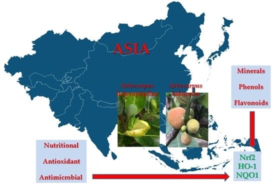
1. Introduction
2. Results
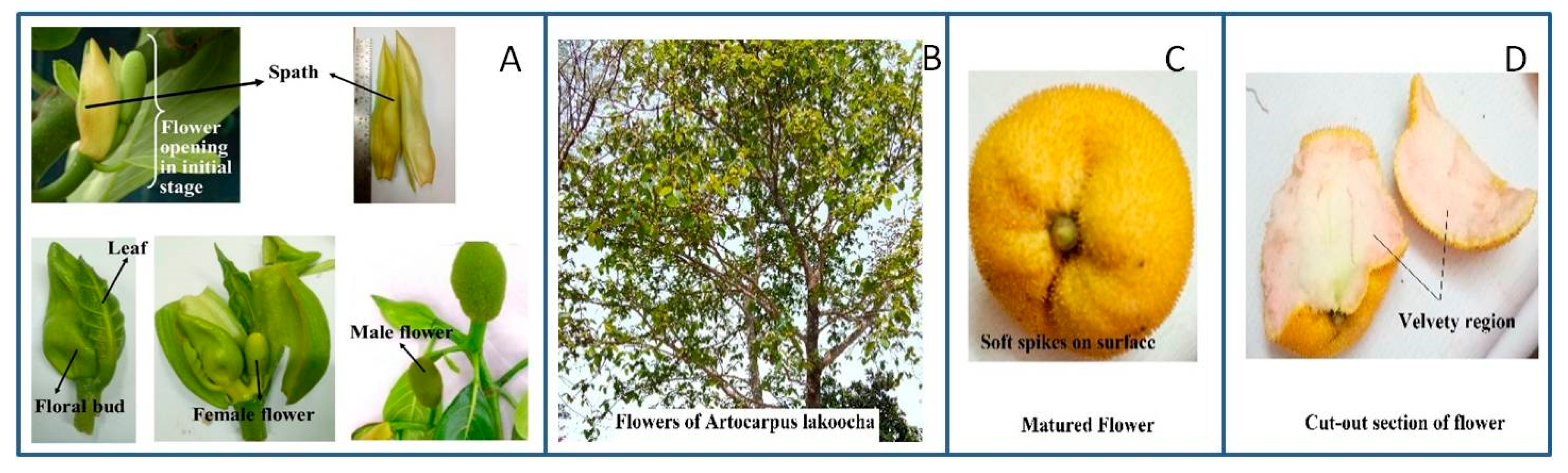
2.1. Chemical Properties of Flowers
2.1.1. Mineral Composition of Flowers
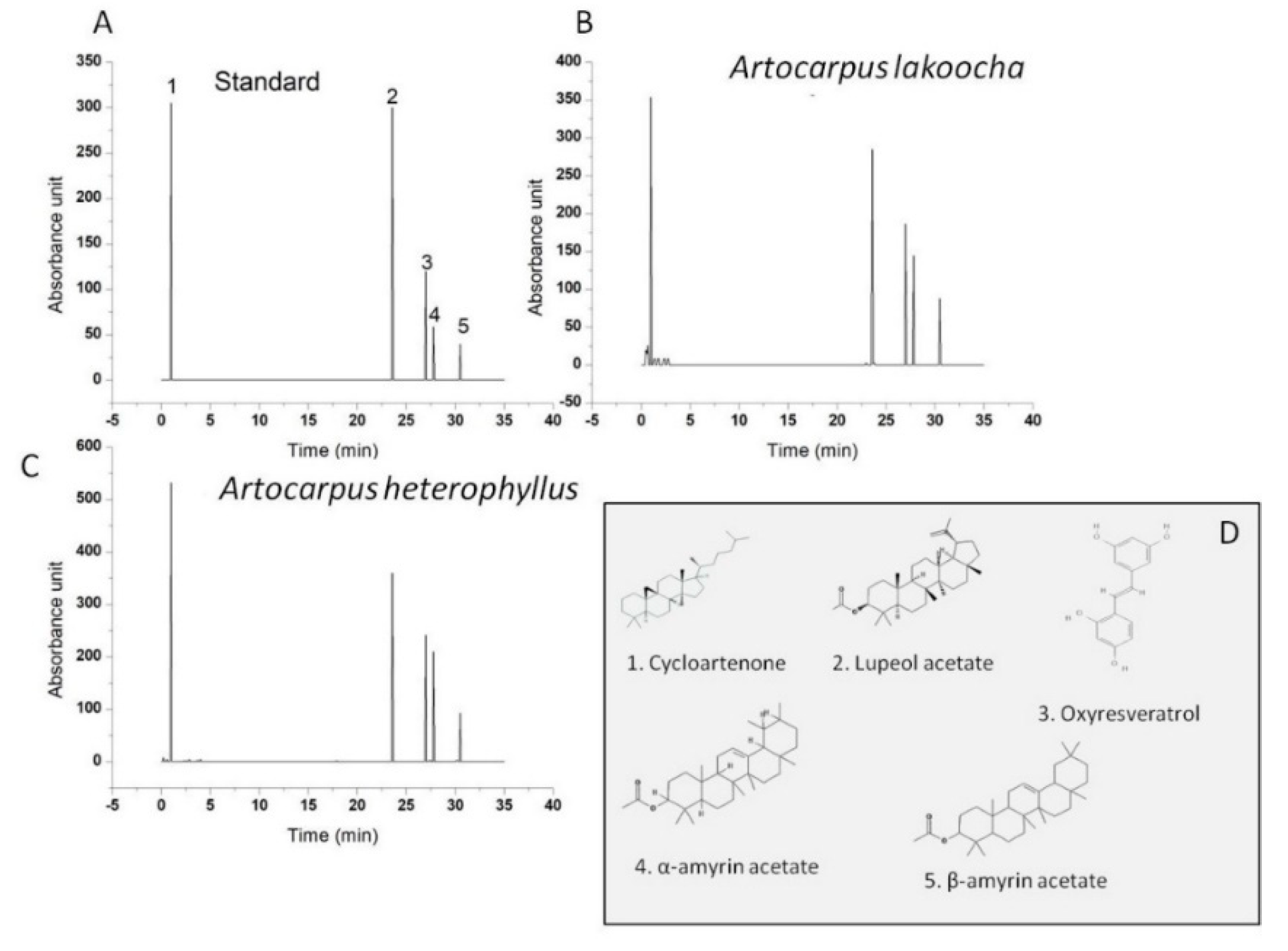
2.1.2. Phytochemical Composition of Flowers
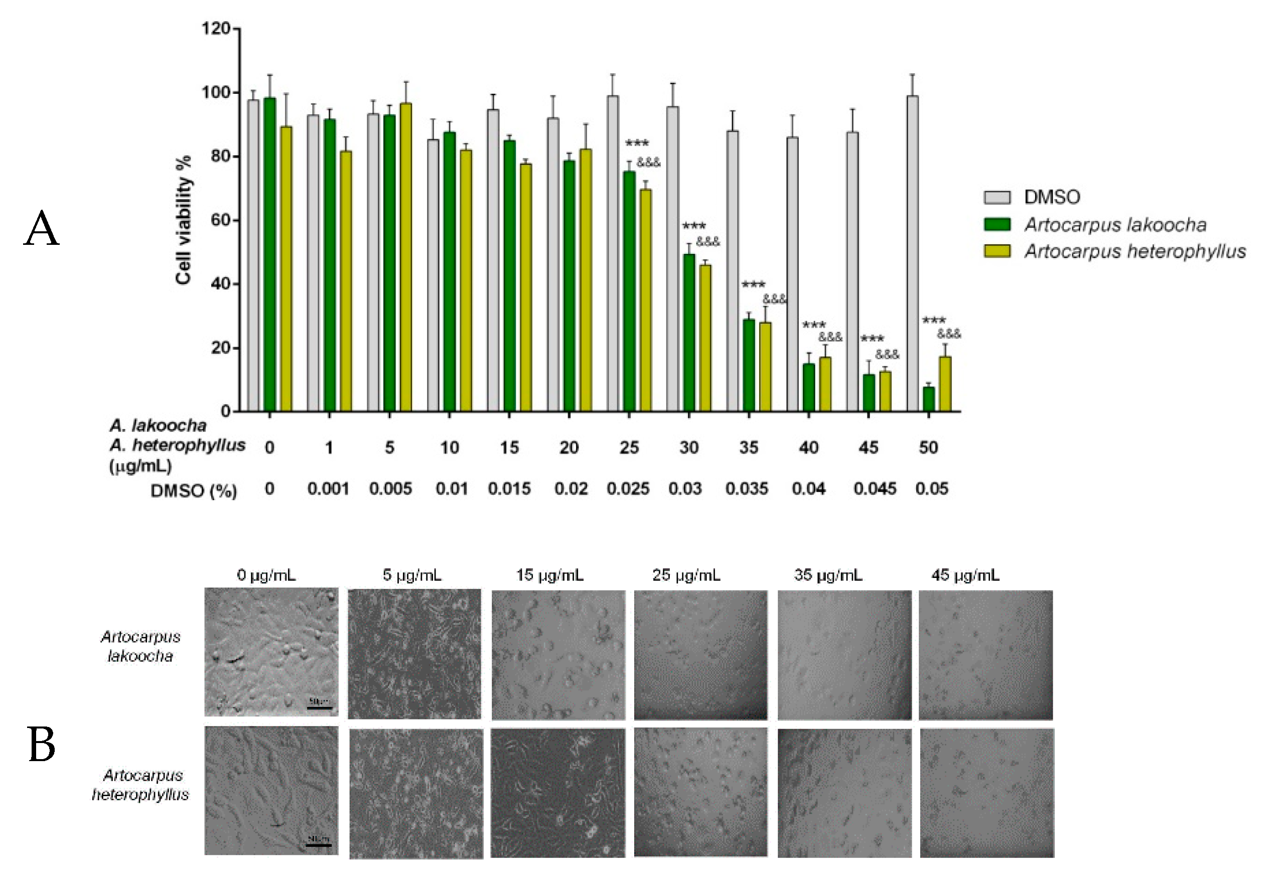
2.2. Cytotoxicity Assay (3-(4,5-Dimethylthiazol-2-yl)-2,5-Diphenyltetrazolium Bromide (MTT))
2.3. Antioxidant Properties
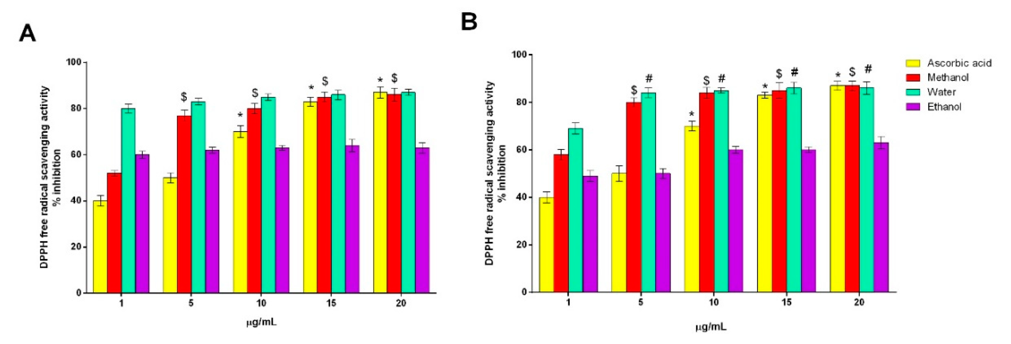
2.3.1. DPPH Free-Radical-Scavenging Activity
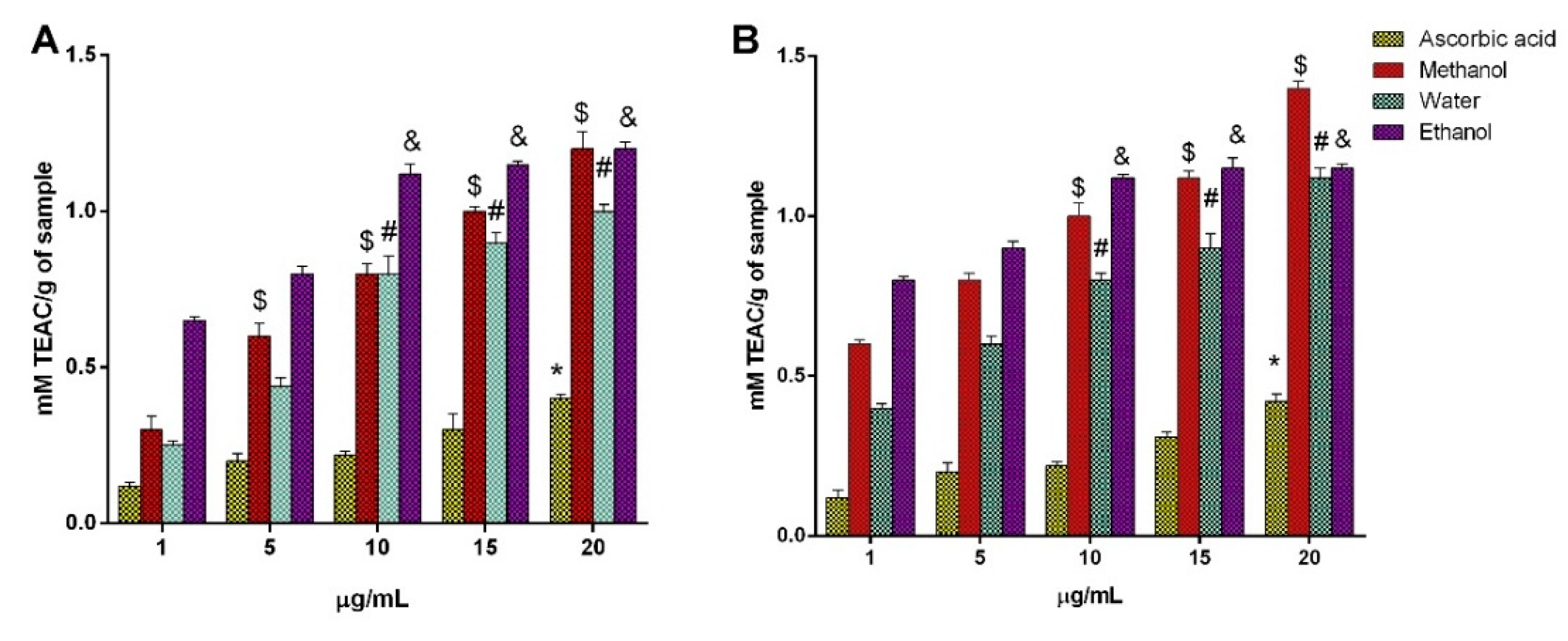
2.3.2. FRAP Assay
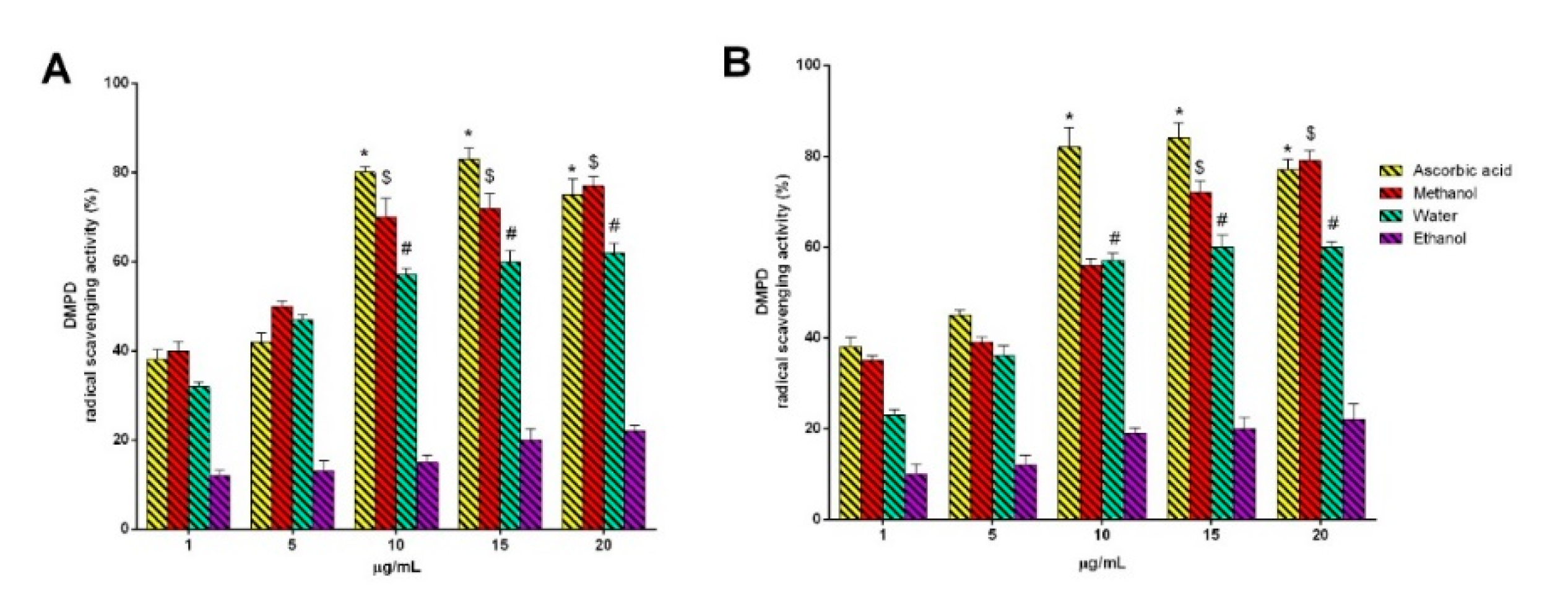
2.3.3. DMPD Radical Cation Decolorization Assay
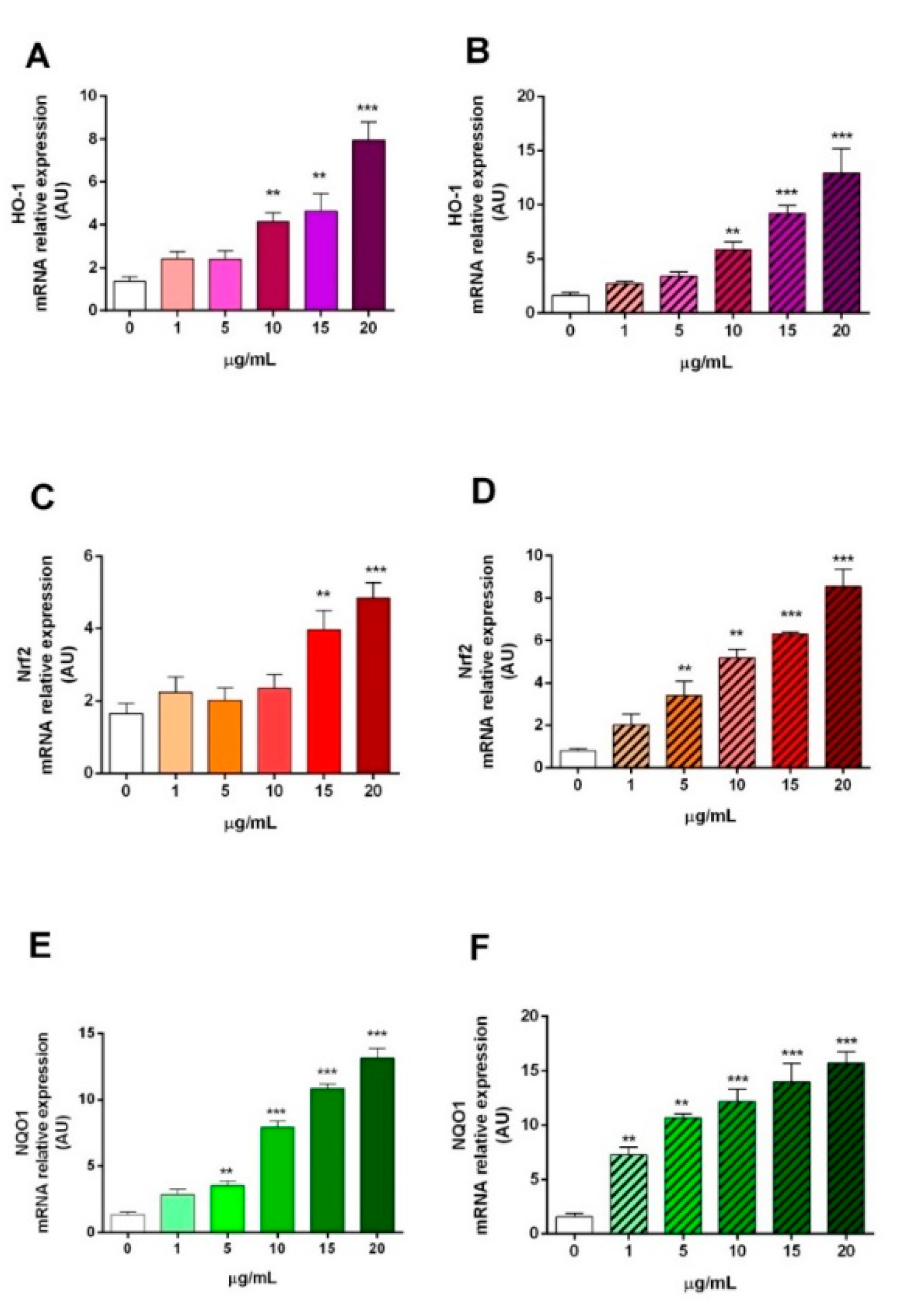
2.3.4. HO-1, Nrf2, and NQO1 Gene Expression
2.4. Antimicrobial Properties
3. Materials and Methods
3.1. Flowers and Chemicals
3.2. Preparation of Flower Extracts
3.3. Mineral Composition
3.4. Spectrophotometric Analysis of Phytochemical Properties
3.5. Cytotoxicity Study
3.6. Antioxidant Potential
3.6.1. DPPH Free-Radical-Scavenging Activity
3.6.2. Ferric-Reducing Antioxidant Power (FRAP)
3.6.3. DMPD (N, N-Dimethyl-p-Phenylenediamine) Radical Cation Decolorization Assay
3.6.4. Expression of Antioxidant Genes
3.7. Antimicrobial Activity
3.8. HPLC Conditions
3.9. Statistical Analysis
4. Conclusions
Author Contributions
Funding
Conflicts of Interest
References
- Burlando, B.; Clericuzio, M.; Cornara, L. Moraceae Plants with Tyrosinase Inhibitory Activity: A Review. Mini Rev. Med. Chem. 2017, 17, 108–121. [Google Scholar] [CrossRef] [PubMed]
- Arung, E.T.; Shimizu, K.; Kondo, R. Artocarpus plants as a potential source of skin whitening agents. Nat. Prod. Commun. 2011, 6, 1397–1402. [Google Scholar] [CrossRef] [Green Version]
- Jagtap, U.B.; Bapat, V.A. Artocarpus: A review of its traditional uses, phytochemistry and pharmacology. J. Ethnopharmacol. 2010, 129, 142–166. [Google Scholar] [CrossRef]
- Zhang, L.; Tu, Z.C.; Xie, X.; Wang, H.; Wang, H.; Wang, Z.X.; Sha, X.M.; Lu, Y. Jackfruit (Artocarpus heterophyllus Lam.) peel: A better source of antioxidants and a-glucosidase inhibitors than pulp, flake and seed, and phytochemical profile by HPLC-QTOF-MS/MS. Food Chem. 2017, 234, 303–313. [Google Scholar] [CrossRef] [PubMed]
- Jagtap, U.B.; Panaskar, S.N.; Bapat, V.A. Evaluation of antioxidant capacity and phenol content in jackfruit (Artocarpus heterophyllus Lam.) fruit pulp. Plant Foods Hum. Nutr. 2010, 65, 99–104. [Google Scholar] [CrossRef] [PubMed]
- Li, J.; Lin, Z.; Tang, X.; Liu, G.; Chen, Y.; Zhai, X.; Huang, Q.; Cao, Y. Oxyresveratrol extracted from Artocarpus heterophyllus Lam. inhibits tyrosinase and age pigments in vitro and in vivo. Food Funct. 2020, 11, 6595–6607. [Google Scholar] [CrossRef] [PubMed]
- Riga, R.; Happyana, N.; Holisotan Hakim, E. Sesquiterpenes produced by Pestalotiopsis microspora HF 12440 isolated from Artocarpus heterophyllus. Nat. Prod. Res. 2020, 34, 2229–2231. [Google Scholar] [CrossRef] [PubMed]
- Ajiboye, B.O.; Ojo, O.A.; Oyinloye, B.E.; Okesola, M.A.; Oluwatosin, A.; Boligon, A.A.; Kappo, A.P. Investigation of the In Vitro Antioxidant Potential Of Polyphenolic-Rich Extract of Artocarpus heterophyllus Lam Stem Bark and Its Antidiabetic Activity In Streptozotocin-Induced Diabetic Rats. J. Evid. Based Integr. Med. 2020, 25, 2515690X20916123. [Google Scholar] [CrossRef]
- Septama, A.W.; Jantan, I.; Panichayupakaranant, P. Flavonoids of Artocarpus heterophyllus Lam. heartwood inhibit the innate immune responses of human phagocytes. J. Pharm. Pharmacol. 2018, 70, 1242–1252. [Google Scholar] [CrossRef]
- Di, X.; Wang, S.; Wang, B.; Liu, Y.; Yuan, H.; Lou, H.; Wang, X. New phenolic compounds from the twigs of Artocarpus heterophyllus. Drug Discov. Ther. 2013, 7, 24–28. [Google Scholar] [CrossRef] [Green Version]
- Hettiaratchi, U.P.; Ekanayake, S.; Welihinda, J. Nutritional assessment of a jackfruit (Artocarpus heterophyllus) meal. Ceylon Med. J. 2011, 56, 54–58. [Google Scholar] [CrossRef] [PubMed] [Green Version]
- Ajayi, I.A. Comparative study of the chemical composition and mineral element content of Artocarpus heterophyllus and Treculia africana seeds and seed oils. Bioresour. Technol. 2008, 99, 5125–5129. [Google Scholar] [CrossRef] [PubMed]
- Shyamalamma, S.; Chandra, S.B.; Hegde, M.; Naryanswamy, P. Evaluation of genetic diversity in jackfruit (Artocarpus heterophyllus Lam.) based on amplified fragment length polymorphism markers. Genet. Mol. Res. GMR 2008, 7, 645–656. [Google Scholar] [CrossRef] [PubMed]
- Shailendra Kumar, M.B.; Rakesh Kumar, M.C.; Bharath, A.C.; Vinod Kumar, H.R.; Prashith Kekuda, T.R.; Nandini, K.C.; Rakshitha, M.N.; Raghavendra, H.L. Screening of selected biological activities of artocarpus lakoocha roxb (moraceae) fruit pericarp. J. Basic Clin. Pharm. 2010, 1, 239–245. [Google Scholar] [PubMed]
- Pandey, A.; Bhatnagar, S.P. Preliminary Phytochemical screening and antimicrobial studies on Artocarpus lakoocha Roxb. Anc. Sci. Life 2009, 28, 21–24. [Google Scholar] [PubMed]
- Maneechai, S.; De-Eknamkul, W.; Umehara, K.; Noguchi, H.; Likhitwitayawuid, K. Flavonoid and stilbenoid production in callus cultures of Artocarpus lakoocha. Phytochemistry 2012, 81, 42–49. [Google Scholar] [CrossRef]
- Boonyaketgoson, S.; Du, Y.; Valenciano Murillo, A.L.; Cassera, M.B.; Kingston, D.G.I.; Trisuwan, K. Flavanones from the Twigs and Barks of Artocarpus lakoocha Having Antiplasmodial and Anti-TB Activities. Chem. Pharm. Bull. 2020, 68, 671–674. [Google Scholar] [CrossRef]
- Adewole, S.O.; Ojewole, J.A. Artocarpus communis Forst. root-bark aqueous extract- and streptozotocin-induced ultrastructural and metabolic changes in hepatic tissues of Wistar rats. Afr. J. Tradit. Complementary Altern. Med. AJTCAM 2007, 4, 397–410. [Google Scholar]
- Mamun, S.; Shaheen, N.; Basak, T.A.; Mohiduzzaman, M.; Banu, C.P.; Takano-Ishikawa, Y. Hydrophilic antioxidant capacities and total phenol content of seasonal fruits of Bangladesh. Malays. J. Nutr. 2012, 18, 355–362. [Google Scholar]
- Povichit, N.; Phrutivorapongkul, A.; Suttajit, M.; Chaiyasut, C.C.; Leelapornpisid, P. Phenolic content and in vitro inhibitory effects on oxidation and protein glycation of some Thai medicinal plants. Pak. J. Pharm. Sci. 2010, 23, 403–408. [Google Scholar]
- Maneechai, S.; Likhitwitayawuid, K.; Sritularak, B.; Palanuvej, C.; Ruangrungsi, N.; Sirisa-Ard, P. Quantitative analysis of oxyresveratrol content in Artocarpus lakoocha and ‘Puag-Haad’. Med Princ. Pract. 2009, 18, 223–227. [Google Scholar] [CrossRef] [PubMed]
- Chuanasa, T.; Phromjai, J.; Lipipun, V.; Likhitwitayawuid, K.; Suzuki, M.; Pramyothin, P.; Hattori, M.; Shiraki, K. Anti-herpes simplex virus (HSV-1) activity of oxyresveratrol derived from Thai medicinal plant: Mechanism of action and therapeutic efficacy on cutaneous HSV-1 infection in mice. Antivir. Res. 2008, 80, 62–70. [Google Scholar] [CrossRef] [PubMed]
- Sritularak, B.; Tantrakarnsakul, K.; Lipipun, V.; Likhitwitayawuid, K. Flavonoids with anti-HSV activity from the root bark of Artocarpus lakoocha. Nat. Prod. Commun. 2013, 8, 1079–1080. [Google Scholar] [CrossRef] [Green Version]
- Puntumchai, A.; Kittakoop, P.; Rajviroongit, S.; Vimuttipong, S.; Likhitwitayawuid, K.; Thebtaranonth, Y. Lakoochins A and B, new antimycobacterial stilbene derivatives from Artocarpus lakoocha. J. Nat. Prod. 2004, 67, 485–486. [Google Scholar] [CrossRef]
- Likhitwitayawuid, K.; Sritularak, B.; Benchanak, K.; Lipipun, V.; Mathew, J.; Schinazi, R.F. Phenolics with antiviral activity from Millettia erythrocalyx and Artocarpus lakoocha. Nat. Prod. Res. 2005, 19, 177–182. [Google Scholar] [CrossRef]
- Saowakon, N.; Tansatit, T.; Wanichanon, C.; Chanakul, W.; Reutrakul, V.; Sobhon, P. Fasciola gigantica: Anthelmintic effect of the aqueous extract of Artocarpus lakoocha. Exp. Parasitol. 2009, 122, 289–298. [Google Scholar] [CrossRef] [PubMed]
- Wongon, M.; Limpeanchob, N. Inhibitory effect of Artocarpus lakoocha Roxb and oxyresveratrol on alpha-glucosidase and sugar digestion in Caco-2 cells. Heliyon 2020, 6, e03458. [Google Scholar] [CrossRef]
- Aneklaphakij, C.; Bunsupa, S.; Sirichamorn, Y.; Bongcheewin, B.; Satitpatipan, V. Taxonomic Notes on the ’Mahat’ (Artocarpus lacucha and A. thailandicus, Moraceae) Species Complex in Thailand. Plants 2020, 9, 391. [Google Scholar] [CrossRef] [PubMed] [Green Version]
- Stevenson, P.C.; Nicolson, S.W.; Wright, G.A.; Manson, J. Plant secondary metabolites in nectar: Impacts on pollinators and ecological functions. Funct. Ecol. 2016, 31, 65–75. [Google Scholar] [CrossRef]
- Esteban, R.; Balaguer, L.; Manrique, E.; Rubio de Casas, R.; Ochoa, R.; Fleck, I.; Pintó-Marijuan, M.; Casals, I.; Morales, D.; Jiménez, M.S.; et al. Alternative methods for sampling and preservation of photosynthetic pigments and tocopherols in plant material from remote locations. Photosynth. Res. 2009, 101, 77–88. [Google Scholar] [CrossRef]
- Li, A.-N.; Li, S.; Li, H.-B.; Xu, D.-P.; Xu, X.-R.; Chen, F. Total phenolic contents and antioxidant capacities of 51 edible and wild flowers. J. Funct. Foods 2014, 6, 319–330. [Google Scholar] [CrossRef]
- González-Barrio, R.; Periago, M.J.; Luna-Recio, C.; Garcia-Alonso, F.J.; Navarro-González, I. Chemical composition of the edible flowers, pansy (Viola wittrockiana) and snapdragon (Antirrhinum majus) as new sources of bioactive compounds. Food Chem. 2018, 252, 373–380. [Google Scholar] [CrossRef] [PubMed]
- Elvira-Torales, L.I.; García-Alonso, J.; Periago-Castón, M.J. Nutritional Importance of Carotenoids and Their Effect on Liver Health: A Review. Antioxidants 2019, 8, 229. [Google Scholar] [CrossRef] [PubMed] [Green Version]
- Baba, S.A.; Malik, S.A. Determination of total phenolic and flavonoid content, antimicrobial and antioxidant activity of a root extract of Arisaema jacquemontii Blume. J. Taibah Univ. Sci. 2018, 9, 449–454. [Google Scholar] [CrossRef] [Green Version]
- Zheng, J.; Meenu, M.; Xu, B. A systematic investigation on free phenolic acids and flavonoids profiles of commonly consumed edible flowers in China. J. Pharm. Biomed. Anal. 2019, 172, 268–277. [Google Scholar] [CrossRef] [PubMed]
- Su, B.-N.; Cuendet, M.; Hawthorne, M.E.; Kardono, L.B.S.; Riswan, S.; Fong, H.H.S.; Mehta, R.G.; Pezzuto, J.M.; Kinghorn, A.D. Constituents of the Bark and Twigs ofArtocarpusdadahwith Cyclooxygenase Inhibitory Activity. J. Nat. Prod. 2002, 65, 163–169. [Google Scholar] [CrossRef]
- Akihisa, T.; Kojima, N.; Kikuchi, T.; Yasukawa, K.; Tokuda, H.; Masters, E.T.; Manosroi, A.; Manosroi, J. Anti-Inflammatory and Chemopreventive Effects of Triterpene Cinnamates and Acetates from Shea Fat. J. Oleo Sci. 2010, 59, 273–280. [Google Scholar] [CrossRef] [Green Version]
- Park, J.; Park, J.H.; Suh, H.-J.; Lee, I.C.; Koh, J.; Boo, Y.C. Effects of resveratrol, oxyresveratrol, and their acetylated derivatives on cellular melanogenesis. Arch. Dermatol. Res. 2014, 306, 475–487. [Google Scholar] [CrossRef]
- Saleem, M. Lupeol, a novel anti-inflammatory and anti-cancer dietary triterpene. Cancer Lett. 2009, 285, 109–115. [Google Scholar] [CrossRef] [Green Version]
- Frankel, E.N.; Meyer, A.S. The problems of using one-dimensional methods to evaluate multifunctional food and biological antioxidants. J. Sci. Food Agric. 2000, 80, 1925–1941. [Google Scholar] [CrossRef]
- Khaled-Khodja, N.; Boulekbache-Makhlouf, L.; Madani, K. Phytochemical screening of antioxidant and antibacterial activities of methanolic extracts of some Lamiaceae. Ind. Crop. Prod. 2014, 61, 41–48. [Google Scholar] [CrossRef]
- Vukics, V.; Kery, A.; Guttman, A. Analysis of Polar Antioxidants in Heartsease (Viola tricolor L.) and Garden Pansy (Viola x wittrockiana Gams.). J. Chromatogr. Sci. 2008, 46, 823–827. [Google Scholar] [CrossRef] [PubMed] [Green Version]
- Njus, D.; Kelley, P.M.; Tu, Y.J.; Schlegel, H.B. Ascorbic acid: The chemistry underlying its antioxidant properties. Free Radic. Biol. Med. 2020, 159, 37–43. [Google Scholar] [CrossRef] [PubMed]
- Kowalczyk, D.; Kazimierczak, W.; Zieba, E.; Mezynska, M.; Basiura-Cembala, M.; Lisiecki, S.; Karas, M.; Baraniak, B. Ascorbic acid- and sodium ascorbate-loaded oxidized potato starch films: Comparative evaluation of physicochemical and antioxidant properties. Carbohydr. Polym. 2018, 181, 317–326. [Google Scholar] [CrossRef]
- Sogi, D.S.; Siddiq, M.; Roidoung, S.; Dolan, K.D. Total phenolics, carotenoids, ascorbic acid, and antioxidant properties of fresh-cut mango (Mangifera indica L., cv. Tommy Atkin) as affected by infrared heat treatment. J. Food Sci. 2012, 77, C1197–C1202. [Google Scholar] [CrossRef]
- Leonard, S.S.; Cutler, D.; Ding, M.; Vallyathan, V.; Castranova, V.; Shi, X. Antioxidant properties of fruit and vegetable juices: More to the story than ascorbic acid. Ann. Clin. Lab. Sci. 2002, 32, 193–200. [Google Scholar]
- Zadra, M.; Piana, M.; Brum, T.; Boligon, A.; Freitas, R.; Machado, M.; Stefanello, S.; Soares, F.; Athayde, M. Antioxidant Activity and Phytochemical Composition of the Leaves of Solanum guaraniticum A. St.-Hil. Molecules 2012, 17, 12560–12574. [Google Scholar] [CrossRef] [Green Version]
- Tai, Z.; Cai, L.; Dai, L.; Dong, L.; Wang, M.; Yang, Y.; Cao, Q.; Ding, Z. Antioxidant activity and chemical constituents of edible flower of Sophora viciifolia. Food Chem. 2011, 126, 1648–1654. [Google Scholar] [CrossRef]
- Loboda, A.; Damulewicz, M.; Pyza, E.; Jozkowicz, A.; Dulak, J. Role of Nrf2/HO-1 system in development, oxidative stress response and diseases: An evolutionarily conserved mechanism. Cell. Mol. Life Sci. 2016, 73, 3221–3247. [Google Scholar] [CrossRef] [Green Version]
- Espinosa-Diez, C.; Miguel, V.; Mennerich, D.; Kietzmann, T.; Sánchez-Pérez, P.; Cadenas, S.; Lamas, S. Antioxidant responses and cellular adjustments to oxidative stress. Redox Biol. 2015, 6, 183–197. [Google Scholar] [CrossRef] [Green Version]
- Mastinu, A.; Bonini, S.A.; Rungratanawanich, W.; Aria, F.; Marziano, M.; Maccarinelli, G.; Abate, G.; Premoli, M.; Memo, M.; Uberti, D. Gamma-oryzanol Prevents LPS-induced Brain Inflammation and Cognitive Impairment in Adult Mice. Nutrients 2019, 11, 728. [Google Scholar] [CrossRef] [PubMed] [Green Version]
- Bhattacharjee, S.; Dashwood, R.H. Epigenetic Regulation of NRF2/KEAP1 by Phytochemicals. Antioxidants 2020, 9, 865. [Google Scholar] [CrossRef] [PubMed]
- Li, L.; Dong, H.; Song, E.; Xu, X.; Liu, L.; Song, Y. Nrf2/ARE pathway activation, HO-1 and NQO1 induction by polychlorinated biphenyl quinone is associated with reactive oxygen species and PI3K/AKT signaling. Chem. Biol. Interact. 2014, 209, 56–67. [Google Scholar] [CrossRef] [PubMed]
- Araujo, J.A.; Zhang, M.; Yin, F. Heme Oxygenase-1, Oxidation, Inflammation, and Atherosclerosis. Front. Pharmacol. 2012, 3, 119. [Google Scholar] [CrossRef] [Green Version]
- Ross, D.; Siegel, D. Functions of NQO1 in Cellular Protection and CoQ10 Metabolism and its Potential Role as a Redox Sensitive Molecular Switch. Front. Physiol. 2017, 8, 595. [Google Scholar] [CrossRef]
- Teanpaisan, R.; Senapong, S.; Puripattanavong, J. In vitro Antimicrobial and Antibiofilm Activity of Artocarpus Lakoocha (Moraceae) Extract against Some Oral Pathogens. Trop. J. Pharm. Res. 2014, 13, 1149–1155. [Google Scholar] [CrossRef] [Green Version]
- Aherne, S.A.; Kerry, J.P.; O’Brien, N.M. Effects of plant extracts on antioxidant status and oxidant-induced stress in Caco-2 cells. Br. J. Nutr. 2007, 97, 321–328. [Google Scholar] [CrossRef] [Green Version]
- Pintus, G.; Jiménez, S.; Gascón, S.; Luquin, A.; Laguna, M.; Ancin-Azpilicueta, C.; Rodríguez-Yoldi, M.J. Rosa canina Extracts Have Antiproliferative and Antioxidant Effects on Caco-2 Human Colon Cancer. PLoS ONE 2016, 11, e0159136. [Google Scholar] [CrossRef] [Green Version]
- Nowak, A.; Sójka, M.; Klewicka, E.; Lipińska, L.; Klewicki, R.; Kołodziejczyk, K. Ellagitannins from Rubus idaeus L. Exert Geno- and Cytotoxic Effects against Human Colon Adenocarcinoma Cell Line Caco-2. J. Agric. Food Chem. 2017, 65, 2947–2955. [Google Scholar] [CrossRef]
- Bonini, S.A.; Mastinu, A.; Maccarinelli, G.; Mitola, S.; Premoli, M.; La Rosa, L.R.; Ferrari-Toninelli, G.; Grilli, M.; Memo, M. Cortical Structure Alterations and Social Behavior Impairment in p50-Deficient Mice. Cereb. Cortex 2016, 26, 2832–2849. [Google Scholar] [CrossRef]
- Gianoncelli, A.; Bonini, S.A.; Bertuzzi, M.; Guarienti, M.; Vezzoli, S.; Kumar, R.; Delbarba, A.; Mastinu, A.; Sigala, S.; Spano, P.; et al. An Integrated Approach for a Structural and Functional Evaluation of Biosimilars: Implications for Erythropoietin. BioDrugs Clin. Immunother. Biopharm. Gene Ther. 2015, 29, 285–300. [Google Scholar] [CrossRef] [PubMed] [Green Version]
- Lazzari, P.; Pau, A.; Tambaro, S.; Asproni, B.; Ruiu, S.; Pinna, G.; Mastinu, A.; Curzu, M.M.; Reali, R.; Emilio Heiner Bottazzi, M.; et al. Synthesis and Pharmacological Evaluation of Novel 4-Alkyl-5-thien-2’-yl Pyrazole Carboxamides. Central Nerv. Syst. Agents Med. Chem. 2012, 12, 254–276. [Google Scholar] [CrossRef] [PubMed]
- Mastinu, A.; Premoli, M.; Ferrari-Toninelli, G.; Tambaro, S.; Maccarinelli, G.; Memo, M.; Bonini, S.A. Cannabinoids in health and disease: Pharmacological potential in metabolic syndrome and neuroinflammation. Horm. Mol. Biol. Clin. Investig. 2018, 36, 20180013. [Google Scholar] [CrossRef] [PubMed]
- Kiran, G.S.; Priyadharshini, S.; Anitha, K.; Gnanamani, E.; Selvin, J. Characterization of an exopolysaccharide from probiont Enterobacter faecalis MSI12 and its effect on the disruption of Candida albicans biofilm. RSC Adv. 2015, 5, 71573–71585. [Google Scholar] [CrossRef]
- Chen, Y.-F.; Ching, C.; Wu, T.-S.; Wu, C.-R.; Hsieh, W.-T.; Tsai, H.-Y. Balanophora spicataand Lupeol Acetate Possess Antinociceptive and Anti-Inflammatory ActivitiesIn VivoandIn Vitro. Evid. Based Complement. Altern. Med. 2012, 2012, 371273. [Google Scholar] [CrossRef] [Green Version]


| Parameters | Artocarpus lakoocha Roxb. | Artocarpus heterophyllus Lam. | p Value |
|---|---|---|---|
| Total phenol content (μg GAE/g) | 217.80 ± 1.25 | 883.20 ± 5.90 | <0.0001 |
| Total flavonoids content (μg QE/g) | 168.26 ± 1.50 | 658.52 ± 5.60 | <0.0001 |
| Total carotenoids (μg/g) | 16.96 ± 0.15 | 27.29 ± 0.25 | <0.0001 |
| Iron (μg/100 g) | 108.35 ± 3.65 | 196.36 ± 8.32 | <0.0001 |
| Copper (μg/100 g) | 597.10 ± 8.96 | 106.62 ± 5.21 | <0.0001 |
| Zinc (μg/100 g) | 421.06 ± 5.12 | 320.22 ± 6.85 | <0.0001 |
| Manganese (μg/100 g) | 250.05 ± 4.68 | 185.62 ± 4.55 | <0.0001 |
| Calcium (mg/100 g) | 20.24 ± 2.13 | 25.35 ± 0.81 | 0.0432 |
| Magnesium (mg/100 g) | 13.40 ± 0.52 | 7.28 ± 0.35 | 0.0074 |
| Potassium (mg/100 g) | 150.00 ± 8.10 | 214.56 ± 4.68 | <0.0001 |
| Phosphorus (mg/100 g) | 11.85 ± 0.06 | 19.63 ± 0.05 | 0.0002 |
| Artocarpus lakoocha Roxb. | Artocarpus heterophyllus Lam. | Streptomycin | ||||
|---|---|---|---|---|---|---|
| 100 μg/mL | 200 μg/mL | 100 μg/mL | 200 μg/mL | 100 μg/mL | 200 μg/mL | |
| Staphylococcus aureus | 8 | 18 | 15 | 21 | 12 | 26 |
| Listeria monocytogenes | 11 | 19 | 12 | 19 | 13 | 23 |
| Salmonella typhimurium | 13 | 24 | 13 | 24 | 17 | 31 |
| Bacillus cereus | 7 | 12 | - | - | 10 | 18 |
| Escherichia coli MTCC 1568 | 12 | 22 | 15 | 22 | 15 | 26 |
| Bacillus subtilis ATCC 6633 | 6.5 | 12 | 5 | 10 | 8 | 15 |
© 2020 by the authors. Licensee MDPI, Basel, Switzerland. This article is an open access article distributed under the terms and conditions of the Creative Commons Attribution (CC BY) license (http://creativecommons.org/licenses/by/4.0/).
Share and Cite
Gupta, A.K.; Rather, M.A.; Kumar Jha, A.; Shashank, A.; Singhal, S.; Sharma, M.; Pathak, U.; Sharma, D.; Mastinu, A. Artocarpus lakoocha Roxb. and Artocarpus heterophyllus Lam. Flowers: New Sources of Bioactive Compounds. Plants 2020, 9, 1329. https://doi.org/10.3390/plants9101329
Gupta AK, Rather MA, Kumar Jha A, Shashank A, Singhal S, Sharma M, Pathak U, Sharma D, Mastinu A. Artocarpus lakoocha Roxb. and Artocarpus heterophyllus Lam. Flowers: New Sources of Bioactive Compounds. Plants. 2020; 9(10):1329. https://doi.org/10.3390/plants9101329
Chicago/Turabian StyleGupta, Arun Kumar, Muzamil Ahmad Rather, Avinash Kumar Jha, Abhinay Shashank, Somya Singhal, Maanas Sharma, Urbi Pathak, Dipti Sharma, and Andrea Mastinu. 2020. "Artocarpus lakoocha Roxb. and Artocarpus heterophyllus Lam. Flowers: New Sources of Bioactive Compounds" Plants 9, no. 10: 1329. https://doi.org/10.3390/plants9101329