
Abdominal Cryptorchidism with Complete Dissociation between the Testis and Deferent Duct Mimicking Testicular Regression Syndrome
 , ,
, ,  , and
, and 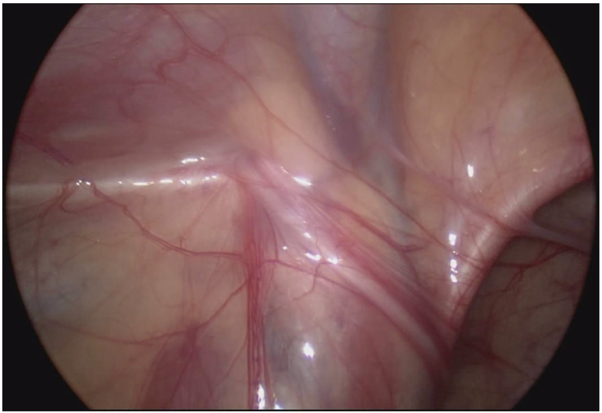
Abstract
:1. Introduction
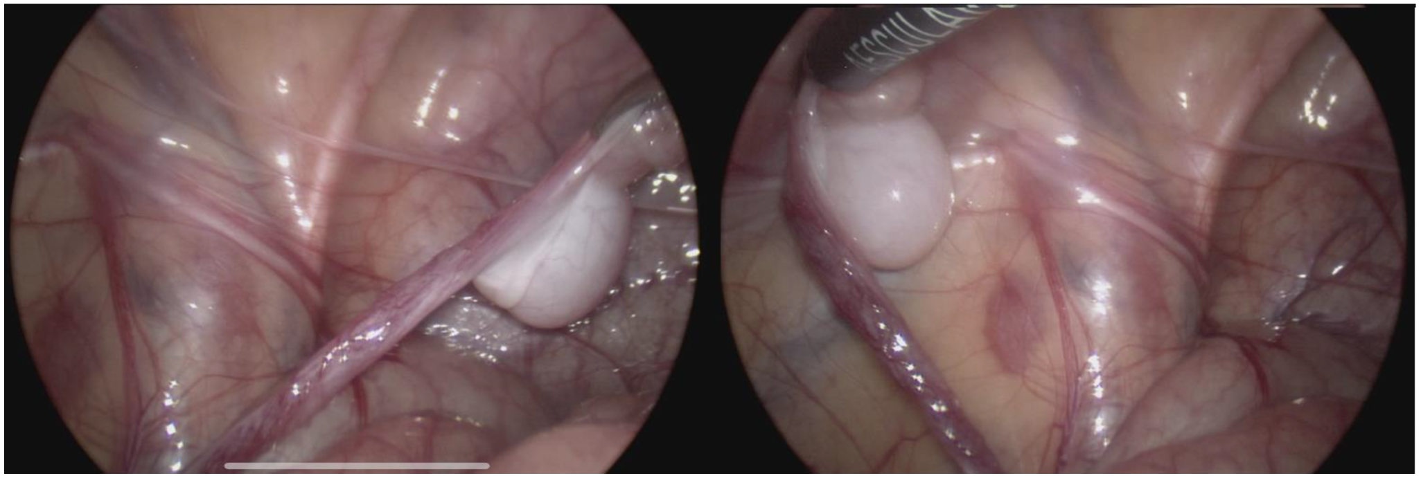
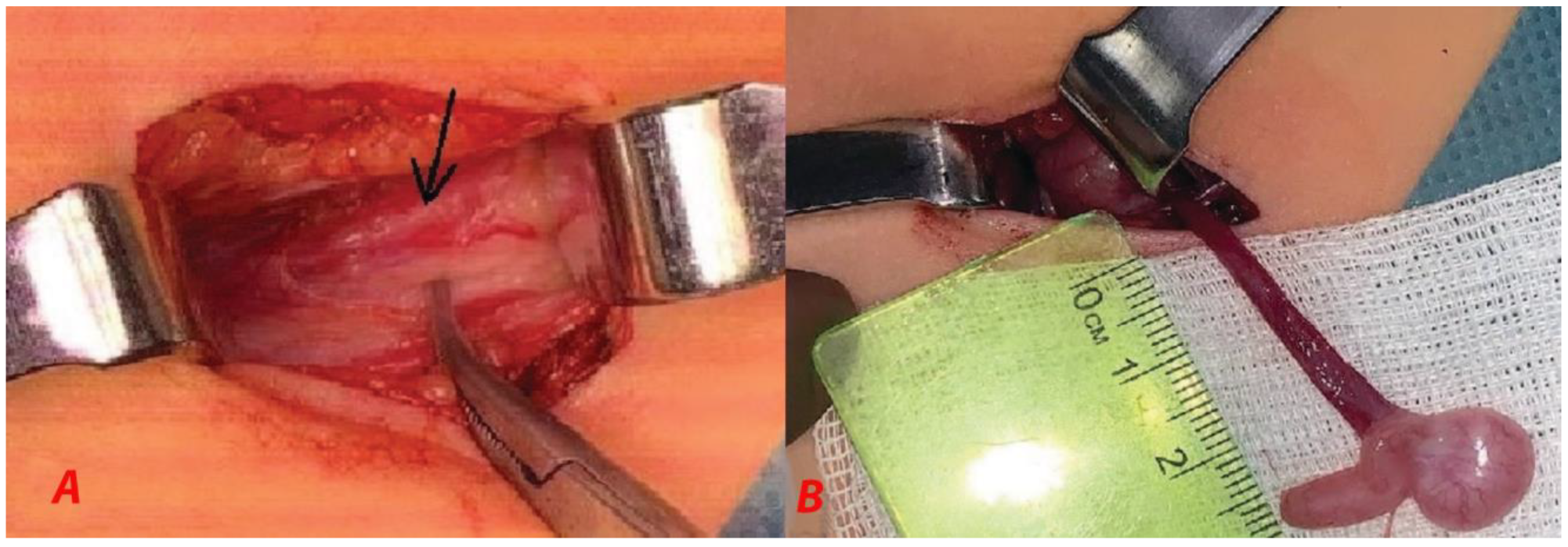
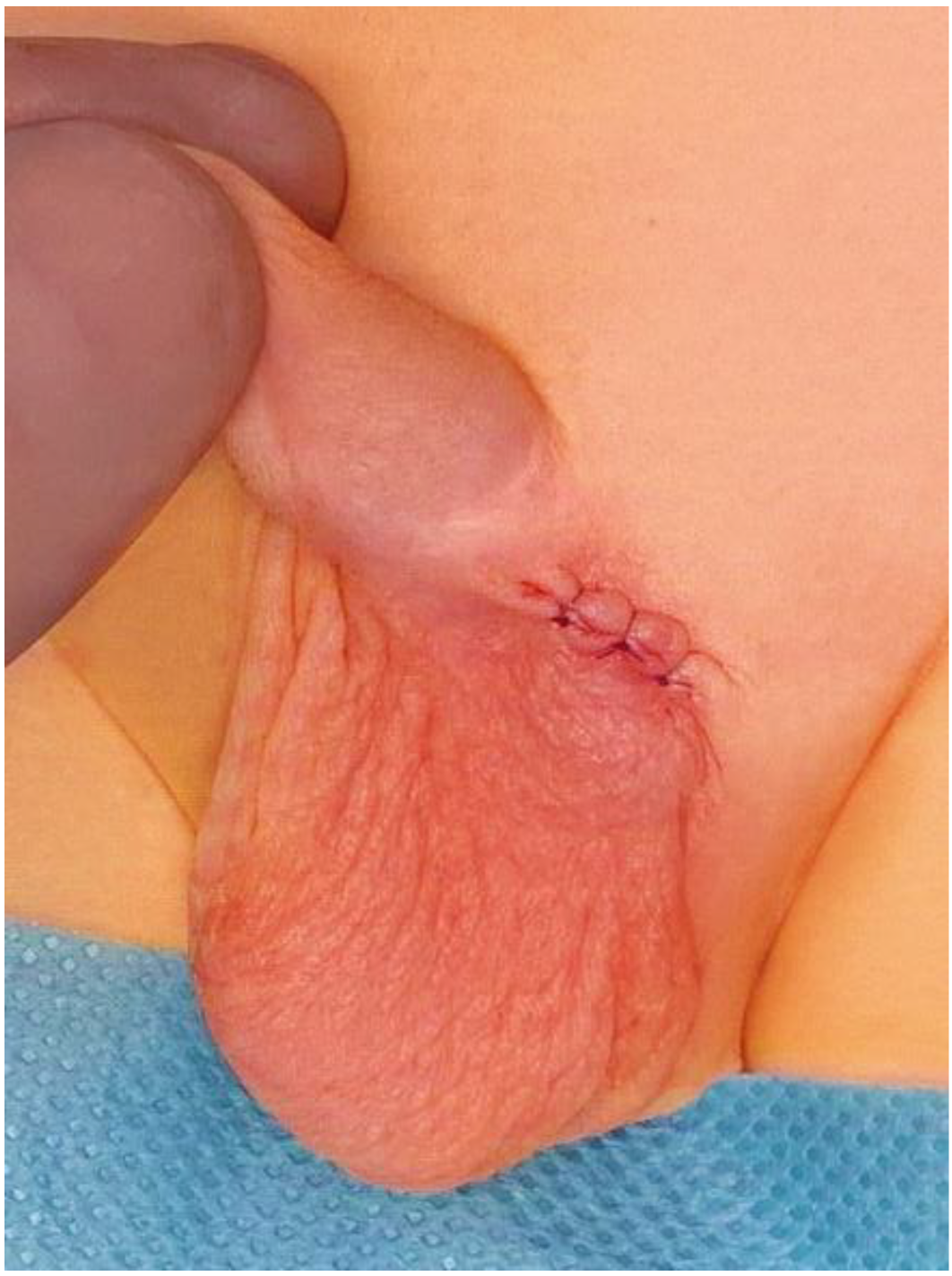
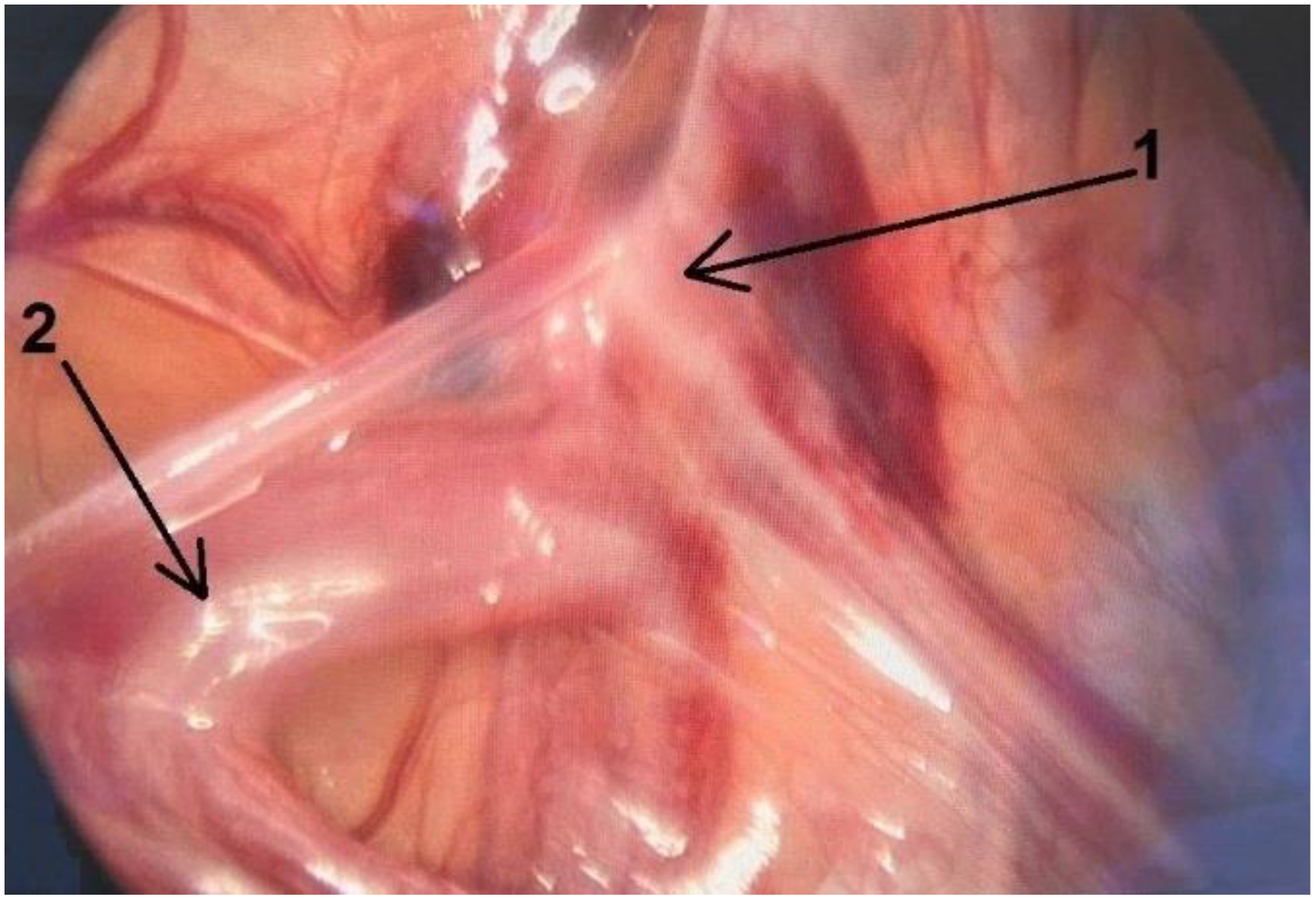
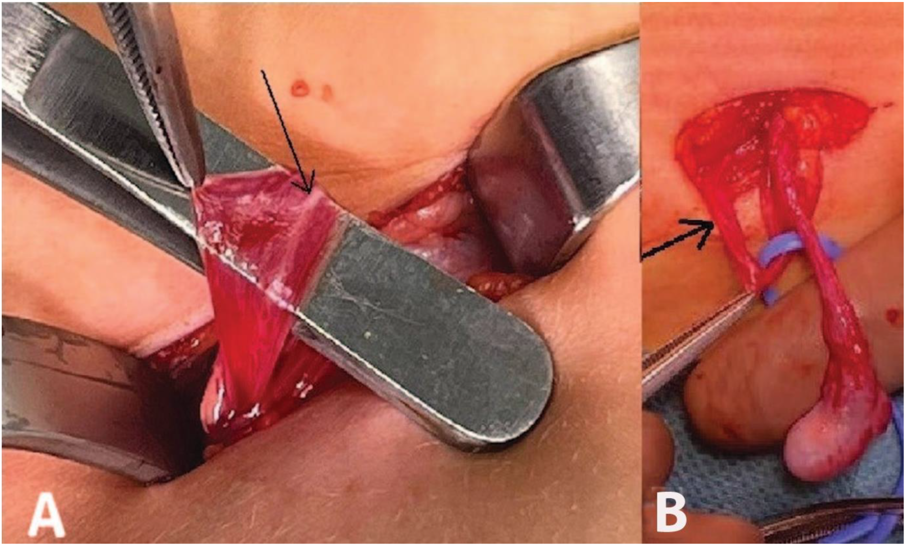
2. Case Description
3. Discussion
4. Conclusions
Author Contributions
Funding
Informed Consent Statement
Data Availability Statement
Acknowledgments
Conflicts of Interest
References
- Sijstermans, K.; Hack, W.W.M.; Meijer, R.W.; Van Der Voort-Doedens, L.M. The frequency of undescended testis from birth to adulthood: A review. Int. J. Androl. 2007, 31, 1–11. [Google Scholar] [CrossRef] [PubMed]
- Acerini, C.L.; Miles, H.L.; Dunger, D.B.; Ong, K.K.; Hughes, I.A. The descriptive epidemiology of congenital and acquired crytorchidism in a UK infant cohort. Arch. Dis. Child. 2009, 94, 868–872. [Google Scholar] [CrossRef] [PubMed]
- Hrivatakis, G.; Astfalk, W.; Schmidt, A.; Hartwig, A.; Kugler, T.; Heim, T.; Clausner, A.; Frunder, A.; Weber, H.; Loff, S.; et al. The Timing of Surgery for Undescended Testistestis—A retrospective multicenter analysis. Dtsch. Arztebl. Int. 2014, 111, 649–657. [Google Scholar] [CrossRef] [PubMed] [Green Version]
- Bergbrant, S.; Omling, E.; Björk, J.; Hagander, L. Cryptorchidism in Sweden: A Nationwide Study of Prevalence, Operative Management, and Complications. J. Pediatr. 2018, 194, 197–203. [Google Scholar] [CrossRef] [PubMed]
- Mavrogenis, S.; Ács, N.; Czeizel, A.E. No increases in the rate of undescended testis in Hungary during the last 50 years: A population-based study. Congenit. Anom. 2015, 55, 145–149. [Google Scholar] [CrossRef]
- Merguerian, P.A.; Mevorach, R.A.; Shortliffe, L.D.; Cendron, M. Laparoscopy for the evaluation and management of the nonpalpable testicle. Urology 1998, 51 (Suppl. 1), 3–6. [Google Scholar] [CrossRef]
- Denes, F.T.; Saito, F.J.; Silva, F.A.; Giron, A.M.; Machado, M.; Srougi, M. Laparoscopic diagnosis and treatment of nonpalpable testis. Int. Braz. J. Urol. 2008, 34, 329–335. [Google Scholar] [CrossRef] [Green Version]
- Smolko, M.J.; Kaplan, G.W.; Brock, W.A. Location and Fate of The Nonpalpable Testis in Children. J. Urol. 1983, 129, 1204–1206. [Google Scholar] [CrossRef]
- Docimo, S.G. The results of surgical therapy for cryptorchidism: A literature review and analysis. J. Urol. 1995, 154, 1148–1152. [Google Scholar] [CrossRef]
- Radmayr, C.; Oswald, J.; Schwentner, C.; Neururer, R.; Peschel, R.; Bartsch, G. Long-Term Outcome of Laparoscopically Managed Nonpalpable Testes. J. Urol. 2003, 170 Pt 1, 2409–2411. [Google Scholar] [CrossRef]
- Hvistendahl, G.; Poulsen, E. Laparoscopy for the impalpable testes: Experience with 80 intra-abdominal testes. J. Pediatr. Urol. 2009, 5, 389–392. [Google Scholar] [CrossRef] [PubMed]
- Ang, C.W.; Forrest, J. Diagnostic laparoscopy and management of the impalpable testis—A review of 10 years’ practice at a non-paediatric specialist centre. J. Pediatr. Urol. 2008, 4, 214–217. [Google Scholar] [CrossRef] [PubMed]
- Vohra, S.; Morgentaler, A. Congenital anomalies of the vas deferens, epididymis, and seminal vesicles. Urology 1997, 49, 313–321. [Google Scholar] [CrossRef]
- Bieth, E.; Hamdi, S.M.; Mieusset, R. Genetics of the congenital absence of the vas deferens. Hum. Genet. 2020, 140, 59–76. [Google Scholar] [CrossRef] [PubMed] [Green Version]
- Merksz, M. Fusional anomalies of the testis and epididymis. Acta Chir. Hung. 1998, 37, 153–170. [Google Scholar]
- Nowak, K. Failure of fusion of epididymis and testicle with complete separation of the vas deferens. J. Pediatr. Surg. 1972, 7, 715–716. [Google Scholar] [CrossRef]
- Bergdahl, L.; Andersson, A. The Importance of a Careful Search for Intra-Abdominal Testes in Cryptorchidism. Report of a case with failure of urogenital union. Scand. J. Urol. Nephrol. 1981, 15, 153–155. [Google Scholar] [CrossRef]
- Caterino, S.; Lorenzon, L.; Cavallini, M.; Cavaniglia, D.; Ferro, F. Epididymal-testicular fusion anomalies in cryptorchidism are associated with proximal location of the undescended testis and with a widely patent processus vaginalis. J. Anat. 2014, 225, 473–478. [Google Scholar] [CrossRef]
- El Gohary, M.A. Failure of Fusion of Epididymis and Testis: A Rare Laparoscopic Finding. Eur. J. Pediatr. Surg. 2009, 19, 108–109. [Google Scholar] [CrossRef]
- Rachmani, E.; Zachariou, Z.; Snyder, H.; Hadziselimovic, F. Complete Testis-Epididymis Nonfusion Anomaly: A Typical Association with Cryptorchid Testis. Urol. Int. 2012, 89, 355–357. [Google Scholar] [CrossRef]
- Kuçukaydin, M.; Özokutan, B.H.; Turan, C.; Okur, H.; Köse, O. Malformation of the epididymis in undescended testis. Pediatr. Surg. Int. 1998, 14, 189–191. [Google Scholar] [CrossRef] [PubMed]
- Koff, W.J.; Scaletscky, R. Malformations of the Epididymis in Undescended Testis. J. Urol. 1990, 143, 340–343. [Google Scholar] [CrossRef] [PubMed]
- Elder, J.S. Epididymal Anomalies Associated with Hydrocele/Hernia and Cryptorchidism: Implications Regarding Testicular Descent. J. Urol. 1992, 148 Pt 2, 624–626. [Google Scholar] [CrossRef]
- Barthold, J.S.; Redman, J.F. Association of epididymal anomalies with patent processus vaginalis in hernia, hydrocele and cryp-torchidism. J. Urol. 1996, 156, 2054–2056. [Google Scholar] [CrossRef] [PubMed]
- Salle, J.L.P.; Langer, J.; Favorito, L.A. Unilateral renal agenesia associated with partial epididymis and vas deferens agenesia in a patient with abdominal testicle. Int. Braz. J. Urol. 2006, 32, 208–210. [Google Scholar] [CrossRef] [PubMed] [Green Version]
- Abdelmohsen, S.M.; Takrouney, M.H.; Osman, M.A.; Abdelmohsen, B.M. Unique anomalies of vas deferens; A case series from Upper Egypt. SM J Surg. 2017, 3, 1013. [Google Scholar]
- Silber, S.J.; Ord, T.; Balmaceda, J.; Patrizio, P.; Asch, R.H. Congenital Absence of the Vas Deferens. The fertilizing capacity of human epididymal sperm. N. Engl. J. Med. 1990, 323, 1788–1792. [Google Scholar] [CrossRef] [PubMed]
- Mickle, J.; Milunsky, A.; Amos, J.; Oates, R. Immunology: Congenital unilateral absence of the vas deferens: A heterogeneous disorder with two distinct subpopulations based upon aetiology and mutational status of the cystic fibrosis gene. Hum. Reprod. 1995, 10, 1728–1735. [Google Scholar] [CrossRef] [PubMed]
- Hurwitz, R.S.; Kaptein, J.S. How Well Does Contralateral Testis Hypertrophy Predict the Absence Of The Nonpalpable Testis? J. Urol. 2001, 165, 588–592. [Google Scholar] [CrossRef]
- Snodgrass, W.T.; Yucel, S.; Ziada, A. Scrotal Exploration for Unilateral Nonpalpable Testis. J. Urol. 2007, 178 Pt 2, 1718–1721. [Google Scholar] [CrossRef]
- Shibata, Y.; Kojima, Y.; Mizuno, K.; Nakane, A.; Kato, T.; Kamisawa, H.; Kohri, K.; Hayashi, Y. Optimal Cutoff Value of Contralateral Testicular Size for Prediction of Absent Testis in Japanese Boys with Nonpalpable Testis. Urology 2010, 76, 78–81. [Google Scholar] [CrossRef] [PubMed]
- Braga, L.; Kim, S.; Farrokhyar, F.; Lorenzo, A. Is there an optimal contralateral testicular cut-off size that predicts monorchism in boys with nonpalpable testicles? J. Pediatr. Urol. 2014, 10, 693–698. [Google Scholar] [CrossRef] [PubMed]
- Hodhod, A.; Capolicchio, J.; Jednak, R.; El-Sherbiny, M. Testicular hypertrophy as a predictor for contralateral monorchism: Retrospective review of prospectively recorded data. J. Pediatr. Urol. 2016, 12, 34.e1–34.e5. [Google Scholar] [CrossRef] [PubMed]


Disclaimer/Publisher’s Note: The statements, opinions and data contained in all publications are solely those of the individual author(s) and contributor(s) and not of MDPI and/or the editor(s). MDPI and/or the editor(s) disclaim responsibility for any injury to people or property resulting from any ideas, methods, instructions or products referred to in the content. |
© 2023 by the authors. Licensee MDPI, Basel, Switzerland. This article is an open access article distributed under the terms and conditions of the Creative Commons Attribution (CC BY) license (https://creativecommons.org/licenses/by/4.0/).
Share and Cite
Sizonov, V.V.; Makarov, A.G.; Mayr, J.M.; Vigera, V.V.; Kogan, M.I. Abdominal Cryptorchidism with Complete Dissociation between the Testis and Deferent Duct Mimicking Testicular Regression Syndrome. Children 2023, 10, 205. https://doi.org/10.3390/children10020205
Sizonov VV, Makarov AG, Mayr JM, Vigera VV, Kogan MI. Abdominal Cryptorchidism with Complete Dissociation between the Testis and Deferent Duct Mimicking Testicular Regression Syndrome. Children. 2023; 10(2):205. https://doi.org/10.3390/children10020205
Chicago/Turabian StyleSizonov, Vladimir V., Alexey G. Makarov, Johannes M. Mayr, Vladimir V. Vigera, and Mikhail I. Kogan. 2023. "Abdominal Cryptorchidism with Complete Dissociation between the Testis and Deferent Duct Mimicking Testicular Regression Syndrome" Children 10, no. 2: 205. https://doi.org/10.3390/children10020205