Article

Clinical Characteristics and Prognosis of the Modified Probable Pneumocystis jirovecii Pneumonia in Korean Children, 2001–2021

1
Department of Pediatrics, Seoul National University Children’s Hospital, Seoul 03080, Korea
2
Department of Pediatrics, Seoul National University College of Medicine, Seoul 03080, Korea
3
Seoul National University Cancer Research Institute, Seoul 03080, Korea
*
Author to whom correspondence should be addressed.
Children 2022, 9(10), 1596; https://doi.org/10.3390/children9101596
Submission received: 25 August 2022 / Revised: 2 October 2022 / Accepted: 19 October 2022 / Published: 21 October 2022
(This article belongs to the Section Pediatric Infectious Diseases)

Abstract

:
There are few data about Pneumocystis jirovecii pneumonia (PCP) in children, particularly in developed countries. This study investigated the clinical characteristics and prognosis of the clinical PCP in non-HIV-infected Korean children. Children with positive results for the staining and/or polymerase chain reaction (PCR) for P. jirovecii between 2001 and 2021 were identified. Patients were grouped into clinical PCP, which comprised proven and modified probable cases, and non-PCP groups. Modified probable PCP (mp-PCP) indicate the case which P. jirovecii was detected by conventional PCR rather than real-time PCR test. The differences in demographic and clinical characteristics were analyzed between the groups. A total of 110 pneumonia cases with positive results for P. jirovecii PCR and/or stain were identified from 107 children. Of these, 28.2% were classified as non-PCP, 12.7% of proven PCP, and 59.1% of mp-PCP. Compared with the non-PCP group, the mp-PCP group had a significantly higher rate of solid organ transplantation (3.2% vs. 24.6%), fever (58.1% vs. 76.9%), tachypnea (25.8% vs. 66.2%), dyspnea (48.4% vs. 83.1%), desaturation (48.4% vs. 80.0%), and bilateral ground-glass opacity on chest radiograph (19.4% vs. 73.8%). However, when the mp-PCP group was compared with the proven PCP group, there was no statistically significant difference. For children with clinical PCP, age under 5 years of age (odds ratio [OR] 10.7), hospital-onset (OR 6.9), and desaturation as initial symptom (OR 63.5) were significant risk factors for death in multivariable analysis. Modified probable PCP might reliably reflect true PCP in terms of patient’s demographic, clinical features, treatment response, and prognosis. Immunocompromised children with hospital-onset pneumonia who are younger than 5 years of age and have desaturation would be more cautiously and aggressively managed for survival through the screening for P. jirovecii by conventional PCR on appropriate lower respiratory specimens.

1. Introduction

Pneumocystis jirovecii pneumonia (PCP) causes substantial morbidity and mortality in immunocompromised patients, which include those infected with human immunodeficiency virus (HIV), patients receiving prolonged corticosteroid therapy, chemotherapy, and/or organ transplantation, patients with hematologic malignancy, primary immune deficiency, or severe malnutrition [1]. In the case of PCP, respiratory symptoms can rapidly develop and lead to intensive care unit (ICU) admission in children as well as the adult population [2,3]. However, few data are available on PCP in non-HIV infected children in developed countries at present. Furthermore, almost all studies only included proven PCP cases; thus, those studies had a very limited number of PCP cases, even for a long period of time [4,5].
The European Organization for Research and Treatment of Cancer and the Mycoses Study Group Education and Research Consortium (EORTC/MSGERC) recently updated the consensus definitions for P. jirovecii disease [6]. The diagnosis of proven PCP is based on the demonstration of P. jirovecii by microscopy using conventional or immunofluorescence (IF) staining in tissue or respiratory tract specimens, particularly bronchoalveolar lavage (BAL) fluid [7]. However, the sensitivity of staining is low and BAL is not a simple procedure, particularly in children. Therefore, PCP is often diagnosed with detection of P. jirovecii from induced sputum or transtracheal aspirate (IS/TTA) by using polymerase chain reaction (PCR). In this regard, EORTC/MSGERC defined probable PCP as the detection of P. jirovecii deoxyribonucleic acid (DNA) by quantitative real-time PCR (qPCR) in respiratory specimens and/or detection of β-d-glucan (BDG) in serum [7]. However, qPCR has not yet been widely distributed and validated for its cut-off value in South Korea. Therefore, in actual clinical practice in this country, it has been common to treat PCP with a modified diagnosis of probable PCP through conventional PCR results performed on IS/TTA. Thus, this study aimed to investigate the clinical features, prognosis, and risk factors of proven and modified probable PCP (mp-PCP) diagnosed by conventional PCR in Korean children at a single institution for the last 21 years.

2. Materials and Methods

From January 2001 to December 2021, patients aged 18 years old or younger who obtained positive results from the direct fluorescent antibody (DFA)/IF and/or conventional nested PCR test at Seoul National University Children’s Hospital were included. After reviewing medical records, only those with proven and mp-PCP were selected. For selected patients, information on demographics, clinical symptoms and signs, laboratory test results, prophylaxis and treatment of PCP, and prognosis were collected. During the study period, the DFA test was used until 2012, and the IF method was used thereafter for P. jirovecii stain. For PCR testing, the extracted DNA was subjected to a nucleic acid amplification process through nested PCR with oligonucleotide primer sets, targeting a large subunit of the mitochondrial ribosomal RNA gene, as previously described [8]. The presence of P. jirovecii was confirmed through electrophoresis using LabChip® GX DNA Assay kit (Caliper Co., Newton, MA, USA). Respiratory samples included only BAL and IS/TTA. Beta-D glucan has been used intermittently in patients with suspected PCP since 2020, so information about the BDG test results was not collected in this study.
Proven and probable PCPs were defined according to EORTC/MSGERC criteria [7]. The definitions were based on the established triad of host factors, clinical features, and microbiological tests. The host factor was the use of therapeutic doses of ≥0.3 mg/kg prednisone equivalent for ≥2 weeks in the past 60 days and low CD4+ lymphocyte counts (observed or expected; <200 cells/mm3) induced by a medical condition, anticancer, anti-inflammatory, and immunosuppressive treatment. Clinical features consisted of two components: first, there symptoms and signs including fever and respiratory symptoms such as cough, dyspnea, or hypoxemia. The other was the radiologic pattern, which appears as bilateral and diffuse ground glass opacity (GGO) on X-ray, or mostly diffuse GGO on computed tomography (CT) scans, which typically has an upper lobe and perihilar predominance.
The diagnosis of proven PCP was based on clinical and radiologic criteria plus a demonstration of P. jirovecii by microscopy using conventional or IF staining in tissue or lower respiratory tract specimens. Probable PCP was defined by the presence of appropriate host factors and clinical-radiologic criteria, plus amplification of P. jirovecii DNA by qPCR in respiratory specimens and/or detection of BDG in serum, provided that another invasive fungal disease and a false-positive result can be ruled out [7]. In this study, we defined mp-PCP as a case in which P. jirovecii detection was performed by conventional nested PCR rather than qPCR while satisfying the criteria of existing probable PCP. Additionally, clinical PCP was defined as a term comprised of proven and mp-PCPs. Non-PCP was defined as a case with positive stain/PCR but not compatible with PCP in terms of host factors, clinical features, or radiologic findings.
The differences in demographic and clinical characteristics between proven PCP, mp-PCP, and non-PCP patients were analyzed with chi square test, Fisher’s exact test, and Kruskal–Wallis test. For the analysis of risk factor of mortality, multiple variable regression analysis was performed by including the variables with p < 0.1 in the univariate analysis as variables. All p-values were two-sided, and values below 0.05 were considered statistically significant. We performed all statistical analyses using SPSS software (v.25.0; IBM Corp., Armonk, NY, USA).

3. Results

3.1. Annual Numbers of P. jirovecii Detection and PCP Diagnosis

A total of 783 children underwent the P. jirovecii detection test (PCR (n = 1565), DFA (n = 410), and IF (n = 804)) during the study period, and 107 (13.7%) of them reported positive results in 110 cases. Among them, 31 (28.2%) cases were classified as non-PCP, and 14 (12.7%) cases of proven PCP and 65 (59.1%) cases of mp-PCP were identified. The main reasons for categorizing children with detected P. jirovecii into the non-PCP group were the presence of an alternative diagnosis and improved disease without PCP-specific treatment (n = 15), no compatible radiologic findings (n = 10) or host factors (n = 5), and another definitive diagnosis with non-compatible clinical presentation to PCP (n = 1). The number of occurrences by year of each case is shown in Figure 1. Until 2015–2017, the number of P. jirovecii detection test and diagnoses showed an upward trend. However, in 2018, there was a very large reduction in cases, despite a similar level of testing. Since then, the level has been gradually increasing again.

3.2. Clinical Characteristics of Children with mp-PCP

The median age of patients with mp-PCP was 7.4 years and the proportion of females was 47.7%. All but one had underlying disease. Solid organ transplant (SOT) recipient was the most common as 24.6%, followed by hematologic malignancy (HM) of 23.1% and hematopoietic cell transplant (HCT) recipient of 20.0%. As initial symptoms, fever and cough were present in 76.9% and 75.4%, respectively. Dyspnea was reported in 83.1% and desaturation was shown in 80.0%. Physical examination revealed rale in 29.2% and wheezing in 6.2%. Chest CT finding showed bilateral GGO pattern in 73.8% and pleural effusion (PE) in 7.7%. Hospital-onset PCP accounted for 36.9%. Twenty-nine percent (n = 19) of patients were receiving PCP prophylaxis within 30 days before the time of PCP diagnosis, and, among them, 42.1% (n = 8) received pentamidine rather than conventional trimethoprim/sulfamethoxazole (TMP/SMX) therapy. PCP-specific treatment was performed in 90.8% of patients with mp-PCP, TMP/SMX alone in 73.8%, and both TMP/SMX and pentamidine in 16.9%. The mortality rate was 27.7% (Table 1).
Compared with the non-PCP group, the mp-PCP group had a significantly higher rate of SOT (3.2% vs. 24.6%, p = 0.033) as underlying disease, fever (58.1% vs. 76.9%, p = 0.026), tachypnea (25.8% vs. 66.2%, p = 0.002), dyspnea (48.4% vs. 83.1%, p = 0.004), and desaturation (48.4% vs. 80.0%, p = 0.006) as initial symptom. There were significantly frequent findings of bilateral GGO pattern on chest CT in mp-PCP compared to non-PCP (73.8% vs. 19.4%, p < 0.001). When the mp-PCP group was compared with the proven PCP group, the bacterial culture positive rate was higher (23.1% vs. 7.1%), and the rate of bilateral GGO pattern (73.8% vs. 92.9%), usage rate of pentamidine for prophylaxis (12.3% vs. 21.4%) and PCP-specific treatment (16.9% vs. 42.9%) and the mortality rate (27.7% vs. 42.9%) were lower, but all these comparisons were statistically not significant (Table 1).

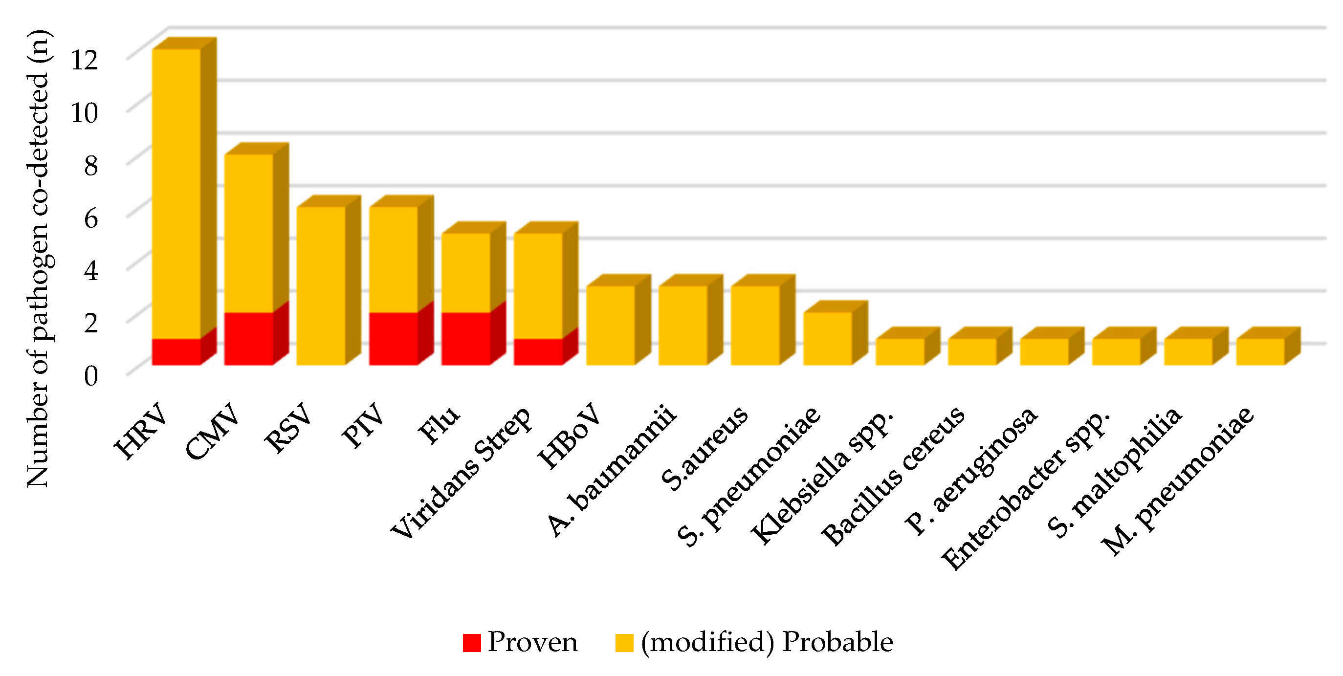
3.3. Co-Detected Respiratory Pathogens in Clinical PCP

Human rhinovirus (HRV) was the most common (15.2%) respiratory pathogen co-detected with P. jirovecii in clinical PCP, followed by cytomegalovirus (CMV) of 10.1%, respiratory syncytial virus (RSV) of 7.6%, and parainfluenza virus (PIV) of 7.6%. Among the bacteria, viridans Streptococcus (6.3%) was the most common, followed by Acinetobacter baumannii (3.8%) and Staphylococcus aureus (3.8%) (Figure 2). Although there were no significant differences, the codetection rate of respiratory pathogen was 7.1% in proven PCP, which tended to be lower than 23.1% in mp-PCP and 35.5% in non-PCP. Among children in mp-PCP group, CMV culture was positive in 12.3%, respiratory viruses were detected by multiplex RT-PCR in 35.4% and pyogenic bacteria was cultured in 23.1% (Table 1).

3.4. Factors Associated with Death in Clinical PCP

As a result of univariate analysis, the proportion of patients under 5 years of age, hospital-onset pneumonia, PCP prophylaxis, and combination of TMP/SMX and pentamidine was significantly higher in the death group than in the survival group (Table 2). In addition, multivariate analysis was performed by adding factors of PCP treatment, desaturation, which presents as an initial symptom, and chest CT findings. As a result, it was analyzed that age under 5 years old (odds ratio [OR] 10.7, 95% confidence interval [CI] 1.8–62.6), hospital-onset PCP (OR 6.9, 95% CI 1.5–32.3), and desaturation as initial symptom (OR 63.5, 95% CI 2.5–1645.5) were significant risk factors for death (Table 3).

4. Discussion

This study investigated the epidemiology, clinical characteristics, and prognostic factors of PCP clinically diagnosed in children aged 18 years old or younger at a single institution for the last 21 years. Modified probable PCP was defined in this study as a positive result of P. jirovecii using conventional PCR, which has been widely used in actual clinical practice in South Korea. This disease entity was comparable to the proven PCP in terms of patient’ demographics, clinical features, laboratory test results, treatment response, and prognosis. Risk factors for mortality in these immunocompromised children with clinical PCP were identified to be younger age, hospital-onset PCP, and desaturation at presentation.
Pneumocystis jirovecii cannot be cultured, and there is no reliable single diagnostic test in PCP diagnosis. The tests recommended by EORTC/MSGERC, IF stain and qPCR, have limitations in practical application due to their low sensitivity, compliance, and universality [9]. Particularly for qPCR, it might be important to set an appropriate target gene and cutoff value to distinguish between colonization and active infection of P. jirovecii [10,11]. For more than 10 years, qPCR has had an important place in the diagnostic strategy for PCP. Recently, serum BDG has been proposed as method to improve diagnostic accuracy for PCP. The sensitivity and specificity in PCP diagnosis can be enhanced when the cut-off value of qPCR is adequately set and used in combination with other tests [12]. However, during the last 21 years in SNUCH, only DFA/IF and nested conventional PCR were used for P. jirovecii detection, and qPCR and BDG were introduced in 2022. The number of proven PCP cases was very small, but it is possible that most children with PCP were diagnosed by conventional PCR alone and successfully treated.
Although the incidence of PCP varies according to the host factors, children with high-risk ALL, high-grade non-Hodgkin lymphomas are at the highest risk. Children with Hodgkin’s lymphoma and children undergoing HCT are also at high risk for developing PCP. Overall, 0.63% of allogeneic and 0.28% of autologous recipients of their first HCT developed PCP [13]. In the current study, all but two patients with clinical PCP had underlying diseases related to immunocompromised status. HCT, SOT, and hematologic malignancy consisted of the majority of the underlying conditions in our patients. However, PID patients were relatively common in the non-PCP group. Therefore, we could more reliably consider PCP in children with hematologic malignancy and/or organ transplantation, whereas we might as well apply more stringent criteria for PCP diagnosis in children with PID, when an immunocompromised children showed a clinical presentation of PCP.
Additionally, in children, typical clinical features of PCP are known to include dry cough, insidious dyspnea, low-grade fever, tachypnea, hypoxia, and bilateral GGO on chest radiograph [3,13]. In the current study, most proven PCPs showed these clinical features, and the mp-PCP group also had very similar clinical features to the proven PCP group, while they were very different from those of the non-PCP group. This finding indicates that the mp-PCP diagnosis, which was temporarily defined in the current study, would reliably include many children with PCP, who could be missed unless PCP PCR is performed. For the clinical purpose, particularly in immunocompromised patients, the diagnostic method should have a higher sensitivity. Therefore, the mp-PCP definition can be used for children in situations where the reliable qPCR test is not available.
In this study, although non-PCP was classified based on the EORTC/MSGERC criteria, since these children underwent work-up for PCP based on the clinical speculation, they might be suspected to be immunosuppressed and/or have unusual clinical and radiologic findings of pneumonia. Therefore, demographic features of children and clinical characteristics of pneumonia in non-PCP group were largely comparable to those in the clinical PCP group. The positive results of P. jirovecii PCR/stain in the non-PCP group might be derived from the airway colonization of P. jirovecii or false positivity of the tests. It might have an advantage as a control group in terms of controlling confounding factors in comparison analyses between PCP groups. However, it is possible that some children with actual PCP could be misclassified into the non-PCP group in situations where we do not have gold-standard diagnostic methods with high sensitivity and specificity. Nevertheless, non-PCP group showed some differentiated clinical characteristics, such as incidence of respiratory distress and C-CT finding, compared to mp-PCP and proven PCP groups.
In the current study, CMV, as well as usual respiratory viruses and bacteria, were frequently co-detected with P. jirovecii from lower respiratory tract samples. CMV might be reactivated and cause lung infection from children with an immunocompromised status, in which PCP can occur [14]. Co-infection with CMV would be associated with more pronounced T-cell immunosuppression and a risk factor for severe disease and poor outcomes in patients with PCP [15,16]. However, in the current study, CMV was not a significant risk factor for mortality in children with clinical PCP, although the small sample size should not confirm this finding.
The mortality rate of children with clinical PCP in this study was 30.4%, which was similar to previous studies on proven PCP [4,17,18]. Previous studies reported that high respiratory and/or pulse rate, elevated C-reactive protein and/or lactate dehydrogenase level, need for mechanical ventilation, hypoxia, combined bacteremia, and preexisting chronic lung disease were more common in mortality cases among adult patients with PCP [19,20]. Although we could not evaluate all these factors for risk analysis in this study, we found that a younger age, hospital-onset pneumonia, and desaturation as initial symptom were the independent risk factors of mortality in children with clinical PCP. These factors would reflect the host’s lower immune and/or worse clinical status at the time of development of PCP, and sever initial presentation. We may use these risk factors to initiate an adequate targeted antimicrobial therapy in children with clinical diagnosis of PCP.
This study has several limitations. First, due to the lack of pediatric studies on a large number of children with PCP, all the definitions and the interpretation of premises and results were based on the studies and guidelines focused on adults. Particularly in respiratory infection, the pathophysiology of disease and available diagnostic methods might differ between children and adults, so more careful interpretation of the findings should be warranted. Second, because we have collected and analyzed data from children with P. jirovecii detected by conventional PCR rather than qPCR, which is officially recommended as a diagnostic tool of probable PCP, some patients could not have true PCP. However, we showed the clinical usefulness and adequacy of the mp-PCP in this study. This could be reinforced with biomarkers for P. jirovecii such as BDG in the near future. Third, we only collected mortality data as the outcome. The need for mechanical ventilation and ICU admission would be valuable for analyzing risk factors for poor outcomes and/or severe disease. However, a large proportion of patients were admitted to the ICU with mechanical ventilation at the time of development of PCP; therefore, we thought it would be difficult to adequately evaluate these factors for risk assessment from PCP in this retrospective design. We may need a large-scale prospective multicenter study. Lastly, this was a single-institution study with a relatively inhomogeneous population (all children were HIV-negative but had several underlying pathologies); thus, the results in this study could not be generalized to other settings.

5. Conclusions

The mp-PCP, which is defined with P. jirovecii detection in patients with classic host factors and clinical features of PCP, might be a good, alternative diagnostic criteria for PCP when qPCR is not available or a reliable cutoff level is not set. In immunocompromised young children with hospital-onset pneumonia showing desaturation as the main initial symptom, the disease could be managed promptly and aggressively based on these new criteria for clinical PCP in children.

Author Contributions

Conceptualization, K.W.Y.; data curation, formal analysis, investigation, and writing—original draft preparation, K.S.Y. and K.W.Y.; resources and validation, B.A., S.H.C., K.T.H., J.Y.C., H.J.K. and E.H.C.; writing—review and editing, K.T.H., J.Y.C., K.W.Y., H.J.K. and E.H.C.; supervision, H.J.K. and E.H.C. All authors have read and agreed to the published version of the manuscript.

Funding

This research received no external funding.

Institutional Review Board Statement

The study was conducted in accordance with the Declaration of Helsinki, and approved by the Institutional Review Board of Seoul National University Hospital (H-2207-002-1336). The approval date is 11 July 2022.

Informed Consent Statement

Patient consent was waived because it was judged that it was practically impossible to obtain informed consent as the patients had already died or there was no follow-up, and the risk to the patients was low if consent was waived.

Data Availability Statement

The data that support the findings of this study are available from the corresponding author, upon reasonable request.

Conflicts of Interest

The authors declare no conflict of interest.

References

  1. Thomas, C.F., Jr.; Limper, A.H. Pneumocystis pneumonia. N. Engl. J. Med. 2004, 350, 2487–2498. [Google Scholar] [CrossRef]
  2. Pyrgos, V.; Shoham, S.; Roilides, E.; Walsh, T.J. Pneumocystis pneumonia in children. Paediatr. Respir. Rev. 2009, 10, 192–198. [Google Scholar] [CrossRef]
  3. Kim, S.Y.R.; Sung, H.; Lee, J. Clinical Characteristics, Prognostic Factors and Influence of Prophylaxis in Children with Pneumocystis jirovecii Pneumonia. Pediatr. Infect. Vaccine 2016, 23, 31–39. [Google Scholar] [CrossRef] [Green Version]
  4. Ling, C.; Qian, S.; Wang, Q.; Zeng, J.; Jia, X.; Liu, J.; Li, Z. Pneumocystis pneumonia in non-HIV children: A 10-year retrospective study. Clin. Respir. J. 2018, 12, 16–22. [Google Scholar] [CrossRef] [PubMed] [Green Version]
  5. Proudfoot, R.H.; Phillips, B. UK paediatric oncology Pneumocystis jirovecii pneumonia surveillance study. Arch. Dis. Child. 2021, 106, 994–998. [Google Scholar] [CrossRef] [PubMed]
  6. Donnelly, J.P.; Chen, S.C.; Kauffman, C.A.; Steinbach, W.J.; Baddley, J.W.; Verweij, P.E.; Clancy, C.J.; Wingard, J.R.; Lockhart, S.R.; Groll, A.H.; et al. Revision and Update of the Consensus Definitions of Invasive Fungal Disease From the European Organization for Research and Treatment of Cancer and the Mycoses Study Group Education and Research Consortium. Clin. Infect. Dis. 2020, 71, 1367–1376. [Google Scholar] [CrossRef] [PubMed] [Green Version]
  7. Lagrou, K.; Chen, S.; Masur, H.; Viscoli, C.; Decker, C.F.; Pagano, L.; Groll, A.H. Pneumocystis jirovecii Disease: Basis for the Revised EORTC/MSGERC Invasive Fungal Disease Definitions in Individuals Without Human Immunodeficiency Virus. Clin. Infect. Dis. 2021, 72 (Suppl. 2), S114–S120. [Google Scholar] [CrossRef] [PubMed]
  8. Choe, P.G.; Kang, Y.M.; Kim, G.; Park, W.B.; Park, S.W.; Kim, H.B.; Oh, M.D.; Kim, E.C.; Kim, N.J. Diagnostic value of direct fluorescence antibody staining for detecting Pneumocystis jirovecii in expectorated sputum from patients with HIV infection. Med. Mycol. 2014, 52, 326–330. [Google Scholar] [CrossRef] [PubMed] [Green Version]
  9. Bateman, M.; Oladele, R.; Kolls, J.K. Diagnosing Pneumocystis jirovecii pneumonia: A review of current methods and novel approaches. Med. Mycol. 2020, 58, 1015–1028. [Google Scholar] [CrossRef] [PubMed]
  10. Alanio, A.; Desoubeaux, G.; Sarfati, C.; Hamane, S.; Bergeron, A.; Azoulay, E.; Molina, J.M.; Derouin, F.; Menotti, J. Real-time PCR assay-based strategy for differentiation between active Pneumocystis jirovecii pneumonia and colonization in immunocompromised patients. Clin. Microbiol. Infect. 2011, 17, 1531–1537. [Google Scholar] [CrossRef] [PubMed]
  11. Perret, T.; Kritikos, A.; Hauser, P.M.; Guiver, M.; Coste, A.T.; Jaton, K.; Lamoth, F. Ability of quantitative PCR to discriminate Pneumocystis jirovecii pneumonia from colonization. J. Med. Microbiol. 2020, 69, 705–711. [Google Scholar] [CrossRef] [PubMed]
  12. Morjaria, S.; Frame, J.; Franco-Garcia, A.; Geyer, A.; Kamboj, M.; Babady, N.E. Clinical Performance of (1,3) Beta-D Glucan for the Diagnosis of Pneumocystis Pneumonia (PCP) in Cancer Patients Tested With PCP Polymerase Chain Reaction. Clin. Infect. Dis. 2019, 69, 1303–1309. [Google Scholar] [CrossRef] [PubMed]
  13. Mantadakis, E. Pneumocystis jirovecii Pneumonia in Children with Hematological Malignancies: Diagnosis and Approaches to Management. J. Fungi. 2020, 6, 331. [Google Scholar] [CrossRef] [PubMed]
  14. Yong, M.K.; Slavin, M.A.; Kontoyiannis, D.P. Invasive fungal disease and cytomegalovirus infection: Is there an association? Curr. Opin. Infect. Dis. 2018, 31, 481–489. [Google Scholar] [CrossRef] [PubMed]
  15. Yu, Q.; Jia, P.; Su, L.; Zhao, H.; Que, C. Outcomes and prognostic factors of non-HIV patients with pneumocystis jirovecii pneumonia and pulmonary CMV co-infection: A Retrospective Cohort Study. BMC Infect. Dis. 2017, 17, 392. [Google Scholar] [CrossRef] [PubMed] [Green Version]
  16. Korkmaz Ekren, P.; Toreyin, Z.N.; Nahid, P.; Doskaya, M.; Caner, A.; Turgay, N.; Zeytinoglu, A.; Toz, S.; Bacakoglu, F.; Guruz, Y.; et al. The association between Cytomegalovirus co-infection with Pneumocystis pneumonia and mortality in immunocompromised non-HIV patients. Clin. Respir. J. 2018, 12, 2590–2597. [Google Scholar] [CrossRef] [PubMed]
  17. Kim, K.R.; Kim, J.M.; Kang, J.M.; Kim, Y.J. Pneumocystis jirovecii pneumonia in pediatric patients: An analysis of 15 confirmed consecutive cases during 14 years. Korean J. Pediatr. 2016, 59, 252–255. [Google Scholar] [CrossRef] [PubMed] [Green Version]
  18. Lee, H.Y.; Lu, C.Y.; Lee, P.I.; Chen, J.M.; Huang, L.M.; Chang, L.Y. Pneumocystis jiroveci pneumonia in Taiwan from 2014 to 2017: Clinical manifestations and outcomes between pediatric and adult patients. J. Microbiol. Immunol. Infect. 2019, 52, 983–990. [Google Scholar] [CrossRef] [PubMed]
  19. Roblot, F.; Godet, C.; Le Moal, G.; Garo, B.; Souala, M.F.; Dary, M.; de Gentile, L.; Gandji, J.A.; Guimard, Y.; Lacroix, C.; et al. Analysis of underlying diseases and prognosis factors associated with Pneumocystis carinii pneumonia in immunocompromised HIV-negative patients. Eur. J. Clin. Microbiol. Infect. Dis. 2002, 21, 523–531. [Google Scholar] [PubMed]
  20. Kim, S.J.; Lee, J.; Cho, Y.J.; Park, Y.S.; Lee, C.H.; Yoon, H.I.; Lee, S.M.; Yim, J.J.; Lee, J.H.; Yoo, C.G.; et al. Prognostic factors of Pneumocystis jirovecii pneumonia in patients without HIV infection. J. Infect. 2014, 69, 88–95. [Google Scholar] [CrossRef] [PubMed]
Figure 1. Annual number of Pneumocystis jirovecii testing performed and clinical PCP diagnosis. PCP, Pneumocystis jirovecii pneumonia; n, number; DFA, direct fluorescent antibody; IF, immunofluorescence; PCR, polymerase chain reaction.
Children 09 01596 g001
Figure 2. Frequency of respiratory pathogens co-detected in children with clinical PCP. PCP, Pneumocystis jirovecii pneumonia; n, number; HRV, human rhinovirus; CMV, cytomegalovirus; RSV, respiratory syncytial virus; PIV, parainfluenza virus; Flu, influenza virus; Strep, Streptococcus; HBoV, human bocavirus; A. baumannii, Acinetobacter baumannii; S. pneumoniae, Streptococcus pneumoniae.; spp., species; P. aeruginosa, Pseudomonas aeruginosa; S. maltophilia, Stenotrophomonas maltophilia; M. pneumoniae, Mycoplasma pneumoniae.
Children 09 01596 g002
Table 1. Clinical characteristics of children with clinical PCP, n (%).
Proven PCP
(n = 14)
mp-PCP
(n = 65)
Non-PCP
(n = 31)
p-Value
Age (y)Median (IQR)5.5 (1.4–11.1)7.4 (2.4–12.8)4.7 (0.9–12.0)0.173
<5 YO6 (42.9)22 (33.8)17 (54.8)0.146
SexFemale6 (42.9)31 (47.7)17 (54.8)0.712
Underlying medical conditionHSCT3 (21.4)13 (20.0)4 (12.9)0.662
SOT2 (14.3)16 (24.6)1 (3.2)0.033 *
HM2 (14.3)15 (23.1)3 (9.7)0.260
SC2 (14.3)7 (10.8)4 (12.9)0.911
PID1 (7.1)7 (10.8)8 (25.8)0.104
None1 (7.1)1 (1.5)3 (9.7)0.178
Symptom/signFever13 (92.9)50 (76.9)18 (58.1)0.026 *
Cough9 (64.3)49 (75.4)19 (61.3)0.437
Sputum7 (50.0)31 (47.7)11 (35.5)0.527
Tachypnea8 (57.1)43 (66.2)8 (25.8)0.002 *
Dyspnea9 (64.3)54 (83.1)15 (48.4)0.004 *
Desaturation11(78.6)52 (80.0)15 (48.4)0.006 *
Breath soundRale4 (28.6)19 (29.2)11 (35.5)0.746
Wheezing0 (0.0)4 (6.2)9 (29.0)0.318
Respiratory pathogen co-detectedCMV culture2 (14.3)8 (12.3)2 (6.5)0.695
Virus RT-PCR5 (35.7)23 (35.4)13 (41.9)0.638
Bacterial culture1 (7.1)15 (23.1)11 (35.5)0.103
C-CT findingsBilateral GGO13 (92.9)48 (73.8)6 (19.4)<0.001 *
Pleural effusion2 (14.3)5 (7.7)3 (9.7)0.675
OnsetHospital onset7 (50.0)24 (36.9)14 (45.2)0.566
PCP PxYes4 (28.6)19 (29.2)7 (22.6)0.786
PMD3 (21.4)8 (12.3)3 (9.7)0.475
PCP TxYes14 (100.0)59 (90.8)13 (41.9)<0.001 *
TMP/SMX only8 (57.1)48 (73.8)11 (35.5)0.119
TMP/SMX + PMD6 (42.9)11 (16.9)1 (3.2)0.060
OutcomeDeath6 (42.9)18 (27.7)8 (25.8)0.470
PCP, P. jirovecii pneumonia; n, number; mp-PCP, modified probable PCP; y, year; IQR, interquartile range; YO, years-old; HSCT, hematopoietic stem cell transplantation; SOT, solid organ transplantation; HM, hematologic malignancy; SC, solid cancer; PID, primary immune deficiency; CMV, cytomegalovirus; RT-PCR, reverse transcriptase polymerase chain reaction; C-CT, chest computerized tomography; GGO, ground glass opacity; Px, prophylaxis; Tx, treatment; PMD, pentamidine; TMP/SMX, trimethoprim/sulfamethoxazole. * p < 0.05 in comparison for three groups.
Table 2. Univariate analyses for risk factors of mortality in clinical PCP, n (%).
Death
(n = 24)
Survival
(n = 55)
p-Value
Age (y)<5 YO13 (54.2)15 (27.3)0.022 *
SexFemale9 (37.5)28 (50.9)0.272
Underlying medical conditionHCT9 (37.5)7 (12.7)0.012 *
SOT2 (8.3)16 (29.1)0.047 *
HM5 (20.8)12 (21.8)1.000
SC3 (12.5)6 (10.9)1.000
PID2 (8.3)6 (10.9)1.000
None2 (8.3)0 (0.0)0.900
Symptom/signFever20 (83.3)43 (78.2)0.532
Cough15 (62.5)43 (78.2)0.536
Sputum10 (41.7)28 (50.9)0.549
Tachypnea14 (58.3)37 (67.3)0.776
Dyspnea19 (79.2)44 (80.0)0.496
Desaturation22 (91.7)41 (74.5)0.055
Breath soundRale9 (37.5)14 (25.5)0.197
Wheezing3 (12.5)6 (10.9)0.713
Respiratory pathogenco-detectedCMV culture1 (4.2)9 (16.4)0.143
Virus RT-PCR8 (33.3)20 (36.4)0.781
Bacterial culture7 (29.2)9 (16.4)0.291
C-CT findingsBilateral GGO14 (58.3)47 (85.5)0.087
Pleural effusion4 (16.7)3 (5.5)0.080
OnsetHospital onset15 (62.5)16 (29.1)0.005 *
PCP prophylaxisYes13 (54.2)10 (18.2)0.001 *
PMD5 (20.8)6 (10.9)0.414
PCP treatmentYes20 (83.3)53 (96.4)0.066
TMP/SMX only11 (45.8)45 (81.8)0.007 *
TMP/SMX + PMD9 (37.5)8 (14.5)0.007 *
PCP, P. jirovecii pneumonia; n, number; y, year; YO, years old; HSCT, hematopoietic stem cell transplantation; SOT, solid organ transplantation; HM, hematologic malignancy; SC, solid cancer; PID, primary immune deficiency; CMV, cytomegalovirus; RT-PCR, reverse transcriptase polymerase chain reaction; C-CT, chest computerized tomography; GGO, ground glass opacity; PMD, pentamidine; TMP/SMX, trimethoprim/sulfamethoxazole; * p < 0.05.
Table 3. Multivariable analysis for mortality in children with clinical PCP, n (%).
Death
(n = 24)
Survival
(n = 55)
OR95% CI
Age (y)<5 YO13 (54.2)15 (27.3)10.660 *1.816–62.570
Underlying conditionHSCT9 (37.5)7 (12.7)5.8610.755–45.524
OnsetHospital15 (62.5)16 (29.1)6.941 *1.491–32.308
Symptom/signDesaturation22 (91.7)41 (74.5)63.547 *2.454–1645.517
C-CT findingBilateral GGO14 (58.3)47 (85.5)0.9760.149–6.406
Pleural effusion4 (16.7)3 (5.5)8.0260.754–85.413
PCP prophylaxisYes13 (54.2)10 (18.2)5.2940.770–36.386
PCP treatmentTMP/SMX + PMD9 (37.5)8 (14.5)3.4720.687–17.533
PCP, P. jirovecii pneumonia; n, number; OR, odds ratio; CI, confidential interval, year; YO, years-old; HSCT, hematopoietic stem cell transplantation; C-CT, chest computerized tomography; GGO, ground glass opacity; PMD, pentamidine; TMP/SMX, trimethoprim/sulfamethoxazole; * p < 0.05.
Publisher’s Note: MDPI stays neutral with regard to jurisdictional claims in published maps and institutional affiliations.

