
First Molecular Confirmation of Equine Ocular Setaria digitata in China
by
 ,
,
Feng Yu
1,†, 
Bo Liu
1,†,
Shulei Chen
1,
Ziwen Yi
1,
Xianyong Liu
2,
Yiping Zhu
1,* and
Jing Li
1,* 1
Equine Clinical Diagnostic Center, College of Veterinary Medicine, China Agricultural University, No. 2 Yuanmingyuan West Road, Beijing 100093, China
2
Department of Veterinary Parasitology, College of Veterinary Medicine, China Agricultural University, Beijing 100193, China
*
Authors to whom correspondence should be addressed.
†
These authors contributed equally to this work.
Vet. Sci. 2021, 8(4), 55; https://doi.org/10.3390/vetsci8040055
Submission received: 23 February 2021
/
Revised: 25 March 2021
/
Accepted: 26 March 2021
/
Published: 28 March 2021
(This article belongs to the Special Issue Advances in Veterinary and Comparative Ophthalmology)
Abstract
:A 5-year-old Mongolian mare (Equus caballus Linnaeus, 1758) was observed to have corneal opacity and excessive ocular discharge. An ophthalmic examination revealed a moving thread-like cylindrical worm in the anterior chamber of the right eye. The parasite was successfully removed surgically. The worm was observed under light microscopy and confirmed as Setaria digitata by 12S rRNA gene amplification and sequencing. Phylogenetic analysis demonstrated similarity with Setaria digitata in the National Center for Biotechnology Information (NCBI) GenBank database isolated from other Asian countries. This report is the first confirmed case of equine ocular setariasis by molecular diagnosis in China, which may indicate its presence in livestock and promote research on its epidemiology.
1. Introduction
Setaria is a genus of roundworms (Nematoda: Filariodea), among which Setaria digitata is mainly found in Asia. Setaria digitata often affects cattle and buffalos, with common predilection site in peritoneal cavity [1]. Within its natural hosts, these nematodes are non-pathogenic in most cases. However, the parasite exhibits migratory behaviors in aberrant hosts including goats, sheep and horses [2]. The most common form of migration in infected horses is ocular migration [3,4]. Infected goats and sheep can experience cerebrospinal nematodiasis, which leads to lumbar paralysis and death [5]. Vectors of S. digitata include biting insects like mosquitoes (genera Aedes, Culex, Anopheles, Hyrcanus and Armigeres) [6]. Setariosis poses a serious threat to susceptible animals in tropical areas. When mosquitoes feed on the blood of infected hosts, they become infected with microfilariae, which develop into infective larvae (L3) inside the mosquitoes within 2 to 3 weeks. Infected mosquitoes transmit L3 to susceptible hosts during blood meals, after which it matures in the definitive host within 8–10 months [7].
Equine ocular setariasis is caused by S. digitata, S. equina or S. marshalli, with most cases reported by countries in south and southeast Asia [8,9,10,11]. According to a retrospective study conducted in India, 57% (138/242) of the regional cases of equine ocular disorders were diagnosed as ocular setariasis [12]. Infected horses displayed multiple ocular signs including lacrimation, photophobia, corneal opacity, conjunctivitis and blindness, especially when treatment was delayed [12,13,14]. Corneal edema, synechia, cataract and retinal detachments may occur with toxins released from worms in the anterior chamber [15].
Setaria digitata infecting horses in China has never been identified. This report is the first time S. digitata has been identified in the eye of an equine patient in China by molecular diagnosis.
2. Materials and Methods
All procedures involving the horse in this study were carried out with a welfare license (AW62201202-2-1) issued by the Animal Care and Use Committee of the China Agriculture University in Beijing.
2.1. Case Report
A 5-year-old Mongolian mare (Equus caballus Linnaeus, 1758) presented with blepharospasm, squinting and photophobia to the Equine Clinical Diagnostic Center of China Agricultural University. A gross ophthalmic examination identified a corneal opacity and ocular discharge in the patient’s right eye (Figure 1). Closer observation revealed a thread-like cylindrical worm, moving in a swirling motion, in the anterior chamber of the right eye. Microfilariae were not detected on a peripheral blood smear, and a complete blood count did not show major abnormalities. Surgical removal was chosen over exclusive medical treatment to remove the worm immediately. The mare was sedated with Xylazole (an α-2 agonist sedative) 1.1 mg/kg intravenously and was induced with and maintained on Tiletamine-Zolazepam 2.0 mg/kg intravenously. The right eye was flushed multiple times with 3% boric acid solution, and the cornea was desensitized with drops of proparacaine hydrochloride ophthalmic solution. Wrapped tightly in sterile medical gauzes and with 2 mm of its tip exposed, a #11 surgical blade was used to puncture open the anterior chamber of the right eye at the 6 o’clock position of the cornea, about 2 mm inside the limbus. The worm moved towards the puncture site and was easily removed with minimal aqueous leakage. The puncture site was left to heal with a 2 mm surgical lesion. Terramycin ophthalmic ointment was administered four times daily for one week as prophylaxis, and flunixin meglumine was injected intravenously at 1.1 mg/kg once daily for three days as an anti-inflammatory medication. The extracted worm was washed three times with phosphate buffer saline (PBS), observed under a light microscope, then preserved in 70% ethanol until its DNA was extracted. The ocular opacity completely cleared approximately 4 weeks after surgery. No other medications were prescribed considering the horse recovered without complications.
2.2. Worm Identification
Light microscopy was used to identify the worm’s species based on referenced morphological criteria [16]. DNA was extracted from the worm by following the instructions specified in the genomic DNA extraction kit (TSINGKE, Beijing, China). The worm was digested with 20 uL Proteinase K and 200 uL distilled water in a 1.5 mL centrifuge tube. The tube was vortexed for 10 s, then 200 uL buffer gA1 was added. The tube was then vortexed for another 10 s. The sample incubated in warm water at 56 °C for 1 to 3 h. After DNA extraction, molecular diagnosis by Polymerase Chain Reaction (PCR) was initiated using the following primer sequence SDF: 5′-AGT CCT CCC TTG TTG CTG GT-3′ and SDR: 5′-GGG TGG TTT GTA CCC CTC CG-3′ [7]. A final PCR volume of 50 μL contained 25 μL 2XTSINGKE Master Mix, 1 μL of each forward and reverse primer and 1 μL of DNA template and 22 μL ddH2O. PCR amplification was performed under the following conditions: initial denaturation at 94 °C for 4 min for 1 cycle, denaturation at 94 °C for 80 s, annealing at 46 °C for 80 s, extension at 72 °C for 60 s for 39 cycles and final extension at 72 °C for 10 min. Amplified products were eletrophoresed on agarose gel and purified for further nucleotide sequences. The resulting sequences were compared with other filarial worms’ sequences stored in GenBank of the National Center for Biotechnology Information (NCBI) using the Basic Local Alignment Search Tool (BLAST) program. Available at: https://blast.ncbi.nlm.nih.gov/Blast.cgi (accessed on 12 January 2021). A multiple sequence alignment was obtained with the ClustalW program within MEGA X (Pennsylvania State University, State College, PA, USA). Phylogenetic analysis was then performed using MEGA X. For distance analysis, the Kimura 2-parameter model was used to construct the distance matrix and the tree was inferred from this using the Maximum Likelihood approach. Bootstrap resampling (1000 pseudoreplicates) was performed, and a bootstrap consensus tree was produced.
3. Results and Discussion
The extracted worm was milky white in color, thread-shaped, tapered at both ends and 7.2 cm long. The tapering posterior end had a spherical knob, which is characteristic of a female S. digitata (Figure 2). The anterior end was round with two equal prominences on the peribuccal crown (Figure 2). Varying lengths of S. digitata from cattle and horses were reported [16,17,18,19].
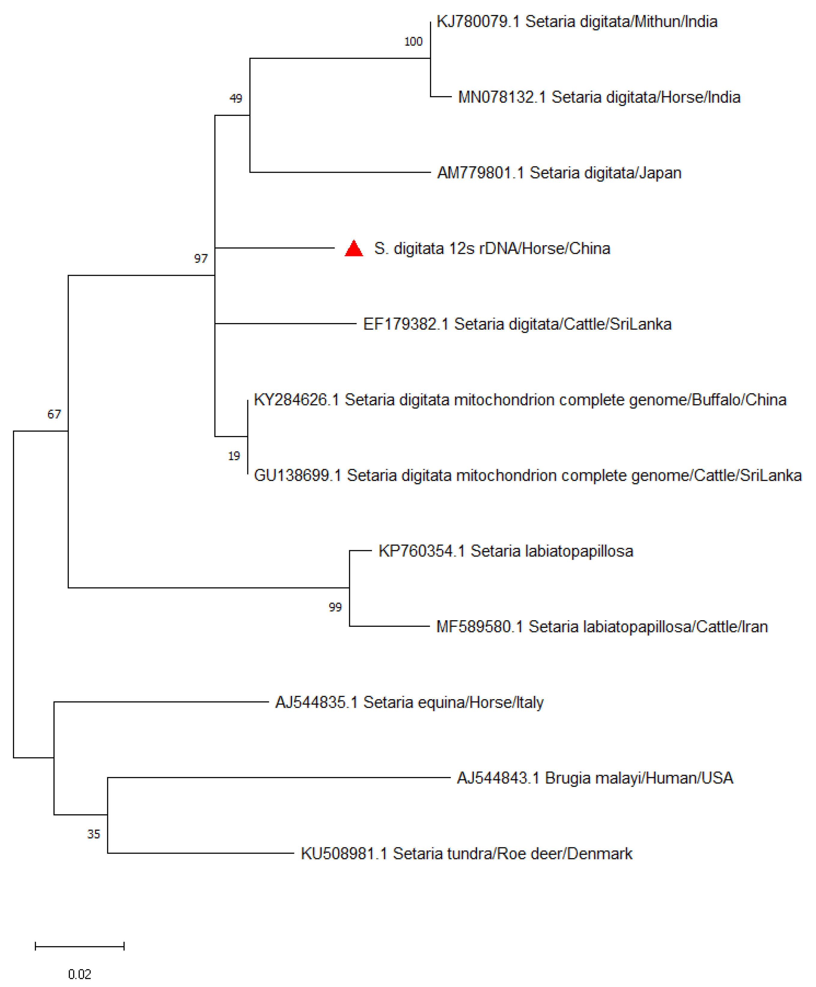
Even though morphology has been traditionally used to identify and differentiate Setaria spp. [7], it was reported to be inadequate since S. digitata and its congeners, including S. digitata, S. equina, S. labiatopapillosa and S. cervi, are similar in morphology [19]. Instead, this study used a nucleic acid-based detection method to confirm the filarial species isolated from the mare. Gel electrophoresis produced a 209 bp band, and the sample was confirmed as S. digitata when compared against other sequences in the NCBI GenBank database using BLAST. The genetic sequence extracted from the worm was 99% similar to that of S. digitata isolated from cattle and buffalos in Sri Lanka and from buffalos in China [20,21]. The mind-point rooted phylogenetic tree further confirmed that the S. digitata isolated from this study belongs to the same major clade isolated from Japan, India and Sri Lanka. S. equina and S. tundra formed in a different cluster. Setaria digitata and S. labiatopapillosa appear to be sister species with a bootstrap value of 67% (Figure 3). The 12S rDNA sequence was a useful marker for phylogenetic analysis due to its slow evolution compared to other protein coding mitochondrial genes [8,20]. Phylogenetic analysis of S. digitata indicated that the isolate sequences from different host species produced a closely related clade with high bootstrap support within the Setaria genus. Minor nucleotide sequence variations were observed. The current findings imply that the isolates of S. digitata detected worldwide have the same molecular characteristics [19].
Different medical therapies have been proposed to treat equine ocular setariasis to avoid complications of surgical treatments such as phthisis bulbi, corneal edema and prolapse of iris. Medical therapies have their inert disadvantages. For example, intraocular reaction to the dead worm body may lead to more severe damage to the intraocular structure [22,23]. Raziq, SA (1989) suggested diethylcarbamazine citrate as a treatment option for microfilaremia, but it was not effective against adult parasites in the eye [24]. Another study demonstrated that ivermectin resolved microfilaremia seven days post-treatment; however, the adult worms took longer to treat [25]. Given the limitations of medical management, surgical treatment was chosen for the immediate removal of the adult worm with minimal risk. In most S. digitata-related ocular cases, only one worm was retrieved from horses even though two worms can be encountered [19,26]. There is no confirmed association between length or load of worms in eyes and the severity of ocular disease. Also, ocular lesions healed well in most cases [8,18,19].
Setaria digitata was mostly reported in tropical Asian countries, where the vectors are prevalent [5,8,9,11,17,19]. South Korea was the only high latitude non-tropical country where S. digitata was reported in horses [18,26]. In this study, S. digitata was identified and confirmed by a molecular method in a horse on a farm located in northern China, where the latitude was close to South Korea. Interestingly, the case occurred during this region’s coldest time of the year (December), which indicates that the mare was infected long before December, when the vectors were active. The occurrence of this incidental case is likely due to the presence of cattle farms in the neighborhood. Therefore, this finding suggests that cattle in northern China may carry S. digitata, and further investigation of its prevalence will be worthwhile. In 2017, S. digitata was first identified by molecular method in the cadavers of buffalos in southern China, yet not in live buffalos or cattle [21]. Setariasis is a well-recognized ophthalmological disorder in equine, with S. digitata being reported as the most common cause [27]. Setaria digitata is considered a major health hazard in animals, with detrimental economic impact on the owners. Therefore, the findings from this study warrant further epidemiological investigation of Setaria digitata in China, especially in the natural hosts.
Author Contributions
Conceptualization, J.L.; methodology, Y.Z.; software, F.Y.; validation, F.Y., Y.Z.; formal analysis, S.C.; investigation, X.L.; resources, S.C.; data curation, Z.Y.; writing—original draft preparation, F.Y.; writing—review and editing, B.L.; visualization, J.L.; supervision, Y.Z.; project administration, J.L.; funding acquisition, J.L. All authors have read and agreed to the published version of the manuscript.
Funding
This research received no external funding.
Institutional Review Board Statement
All procedures involving the horse in this study were carried out with welfare license (AW62201202-2-1) issued by the Animal Care and Use Committee of the China Agriculture University.
Informed Consent Statement
Not applicable.
Data Availability Statement
Data sharing not applicable.
Acknowledgments
The authors would like to thank Academician Jianzhong Shen and the China Horse Industry Association for their support at the Equine Clinical Diagnostic Center in China Agriculture University. The author would also like to thank June Barrera (College of Veterinary Medicine, University of California, Davis) for her language refining for this paper.
Conflicts of Interest
The authors declare no conflict of interest.
References
- Azawi, A.K.A.L.; Fadhl, R.A.; Fadhl, R.S. Epidemiological study of Setaria equina infection in donkeys. Iraq Vet. J. 2012, 36, 93–97. [Google Scholar]
- Bazargani, T.; Eslami, A.; Gholami, G.R.; Molai, A.; Ghafari-Charati, J.; Dawoodi, J.; Ashrafi, J. Cerebrospinal nematodiasis of cattle, sheep and goats in Iran. Iran. J. Parasitol. 2008, 3, 16–20. [Google Scholar]
- Jayakumar, K.; Dharmaceelan, S.; Rajendran, N.; Senthilkumar, S.; Kathirvel, S.; Nagarajan, L.; Kumaresan, A. Ocular setariasis in a pony. Indian Vet. J. 2012, 89, 64–66. [Google Scholar]
- Radwan, A.M.; Ahmed, N.E.; Elakabawy, L.M.; Ramadan, M.Y.; Elmadawy, R.S. Prevalence and pathogenesis of some filarial nematodes infecting donkeys in Egypt. Vet. World 2016, 9, 888–892. [Google Scholar] [CrossRef] [PubMed]
- Voronin, D.; Abeykoon, A.M.; Gunawardene, Y.I.; Dassanayake, R.S. Absence of Wolbachia endobacteria in Sri Lankan isolates of the nematode parasite of animals Setaria digitata. Vet. Parasitol. 2015, 207, 350–354. [Google Scholar] [CrossRef] [PubMed]
- Tung, K.C.; Cheng, F.P.; Lai, C.H.; Wang, K.S.; Wang, J.S.; Lee, W.M. Demonstration of vector competence of Culex quinquefasciatus (Diptera: Culicidae) for Setaria digitata. Vet. Parasitol. 2004, 123, 279–284. [Google Scholar] [CrossRef] [PubMed]
- Perumal, A.N.I.; Gunawardene, Y.I.N.S.; Dassanyake, R.S. Setaria digitata in advancing our knowledge of human lymphatic filariasis. J. Helminthol. 2016, 90, 129–138. [Google Scholar] [CrossRef] [PubMed]
- Peng, T.L.; Armiladiana, M.; Ruhil, H.H.; Maizan, M.; Choong, S.S. First report of equine Setaria digitata (von Linstow 1906) infestation in Malaysia. Vet. Parasitol. Reg. Stud. Reports. 2019, 17, 100310. [Google Scholar] [CrossRef] [PubMed]
- Sathu, S. Intraocular parasites in horses. A report of five cases. Indian Vet. J. 1974, 5, 225. [Google Scholar]
- Ladoucer, C.A.; Kazacos, K.R. Thelazia lacrimalis in horses in India. J. Am. Vet. Med. Assoc. 1981, 178, 301–302. [Google Scholar]
- Parrah, J.D.; Buchoo, B.A.; Moulvi, B.A. Ocular filariasis in equines: A study of 9 cases. Centaur 2004, 4, 70–71. [Google Scholar]
- Tamilmahan, P.; Zama, M.M.S.; Pathak, R.; Muneeswaran, N.S.; Karthik, K. A retrospective study of ocular occurrences in domestic animals: 799 cases. Vet. World 2013, 6, 274–276. [Google Scholar] [CrossRef]
- Shin, S.S.; Cho, K.O.; Wee, S.H. Ocular infection of cattle with Setaria digitata. J. Vet. Med. Sci. 2002, 64, 7–10. [Google Scholar] [CrossRef] [PubMed] [Green Version]
- Basak, S.K.; Hazra, T.K.; Bhattacharya, D. Persistent corneal oedema secondary to presumed dead adult worm in the anterior chamber. Indian J. Ophthalmol. 2007, 55, 679. [Google Scholar] [CrossRef]
- Paglia, D.T.; Miller, P.E.; Dubielzig, R.R. James Wardrop and equine recurrent uveitis. Arch. Ophthalmol. 2004, 122, 1218–1223. [Google Scholar] [CrossRef] [PubMed] [Green Version]
- Rhee, J.K.; Choi, E.Y.; Park, B.K.; Jang, B.G. Application of scanning electron microscopy in assessing the prevalence of some Setaria species in Korean cattle. Korean J. Parasitol. 1994, 32, 1–6. [Google Scholar] [CrossRef]
- Sundar, S.T.B.; D’Souza, P.E. Morphological characterization of Setaria worms collected from cattle. J. Parasit. Dis. 2015, 39, 572–576. [Google Scholar] [CrossRef] [Green Version]
- Shin, J.; Ahn, K.-S.; Jeong, H.-S.; Kim, B.-S.; Choi, E.; Shin, S.-S.; Suh, G.-H.; Kim, H.-J. First blindness cases of horses infected with Setaria digitata (Nematoda: Filarioidea) in the Republic of Korea. Korean J. Parasitol. 2017, 55, 667–761. [Google Scholar] [CrossRef]
- Maharana, B.R.; Potliya, S.; Ganguly, A.; Bisla, R.S.; Mishra, C.; Ganguly, I. First report of the isolation and phylogenetic characterization of equine Setaria digitata from India based on mitochondrial COI, 12S rDNA, and nuclear ITS2 sequence data. Parasitol. Res. 2020, 119, 473–481. [Google Scholar] [CrossRef] [PubMed]
- Yatawara, L.; Wickramasinghe, S.; Rajapakse, R.P.; Agatsuma, T. The complete mitochondrial genome of Setaria digitata (Nematoda: Filarioidea): Mitochondrial gene content, arrangement and composition compared with other nematodes. Mol. Biochem. Parasitol. 2010, 173, 32–38. [Google Scholar] [CrossRef]
- Liu, G.; Li, J.; Zhu, X. Characterization of the complete mitochondrial genome of Setaria digitata (Nematoda: Setariidae) from China. J. Helminthol. 2017, 91, 772–776. [Google Scholar] [CrossRef]
- Lavach, J.D. Parasitic diseases. In Large Animal Ophthalmology; Mosby: Philadelphia, PA, USA, 1990; pp. 260–263. [Google Scholar]
- Moore, C.P.; Sarazan, R.D.; Whitley, R.D.; Jackson, W.F. Equine ocular parasites: A review. Equine Vet. J. Suppl. 1983, 2, 76–85. [Google Scholar] [CrossRef]
- Raziq, S.A. A preliminary clinical trial on the use of diethylcarbamazine citrate for the treatment of equine filariasis. Pak. Vet. J. 1989, 9, 93–96. [Google Scholar]
- Muhammad, G.; Saqib, M. Successful treatment of ocular equine microfilariasis (Setaria species) with ivermectin. Vet. Rec. 2007, 160, 25–26. [Google Scholar] [CrossRef] [PubMed]
- Kim, H.; Ahn, D.C.; Jin, H.P.; Yu, D.; Chae, J.; Yoo, J.; Sim, C.H.; Choi, K.-S.; Park, Y.-J.; Park, B.-K. Ocular setariasis by Setaria digitata in a horse in Korea. Korean J. Vet. Serv. 2018, 41, 15–19. [Google Scholar]
- Nabie, R.; Spotin, A.; Rouhani, S. Subconjunctival setariasis due to Setaria equina infection; a case report and a literature review. Parasitol. Int. 2017, 66, 930–932. [Google Scholar] [CrossRef] [PubMed]
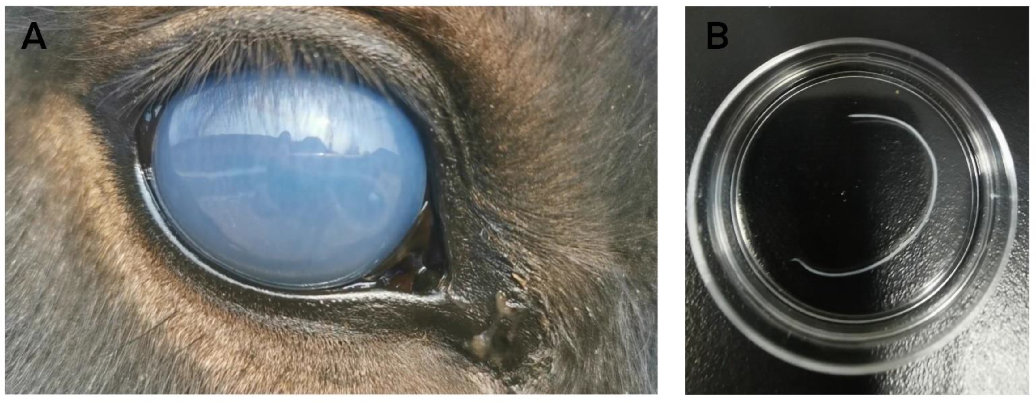
Figure 1.
Corneal edema and ocular discharge in the right eye upon examination (A). Extracted S. digitata worm from the eye (B).
Figure 1.
Corneal edema and ocular discharge in the right eye upon examination (A). Extracted S. digitata worm from the eye (B).

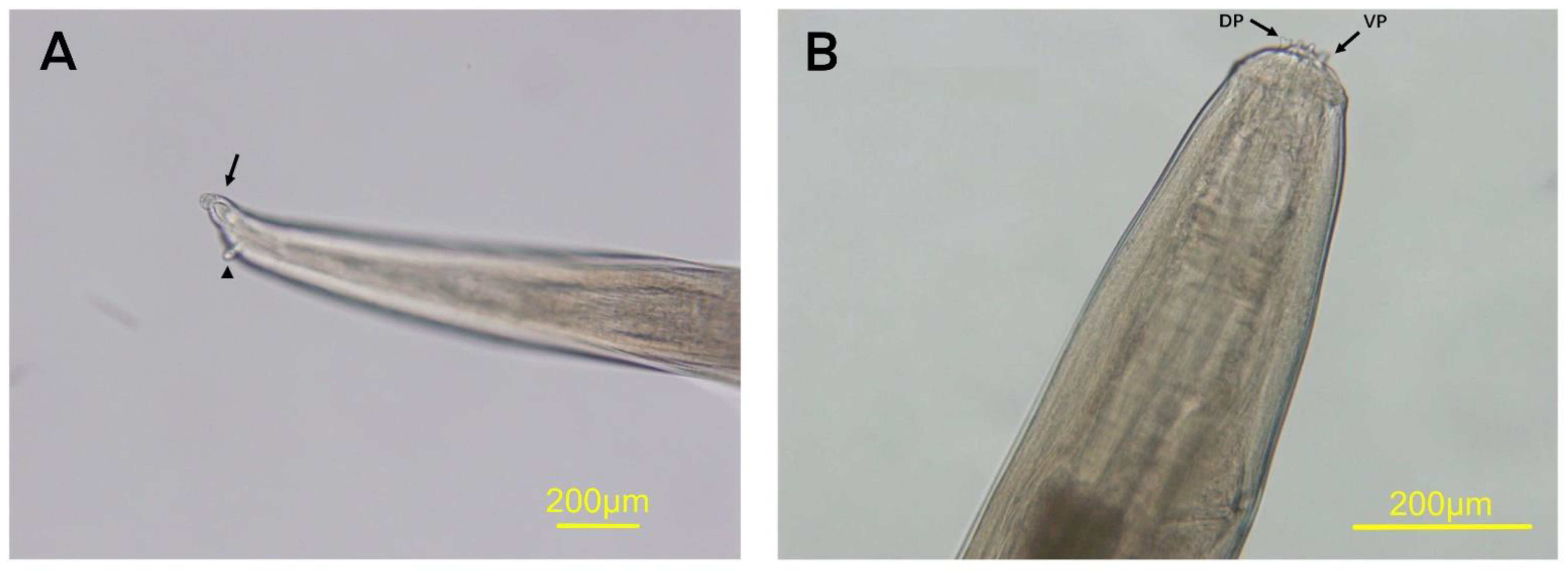
Figure 2.
Posterior end of female adult of Setaria digitata has spherical terminal knob (arrow) and lateral appendages (arrowhead) (A). Anterior region of S. digitata shows the dorsal projection (DP) and ventral projections (VP) on peribuccal crown (B).
Figure 2.
Posterior end of female adult of Setaria digitata has spherical terminal knob (arrow) and lateral appendages (arrowhead) (A). Anterior region of S. digitata shows the dorsal projection (DP) and ventral projections (VP) on peribuccal crown (B).

Figure 3.
A mind-point rooted phylogenetic tree, inferred from 12SrDNA nucleotide sequences using the Maximum Likelihood method in MEGA X after Kimura-2 correction. Scale bar indicates the proportion of sites changing along each branch. Accession numbers of all sequences used for tree analyses are given. The sequence generated in this study was marked as red rectangular.

Publisher’s Note: MDPI stays neutral with regard to jurisdictional claims in published maps and institutional affiliations. |
© 2021 by the authors. Licensee MDPI, Basel, Switzerland. This article is an open access article distributed under the terms and conditions of the Creative Commons Attribution (CC BY) license (http://creativecommons.org/licenses/by/4.0/).
Share and Cite
MDPI and ACS Style
Yu, F.; Liu, B.; Chen, S.; Yi, Z.; Liu, X.; Zhu, Y.; Li, J. First Molecular Confirmation of Equine Ocular Setaria digitata in China. Vet. Sci. 2021, 8, 55. https://doi.org/10.3390/vetsci8040055
AMA Style
Yu F, Liu B, Chen S, Yi Z, Liu X, Zhu Y, Li J. First Molecular Confirmation of Equine Ocular Setaria digitata in China. Veterinary Sciences. 2021; 8(4):55. https://doi.org/10.3390/vetsci8040055
Chicago/Turabian StyleYu, Feng, Bo Liu, Shulei Chen, Ziwen Yi, Xianyong Liu, Yiping Zhu, and Jing Li. 2021. "First Molecular Confirmation of Equine Ocular Setaria digitata in China" Veterinary Sciences 8, no. 4: 55. https://doi.org/10.3390/vetsci8040055
Note that from the first issue of 2016, this journal uses article numbers instead of page numbers. See further details here.