Article

Transcriptome Analysis of Fusarium–Tomato Interaction Based on an Updated Genome Annotation of Fusarium oxysporum f. sp. lycopersici Identifies Novel Effector Candidates That Suppress or Induce Cell Death in Nicotiana benthamiana

1
State Key Laboratory of North China Crop Improvement and Regulation, Hebei Agricultural University, Baoding 071001, China
2
Division of Plant Science, Research School of Biology, the Australian National University, Canberra 2601, Australia
3
Hebei Key Laboratory of Plant Physiology and Molecular Pathology, College of Life Science, Hebei Agricultural University, Baoding 071001, China
4
State Key Laboratory of Grassland Agro-Ecosystems, Key Laboratory of Grassland Livestock Industry Innovation, Ministry of Agriculture and Rural Affairs, College of Pastoral Agriculture Science and Technology, Lanzhou University, Lanzhou 730020, China
5
College of Horticulture, Hebei Agricultural University, Baoding 071001, China
*
Authors to whom correspondence should be addressed.
J. Fungi 2022, 8(7), 672; https://doi.org/10.3390/jof8070672
Submission received: 25 May 2022 / Revised: 15 June 2022 / Accepted: 23 June 2022 / Published: 26 June 2022
(This article belongs to the Section Fungal Pathogenesis and Disease Control)

Abstract

:
Fusarium oxysporum f. sp. lycopersici (Fol) causes vascular wilt disease in tomato. Upon colonization of the host, Fol secretes many small effector proteins into the xylem sap to facilitate infection. Besides known SIX (secreted in xylem) proteins, the identity of additional effectors that contribute to Fol pathogenicity remains largely unexplored. We performed a deep RNA-sequencing analysis of Fol race 2-infected tomato, used the sequence data to annotate a published genome assembly generated via PacBio SMRT sequencing of the Fol race 2 reference strain Fol4287, and analysed the resulting transcriptome to identify Fol effector candidates among the newly annotated genes. We examined the Fol-infection expression profiles of all 13 SIX genes present in Fol race 2 and identified 27 new candidate effector genes that were likewise significantly upregulated upon Fol infection. Using Agrobacterium-mediated transformation, we tested the ability of 22 of the new candidate effector genes to suppress or induce cell death in leaves of Nicotiana benthamiana. One effector candidate designated Fol-EC19, encoding a secreted guanyl-specific ribonuclease, was found to trigger cell death and two effector candidates designated Fol-EC14 and Fol-EC20, encoding a glucanase and a secreted trypsin, respectively, were identified that can suppress Bax-mediated cell death. Remarkably, Fol-EC14 and Fol-EC20 were also found to suppress I-2/Avr2- and I/Avr1-mediated cell death. Using the yeast secretion trap screening system, we showed that these three biologically-active effector candidates each contain a functional signal peptide for protein secretion. Our findings provide a basis for further understanding the virulence functions of Fol effectors.

1. Introduction

Plants continuously face biotic stresses associated with fungal pathogens. To combat fungal infections, plants have evolved multiple cell-surface and intracellular receptors able to perceive pathogen molecules and activate plant immune responses [1]. Conversely, adapted fungal pathogens must avoid or suppress plant immune responses to establish compatible infections [2]. One of the strategies employed by fungal pathogens is the secretion of effectors that can suppress plant immune responses and manipulate plant cell physiology to facilitate infection and fungal proliferation [2,3]. However, some effectors can be recognized by plant receptors to activate effector-triggered immunity (ETI) [2,4]. ETI often results in a rapid and localized cell death that can impede the proliferation of biotrophic fungal pathogens [5]. In turn, fungal pathogens have evolved strategies to avoid ETI via genetic changes in effector genes (also termed Avr or avirulence genes) recognized by corresponding R (resistance) genes in the host or to suppress ETI by deploying other effectors. For example, the hemibiotrophic fungal pathogen Leptosphaeria maculans has been shown to use AvrLm3 and AvrLm5-9 effectors to suppress race-specific resistance mediated by Rlm4-7, which perceives the intracellularly localized AvrLm4-7 effector [6,7,8]. The wheat powdery mildew effector SvrPm3a1/f1 suppresses Pm3-mediated race-specific resistance triggered by the distantly related AvrPm3s [9]. Similarly, the Fusarium oxysporum f. sp. lycopersici Avr1 effector contributes to pathogenicity by suppressing resistance mediated by the I-2 and I-3 R proteins [10]. Nine wheat rust effectors have been shown to suppress the cell-death response caused by the transient co-expression of different Avr/R gene combinations in Nicotiana benthamiana, implying a possible role in suppressing ETI [11].
The large-scale identification of putative fungal effectors remains challenging due to a lack of sequence conservation among known fungal effectors. However, the availability of fungal genome sequences and RNA-seq techniques have facilitated high throughput and accurate identification of putative effectors, thereby accelerating our understanding of the molecular interactions between plants and pathogens [12]. Comprehensive and enhanced bioinformatics pipelines have been developed to successfully predict effector candidates from plant pathogen genomes. For example, a bioinformatics pipeline was used to identify 725 candidate effectors from the genome and infection transcriptome of the flax rust pathogen Melampsora lini [13]. With a similar strategy and additional selection criteria, 78 effector candidates were identified from the necrotrophic plant pathogen Sclerotinia sclerotiorum [14]. Similarly, 80 effector candidates were identified via a transcriptomic analysis of the hemibiotrophic fungal pathogen Leptosphaeria maculans during the colonization of Brassica napus cotyledons [15] and 32 during infection of Arabidopsis thaliana by Plasmodiophora brassicae [16].
Unlike bacterial effectors, large-scale determinations for the virulence roles of putative fungal effectors remain limited owing to the difficulties in obtaining sufficient or multiple gene knockout mutants [17]. Because host cell death plays an important role in plant–pathogen interaction, the identification of effectors with the ability to suppress or induce cell death has been of long-standing interest. Agrobacterium-mediated transient expression in planta has been demonstrated as an effective high-throughput tool to rapidly identify fungal proteins with the ability to suppress host cell death associated with plant immunity or to induce cell death [18]. Cell death triggered by the pro-apoptotic mouse protein Bax in plants represents a similar defense-related cell-death response [19]. The suppression of Bax-induced cell death by co-expression of effectors in N. benthamiana with Bax has been widely used as an efficient screening tool to identify many putative fungal effectors [11,15,20,21,22,23,24,25]. A growing number of fungal effectors have also been found to induce cell death by transient expression in planta, and they can be initially considered “elicitors” or “toxins” or potential avirulence determinants recognized by plant resistance proteins [26,27]. However, their roles in plant–fungal interactions remain controversial.
Fusarium oxysporum f. sp. lycopersici (Fol) is a soil-borne hemibiotrophic fungal pathogen that causes vascular wilt of tomato and severe yield losses in tomato production [28]. Fol enters the tomato root through natural wounds or by direct penetration of epidermal cells. Following the colonization of the root, Fol invades the xylem where it produces microconidia and rapidly proliferates to spread throughout the xylem. The accumulation of fungal biomass results in typical wilt disease symptoms [29]. Because the xylem serves as the primary interface between the host and pathogen, 14 small secreted Fol proteins (<25 kDa), named SIX (secreted in xylem) proteins, have been identified in the xylem sap of Fol-infected tomato plants by proteomic analysis [30,31,32]. Most of the SIX genes are located on the Fol pathogenicity chromosome 14 [32,33,34]. Knockout mutants of five SIX genes, namely SIX1 (Avr3) [30], SIX2 [35], SIX3 [36], SIX5 [37], and SIX6 [38], are compromised in pathogenicity on susceptible tomato plants, showing that they are required for virulence. Further studies showed that the complete loss of the long arm of chromosome 14, containing SIX6/9/11, and part of the short arm, including SIX7/10/12, did not abolish Fol pathogenicity [34,39]. Recently, a conserved SIX8-PSE1 (pair with SIX eight 1) gene pair in F. oxysporum isolates infecting Arabidopsis was found to suppress phytoalexin-based immunity [40]. A PSE1 homolog designated PSL1 (PSE1-like 1) has also been found paired with SIX8 in Fol [40]. PSL1 also encodes a small, secreted protein likely to be secreted into the xylem of Fol-infected tomato plants.
Currently, four resistance genes have been introgressed into commercial tomato cultivars to protect against Fol infection and they have been named I (for Immunity), I-2, I-3, and I-7 [41]. Avr2 (Six3) is recognized by the intracellular coiled-coil nucleotide-binding leucine-rich repeat (CC-NB-LRR) receptor I-2 [36,42], whereas Avr1 (Six4) and Avr3 (Six1) are recognized by the cell-surface receptors I (a LRR receptor protein; LRR-RP) and I-3 (an S-receptor-like kinase; SRLK), respectively [10,30,43,44]. The heterologous co-expression of I-2/Avr2 triggers cell death in N. benthamiana and tomato [36]. Similarly, heterologous co-expression of I/Avr1 triggers cell death in N. benthamiana [44]. The arms race between Fol and tomato results in the emergence of new pathogenic races able to overcome the resistance conferred by I genes and is driven by the evolutionary adaption of Fol effector genes to the host [28,45]. Therefore, the discovery of novel Fol effectors is required to completely understand the virulence mechanisms evolved by Fol and counter this threat. Newly identified effectors can be utilized as probes to identify new resistance genes or to determine susceptibility loci in vulnerable crops [46,47].
To identify new effectors contributing to Fol virulence and to obtain a comprehensive expression profile of known SIX genes and new effector candidates during Fol infection, we performed a genome-wide transcriptomic analysis to identify genes encoding small, secreted proteins upregulated during infection of tomato at 2, 4, and 6 days post inoculation (dpi). We identified 40 effector candidates and obtained expression profiles for known SIX genes and novel effector candidates. We validated the secretion signal peptide of selected effector candidates and characterized the ability of effector candidates to suppress Bax-, Avr2/I-2-, and Avr1/I-induced cell death in N. benthamiana. In addition, we examined their ability to induce cell death in N. benthamiana. Our findings provide strong evidence that some of the new effector candidates contribute to Fol pathogenicity.

2. Materials and Methods

2.1. Plant Material and Growth Conditions

The tomato cultivar Moneymaker, which is susceptible to all Fol races, was used in this study. Moneymaker seedlings were germinated and grown in a growth chamber at 22 °C, 16 h days (100 μE m−2 s−1), and 18 °C, 8 h nights. Nicotiana benthamiana plants were grown in a growth chamber at 25 °C with a 16 h (100 μE m−2 s−1) day length.

2.2. Fol Strain and Inoculations

Fol race 2 isolate Fol007 (carrying Avr2 and Avr3) was used in this study. Ten-day-old Moneymaker seedlings were inoculated with Fol spores according to the root dip method [48]. After inoculation, the plants were kept in a growth chamber at 22 °C with a 16 h day length, as described above.

2.3. Extraction of RNA for RNA-Seq and qRT-PCR Analysis

For RNA extraction, mock- or Fol-inoculated Moneymaker seedlings were grown on vermiculite supplemented with nutrients. The roots of six plants per treatment at 2, 4, and 6 dpi were harvested and frozen in liquid nitrogen. This experiment was repeated three times to generate biological replicates. For RNA extraction from mycelium, Fol007 mycelia were grown in minimal growth medium (3% w/v sucrose, 1% w/v KNO3, and 0.17% w/v yeast nitrogen base without amino acids or ammonia) at 25 °C and 175 rpm for five days, and mycelium was collected, quickly dried, and frozen in liquid nitrogen. RNA was extracted as described previously [15]. Briefly, six roots per sample or 1 mg of mycelium were ground in liquid nitrogen. The ground samples were extracted using TRIzol LS reagent (Invitrogen, Carlsbad, CA, USA) and purified using the PureLink™ RNA Mini Kit following the manufacturer’s instructions (Invitrogen). DNA was removed with PureLink DNase Set (Invitrogen) using the on-column approach. RNA was quantified by a Qubit fluorometer (Invitrogen) and checked for quality by an Agilent Bioanalyzer 2100 (Agilent Technologies, Santa Clara, CA, USA) following the manufacturer’s protocol. Samples with integrity numbers above 8.0 were used for RNA-seq.
For qRT-PCR analysis, cDNA was synthesized using the iScript™ cDNA Synthesis Kit (Bio-Rad, Hercules, CA, USA). qRT-PCR primers were designed using Primer-Blast [49] and checked against the Fol genome for specificity (Table S1). qRT-PCR was performed using Applied Biosystems ViiA™ 7 Real-Time PCR System (Applied Biosystems, Waltham, MA, USA) and SYBR Green Master Mix (Applied Biosystems). The Fol ACTIN gene was used as a reference. The quantification of relative gene expression levels was performed using the 2−△△Ct method [50].

2.4. RNA-Seq-Guided Genome Annotation and Genome Alignment

RNA-seq libraries were prepared and sequenced by Novogene (Beijing, China) using a HiSeq 2500 platform (Illumina, Inc., San Diego, CA, USA). To predict and annotate genes, we aligned RNA-seq reads (150 bp paired end) to the recent assembly of the Fol4287 genome generated by SMRT (single molecule real time) PacBio sequencing (GCA_001703175.2 [34]) using HISAT2.2 with the parameter exon [51]. Scallop v0.10.4 [52] was used to assemble the paired reads into transcripts. All assembled transcripts were used for gene prediction by CodingQuarry v2.0 (-d parameter) [53] and aligned reads were used for gene prediction by BRAKER v2.1.6 with nondefault parameters: fungus–species = “fusarium_oxysporum” [54]. These two gene prediction outputs were combined using funannotate pipeline v 1.8.7 (https://github.com/nextgenusfs/funannotate/ accessed on 10 July 2021), which passes the combined gene predictions onto EVidenceModeler v.1.1.1 to generate a consensus annotation of protein-coding genes (https://zenodo.org/record/4054262#.X8MXPM0zaHs accessed on July 2021). The completeness of gene prediction was examined using BUSCO v.5.2.1 (benchmarking universal single-copy ortholog) with the “fungi_odb10” library [55].
MCScanX toolkit was used to distinguish between collinear and lineage-specific (LS) regions in the Fol4287 genome assembly generated via SMRT PacBio sequencing (GCA_001703175.2) relative to the Fusarium verticillioides (Fv) genome [56]. Chromosome regions with at least ten orthologous gene pairs shared between the Fol genome and Fv genomes were identified as collinear. Orthologous genes were identified via BLASTp with an E-value cut-off of 1 × 10⁻¹⁰ [57].
The Fol4287 genome assembly generated via SMRT PacBio sequencing (GCA_001703175.2) was aligned with the previous Fol4287 genome assembly generated via Sanger sequencing (GCA_000149955.2 [33]) using MUMmer 3.23 with maxmatch [58]. LS regions, the locations of candidate effector genes, and the relationship between the two genome assemblies were visualized via Circos plots constructed using Advanced Circos in TBtools [59].

2.5. Secretome Prediction

The pipeline used in this study for the prediction of the Fol secretome was modified from previous studies [13,15,60]. Briefly, SignalP 5.0 [61] was used to predict signal peptides with a cut-off of 0.8. Mature protein sequences (with signal peptides removed) were used as inputs for TMHMM v2.0c [62] to identify proteins with transmembrane domains, which were then removed. All protein sequences remaining after this step were checked by TargetP 1.1 [63] to remove proteins targeted to mitochondria. ScanProsite [64] was then used to remove proteins targeted to the endoplasmic reticulum. Finally, PredGPI [65] was used to remove proteins with glycosylphosphatidylinositol anchors with a false positive rate ≤ 0.01.

2.6. Gene Expression Analysis

The expression of newly assembled transcripts was quantified via Salmon 1.5.1 [66], which was set with the argument KeepDuplicates in the indexing step and validateMappings and numBootstraps 100 in the quantification step. Differential gene expression analysis was performed using the Bioconductor package DESeq2 v. 1.6.3 [67] by comparing the gene expression at different time points in Fol007-inoculated samples with gene expression in the mycelium sample. Clustered heatmaps showing the expression profiles of candidate effector genes were generated using TBtools [59].

2.7. Plant Transformation Vector Construction

To generate constructs for transient expression in N. benthamiana, 22 effector candidate genes were amplified using primers listed in Supplementary Table S1. The resulting PCR products were cloned into the in-house ligation-independent cloning (LIC) vector pSL, which was derived from pGreenII [68] and carries a CaMV 35S promoter and tobacco PR1a signal peptide sequence, using the protocol described previously [69]. The resulting pSL: effector–candidate–gene constructs were used for Agrobacterium transformation, as described previously [70].

2.8. Agrobacterium-Mediated Transient Assays in N. benthamiana

The Agrobacterium-mediated transient transformation of N. benthamiana was performed according to methods described previously [70]. Briefly, Agrobacterium cultures were grown to an absorbance of 0.8 at 600 nm in LB-mannitol medium supplemented with 20 µM acetosyringone and 10 mM MES pH 5.6. Cells were pelleted by centrifugation at 2800× g for 20 min and then resuspended in infiltration medium (10 mM MES pH 5.6, 2% w/v sucrose, 200 µM acetosyringone) to an absorbance at 600 nm of 1. Infiltrations were conducted on leaves of 4–5-week old N. benthamiana plants. The resulting responses were photographed 5 days after infiltration.

2.9. Yeast Secretion Trap Assay

The predicted coding sequences of the signal peptides of selected effector candidates were amplified from cDNA using the primers listed in Table S1 and cloned into the yeast secretion trap vector pSUC2 using EcoRI and XhoI restriction sites. The signal peptide coding sequence of N. benthamiana pathogenesis-related protein 1a (PR1a) was also cloned into pSUC2 for use as a positive control. The resulting constructs were transformed into the yeast strain YTK12 to examine secretion following the method described previously [71,72]. Briefly, yeast transformants able to grow on selective CMD-W media (6.7 g yeast nitrogen base without amino acids, 20 g sucrose, 1 g glucose, and 0.74 g minus tryptophan dropout supplement in 1000 mL distilled water with 15 g agar) [72] were transferred to fresh CMD-W and YPRAA plates (1% w/v yeast extract, 2% w/v peptone, 2% w/v raffinose, and 2 µg/mL antimycin A). The secretory function of a putative signal peptide is determined by the growth of colonies on YPRAA plates after 3 days incubation at 30 °C and the reduction of colorless 2,3,5-triphenyltetrazolium chloride (TTC) to red-colored insoluble triphenylformazan, as described by Yin et al. [72].

3. Results

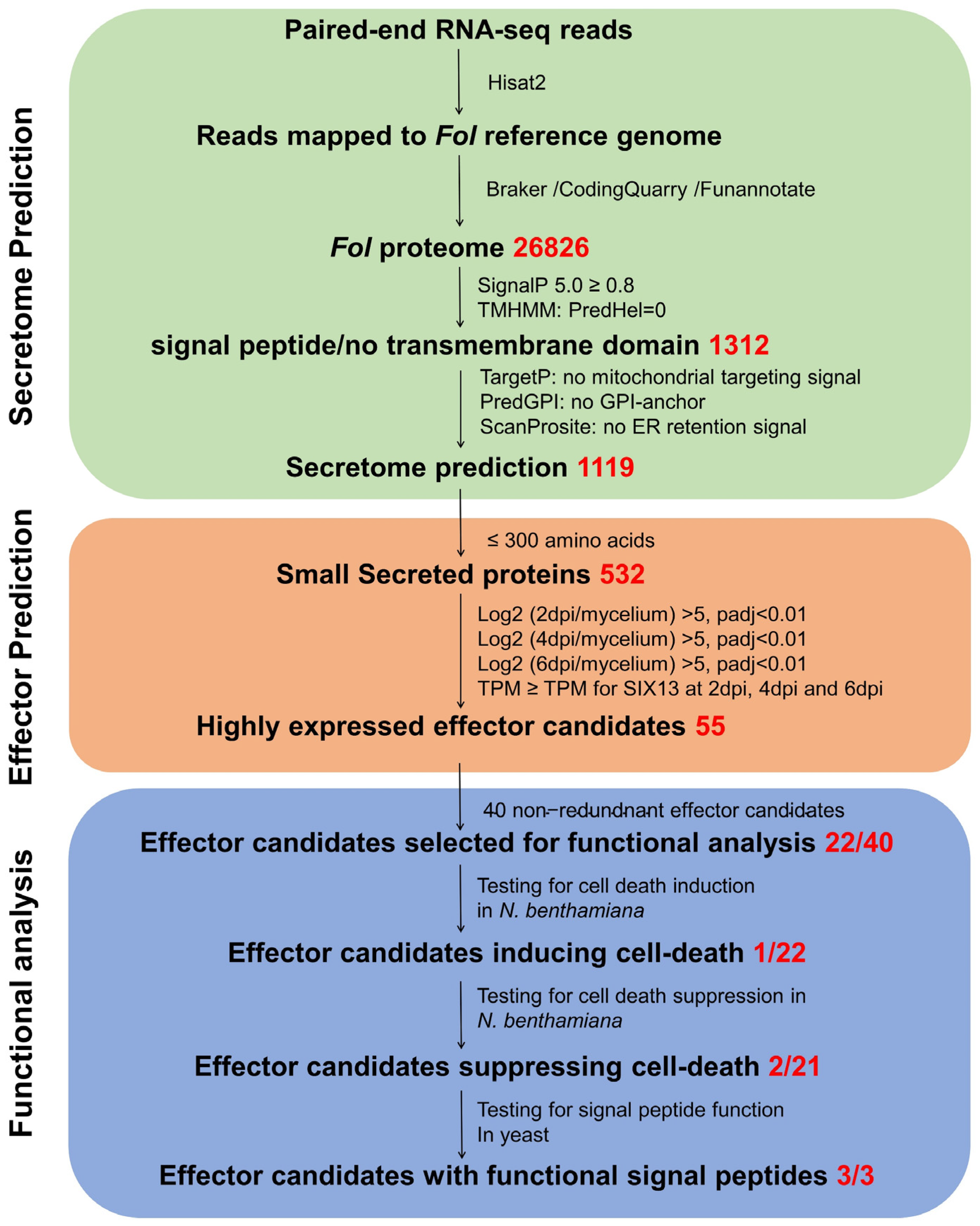
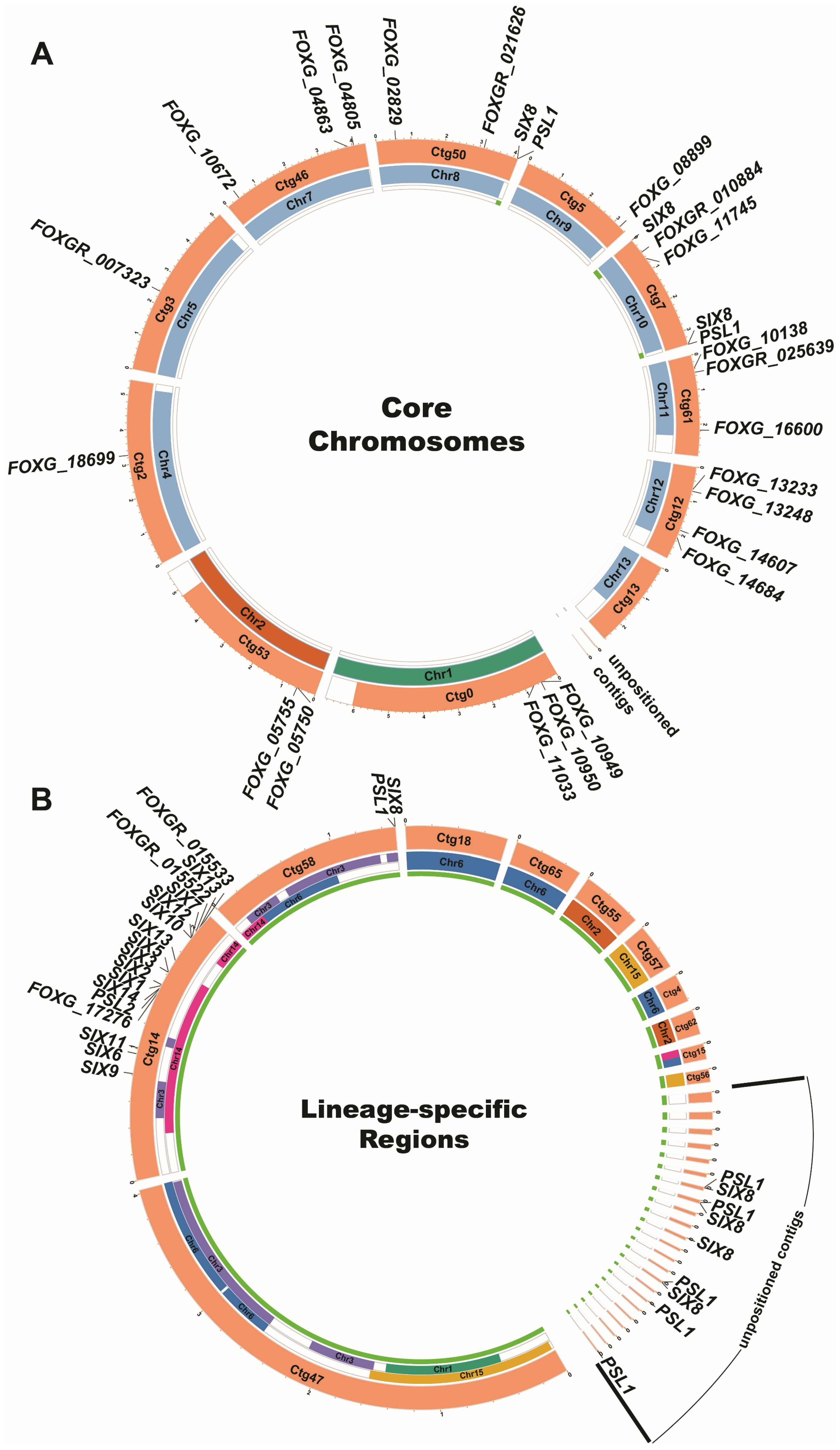
3.1. Identifying Candidate Effector Genes Expressed during Fol Infection

To identify novel Fol effector candidates expressed during Fol infection, we performed RNA-seq analysis using RNA samples prepared from Moneymaker tomato plants at 2, 4 and 6 dpi with Fol race 2 and from mycelium grown in vitro. The Fol4287 reference genome was recently re-sequenced using PacBio SMRT sequencing to improve the quality of the genome sequence and assembly [34]. We aligned the previous Fol4287 genome assembly (GCA_000149955.2 [33]) based on Sanger sequencing, here designated as the Fol 2010 genome, with the newly assembled genome (GCA_001703175.2), here designated as the Fol 2020 genome, and found the assembly of LS chromosomes differed markedly between the two assemblies (Figures S1 and S2). Based on this observation, we decided to map RNA-seq reads to the Fol 2020 genome. For RNA samples prepared from Fol infected tomato, 80–97 million raw reads were produced and after filtering out small reads, 0.35–1.31 million paired-end reads were mapped to the Fol 2020 genome, accounting for 0.39%–1.35% of the reads (Table S2). For RNA prepared from Fol growing in vitro, a total of 30.2 million raw reads were produced and 29.2 million paired-end reads were mapped to the Fol 2020 genome. Next, we annotated the Fol 2020 genome using our RNA-seq data, resulting in 26,826 gene models (Table S3). Genome annotation quality was assessed using the BUSCO tool, which produced 751 complete, 3 fragmented, and 4 missing BUSCOs from a total of 758 BUSCO groups based on the fungi_obd10 database, resulting in a 99.5% BUSCO completeness score (Table S4). The annotated protein-coding sequences were assigned FOXGR gene IDs and used to search against the old Fol 2010 proteome. Identical sequences with their old FOXG gene IDs were also listed (Table S3). In total, 26,826 predicted proteins were used as the basis for the prediction of the Fol secretome.
Based on the effector prediction pipelines employed previously for other plant pathogens and the characteristics of known effectors of plant pathogenic fungi, e.g., less than 300 amino acids in length [13,15,60,73], a bioinformatics pipeline was adopted to identify the Fol effector candidates expressed during infection (Figure 1) This pipeline involved three main processes: secretome prediction, effector prediction, and functional analysis (Figure 1). As shown in Figure 1, 1312 proteins were identified with a signal peptide and no transmembrane domain, consistent with secretion via the classical secretory pathway. After removing proteins predicted to target the ER or mitochondria, and GPI anchored proteins, a total of 1119 proteins were included in the predicted Fol 2020 secretome, accounting for 4.17% of the predicted Fol 2020 proteome. Of these 1119 proteins, 532 were ≤300 amino acids and of these 77 proteins were encoded by genes that were significantly up-regulated at 2, 4, and 6 dpi during Fol infection in comparison to their expression in mycelium, including all of the SIX genes present in Fol race 2. As SIX13 exhibited the lowest transcript per million (TPM) values among all SIX genes at 2, 4, and 6 dpi, we selected 55 secreted-protein-coding genes that had TPM values equal to or greater than that of SIX13 at each time point as a set of Fol effector candidates (Table 1). These included two identical copies of SIX13 and eight identical copies each of SIX8 and PSL1, leaving a non-redundant total of 40 candidate effectors. Next, the sequence annotations of these candidate effector genes were validated by the alignment of reads to the reference genome.
In addition, we generated Circos plots to visualise the chromosomal distribution of Fol effector candidates. As shown in Figure 2A, 22 effector candidates are located on Fv-collinear regions of the 11 core chromosomes in the Fol genome. However, effector candidate FOXGR_025639 had no ortholog in Fv despite having orthologs in other Fusarium species, including F. graminearum. Four of these 22 effector candidates, FOXGR_007323, FOXGR_010884, FOXGR_021626, and FOXGR_025639, were either not predicted or predicted incorrectly in the annotation of the Fol 2010 genome assembly. As shown in Figure 2B, 33 effector candidates were located in LS regions compared to the Fv genome, most on chromosome 14. Two SIX8/PSL1 gene pairs and one lone copy of SIX8 were located in LS regions near the ends of core chromosomes 8 and 10 in the Fol 2020 assembly; one SIX8/PSL1 gene pair on contig 58; a large chromosome-sized contig similar to LS chromosome 3 in the Fol 2010 genome assembly; and three SIX8/PSL1 gene pairs, one lone copy of SIX8 and two lone copies of PSL1 on small unpositioned contigs (Figure 2). Four new effector candidates were identified on chromosome 14, including FOXGR_015533, which was completely absent from the Fol 2010 genome sequence; FOXGR_015322 (a homologue of PSE1 here designated PSL2), which was not predicted in the annotation of the Fol 2010 genome assembly; and FOXGR_015522, which was predicted incorrectly. All 13 of the SIX genes present in Fol race 2 and PSL1 were identified in the Fol 2020 assembly, whereas SIX7, SIX8, SIX10, SIX11, SIX12, and PSL1 were either not predicted or predicted incorrectly in the annotation of the Fol 2010 genome assembly.

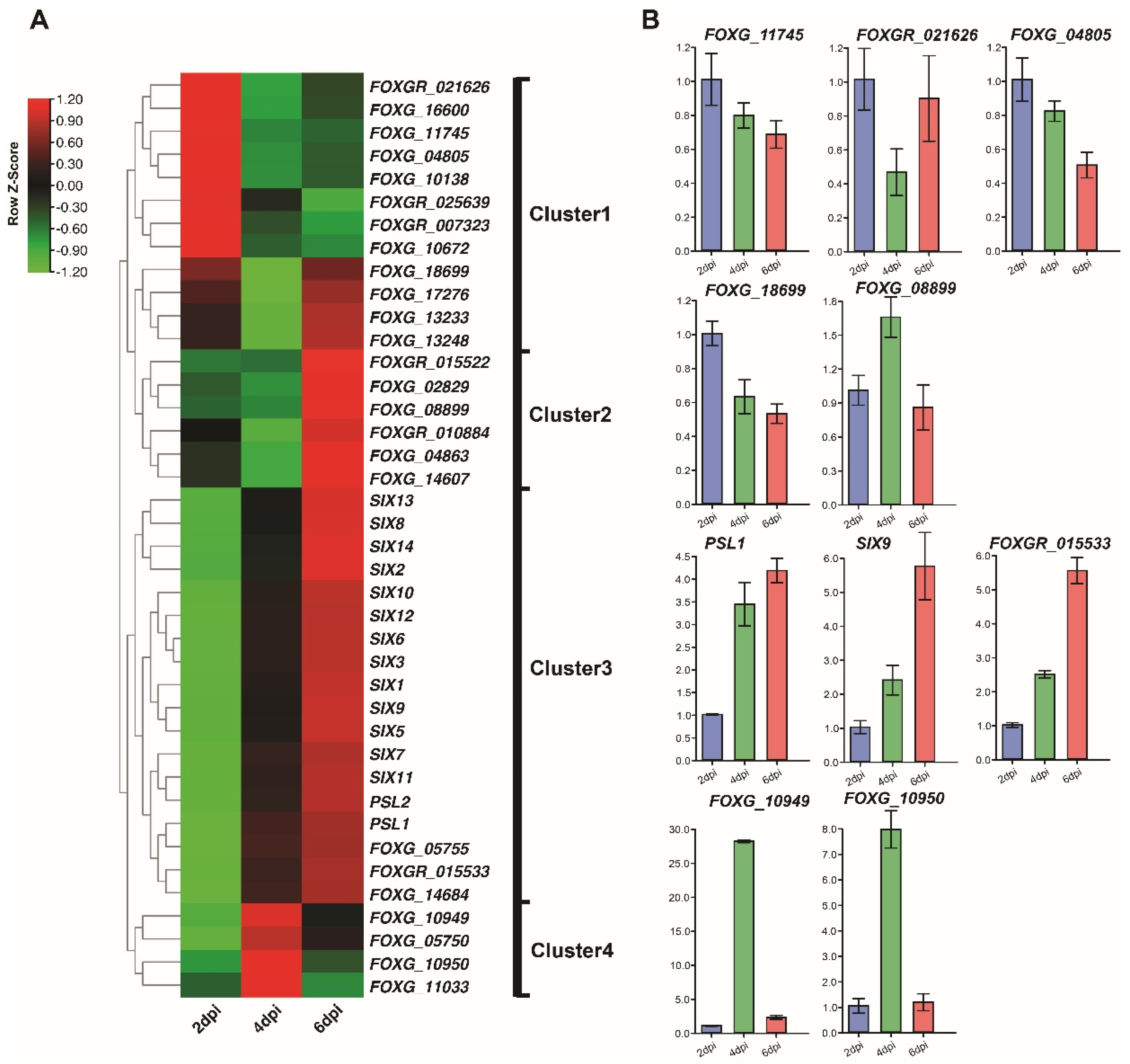
3.2. Expression Profiles of Fol Effector Candidates during Infection

To explore the expression profiles of Fol effector candidates upon infection, we performed a hierarchical clustering heatmap analysis of the 40 non-redundant effector candidates using their TPM values. Figure 3A shows that they grouped into four clusters correlated with their expression at three different Fol infection time points. Cluster 1 contains genes that were highly up-regulated at 2 dpi but less so at 4 and 6 dpi. Cluster 2 represents genes that were highly up-regulated at 2 and 6 dpi but less so at 4 dpi. All 13 SIX genes were included in Cluster 3, which contains genes up-regulated progressively from 2 to 6 dpi. Genes in Cluster 4 were highly up-regulated at 4 dpi but less so at 2 and 6 dpi. Our results indicate that all identified Fol effector candidate genes exhibit high expression upon Fol infection and that candidates in Clusters 2 and 3, which were most up-regulated at 6 dpi, might play important roles during Fol infection, given that all SIX genes that contribute to Fol pathogenicity were highly up-regulated at 6 dpi.
To validate the quantitative gene expression detected in the RNA-seq analysis (Figure 3B), a total of ten differentially expressed effector candidates, including two or three genes from each cluster, were selected for validation. Their relative expression levels at 2, 4, and 6 dpi were quantified by qRT-PCR. The qRT-PCR expression profiles of all ten genes confirmed their upregulation during infection and seven of the ten showed similar patterns of expression to those evident from the RNA-seq data (Figure 3B).

3.3. Identification of Cell Death-Inducing Effector Candidates

Host cell death is a hallmark of defense against a biotrophic fungal pathogen but could contribute to pathogenicity for a hemi-biotrophic pathogen like Fol. To identify effector candidates inducing cell death, 22 effector candidate genes were selected for cloning and transient expression via agroinfiltration of N. benthamiana leaves (Table 1). GFP was used as a negative control and Bax was used as a positive control in these experiments. We found that one of the 22 effector candidates, Fol-EC19, triggered cell death at 3 dpi (Figure 4). In addition, we used RT-PCR to show that all 22 candidate genes were expressed in N. benthamiana. (Figure S3).

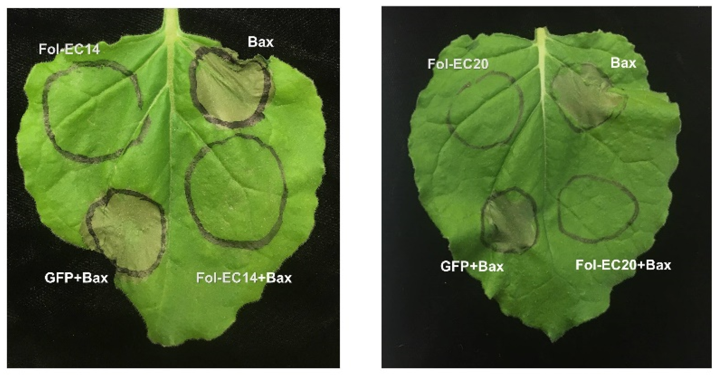
3.4. Identification of Effector Candidates That Suppress Bax-Induced Cell Death Using a Transient Expression Assay

Many fungal effectors suppress host cell-death to facilitate pathogen infection [74]. To identify effector candidates that can suppress Bax-induced cell death, we co-agroinfiltrated N. benthamiana leaves with Bax and each of the remaining 21 effector genes used for the cell-death experiments described above. GFP was used as a negative control. Bax triggers rapid cell death in N. benthamiana leaves at 2 dpi (Figure 5). Fol-EC14 and Fol-EC20 were found to completely suppress Bax-induced cell death whereas leaf regions co-expressing GFP and Bax developed pronounced cell death (Figure 5).

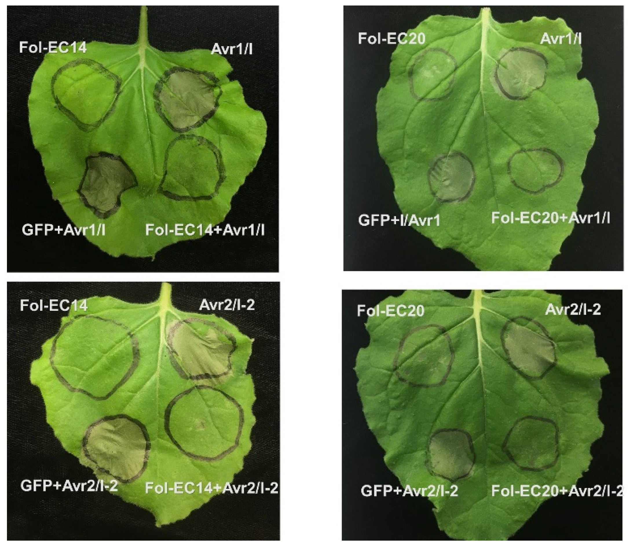
3.5. Suppression of I/Avr1- and I-2/Avr2-Induced Cell Death

To investigate whether Fol-EC14 and Fol-EC20 function as suppressors of cell death triggered by tomato proteins conferring Fol-resistance, we transiently co-expressed Fol-EC14 and Fol-EC20 with I/Avr1 or I-2/Avr2 in N. benthamiana leaves, using GFP as a negative control. No cell death was observed in leaf regions co-expressing Fol-EC14 or Fol-EC20 with I/Avr1 or I-2/Avr2 (Figure 6). In contrast, co-expression of I/Avr1 or I-2/Avr2 with GFP triggered rapid cell death at 3 dpi. These findings indicated that Fol-EC14 and Fol-EC20 inhibited cell death triggered by both a CC-NB-LRR resistance protein (I-2 in this instance) and an LRR-RP resistance protein (I in this instance).
To further explore whether Fol-EC14 and Fol-EC20 are able to suppress cell death triggered by other plant R genes, we transiently co-expressed them with L6TIR in N. benthamiana leaves. The TIR (Toll Interleukin-1 Receptor) domain of the flax L6 TIR-NB-LRR resistance protein is able to homodimerize in the absence of its NB and LRR domains to trigger plant cell death [75]. L6TIR-induced cell death was not inhibited by either Fol-EC14 or Fol-EC20 (Figure S4). In addition, we examined their ability to suppress Fol-EC19-induced cell death. Neither Fol-EC14 nor Fol-EC20 was able to suppress cell death induced by Fol-EC19 (Figure S4).

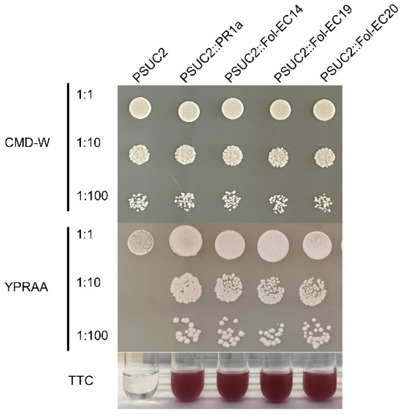
3.6. Functional Characterization of the Signal Peptides of the Fol-EC14, Fol-EC19, and Fol-EC20 Effectors

To examine the function of the predicted signal peptide sequences of Fol-EC14, Fol-EC19, and Fol-EC20, a yeast signal sequence trap system was employed. The predicted signal peptide coding sequences of Fol-EC14, Fol-EC19, and Fol-EC20 were amplified and cloned in frame with a truncated SUC2 gene, which encodes yeast invertase, lacking its signal peptide. The resulting constructs and positive control pSUC2::SPPR1a (carrying the N. benthamiana PR1a signal-peptide coding sequence) were transformed into yeast strain YKT12, which cannot grow on medium without a simple sugar due to a deficiency of the yeast invertase gene. Consistent with the positive control, YTK12 strains carrying the predicted signal-peptide coding sequences of Fol-EC14, Fol-EC19, and Fol-EC20 fused to SUC2 were able to grow on YPRAA plates (Figure 7). As expected, they were also able to catalyze the conversion of colorless TTC to red-colored TFP (Figure 7). Conversely, the negative control (YTK12 with empty vector) was not able to grow on a YPRAA plate and did not induce a TTC to TFP color change (Figure 7). These results demonstrated that the predicted signal peptides of Fol-EC14, Fol-EC19, and Fol-EC20 are functional.

4. Discussion

The effectors secreted by plant pathogenic fungi determine the outcome of infection by either suppressing plant immune response or interfering with plant physiological processes to facilitate pathogen infection [2,76,77]. The study presented here identified a repertoire of newly assembled Fol transcripts and 40 non-redundant effector candidates whose expression is highly induced during tomato infection, including all 13 SIX genes present in Fol race 2. Functional analysis of 22 of the 27 novel effector candidates revealed that one effector candidate induced cell death and two effector candidates suppressed R-protein-mediated cell death. We further demonstrated that these three effector candidates have a functional signal peptide. Together, these findings enrich the genome annotation of Fol, deliver genome-wide expression profiles of genes encoding small, secreted proteins, including the SIX genes, and provide a basis for further exploring Fol pathogenic mechanisms.
Previously, the annotation of the published Fol4287 reference genome was based on gene prediction without transcriptome analysis [33]. Thus, inevitable inaccuracies occurred, and some still remain. For example, several SIX genes, including SIX7, SIX8, SIX11, SIX12, and SIX14 were not annotated in the reference genome but were subsequently submitted as separate NCBI accessions [32]. We have been able to assemble 26,826 transcripts from transcriptome sequencing data mapped to a genome assembly based on PacBio SMRT genome sequencing (Figure 1), including all of the previously unannotated SIX genes. In addition, eight effector candidate genes were identified that were absent from the Fol 2010 reference genome annotation (Table 1). Of the non-redundant total of 40 candidate effector genes, 18, including all 13 SIX genes present in Fol race 2, are located on LS regions and the remaining 22 on core chromosomes (Table 1 and Figure 2). Importantly, three of the newly annotated genes, PSL2, FOXGR_015522 and FOXGR_015533, and one previously annotated gene, FOXG_17276, are located on the LS pathogenicity chromosome 14, providing a strong indication that they might be involved in Fol infection. Multiple copies of SIX8 and the newly annotated PSL1 gene are located in other LS regions, mostly as divergently oriented pairs (Table 1 and Figure 2). None of the 18 candidate LS effectors had homologs with a known function, although FOXG_17276 has a LysM domain, suggesting a role in carbohydrate binding. In contrast, eight of the 22 candidate core region effectors had homology to proteins of known function, including two hydrophobins, a glucanase, a phospholipase, a ribonuclease, and three proteases (Table 1). Cell wall-degrading, membrane-attacking, and proteolytic enzymes are well-known parts of a plant pathogen’s arsenal, and it is not surprising that degradative enzymes falling below the 300 amino acid threshold should be captured by this analysis as potential effectors. However, roles for a glucanase and trypsin in suppression of defence-related cell death and a ribonuclease in the induction of cell death were unexpected.
FOXGR_021626, here designated Fol-EC14, encodes a glucanase containing a GH131 glycosyl hydrolase domain, which was first described in the ascomycete Podospora anserina as PaGluc131A [78] and in the basidiomycete Coprinopsis cinerea as CcGH131A [79]. PaGluc131A has been reported to have β-1,3- and β-1,6-exoglucanase and β-1,4 endoglucanase activities [78], and a later study also reported β-1,3-endoglucanase activity, but could not confirm β-1,6-exoglucanase activity [80]. Despite comprising almost entirely a GH131 domain, Fol-EC14 only has 23% and 24% amino acid sequence identity with the N-terminal glycosyl hydrolase domains of PaGluc131A and CcGH131A, respectively. GH131 proteins are widespread among the Ascomycota and Basidiomycota and an extensive phylogenetic analysis identified two clades, each containing ascomycete and basidiomycete sequences suggesting divergence of the two GH131 clades prior to the divergence of the Ascomycota from the Basidiomycota [80]. Fol-EC14 falls in one clade and PaGluc131A and CcGH131A fall in the other. Interestingly, Fusarium is completely absent from the PaGluc131A/CcGH131A clade, although other members of the Sordariomycetes are represented in both (Figures S5 and S6).
Two members of the Fol-EC14 clade, Colletotrichum higginsianum ChGluc131A and ChGluc131B (Figures S7 and S8), have also been shown to have β-1,3-exoglucanase and β-1,3- and β-1,4 endoglucanase activities [80]. Fol-EC14 and ChGluc131B and most of their orthologs lack three of the four residues thought to be important for PaGluc131A and CcGH131A catalytic activity [79], whereas ChGluc131A and its orthologs lack only one of these residues (Figures S6–S8). They also differ from members of the PaGluc131A/CcGH131A clade by the presence of two conserved cysteine residues flanking the GH131 domain, which could potentially form a stabilizing disulfide bond (Figures S6–S8). Fol-EC14 has only 43% sequence identity with ChGluc131A and 48% with ChGluc131B, which in turn have only 43% sequence identity with one another. Consistent with these differences, Fol-EC14, ChGluc131A, and ChGluc131B are each member of distinct phylogenetic subgroups within the Fol-EC14 clade (Figures S6–S8), with ChGluc131B and its orthologs limited to the genus Colletotrichum (Figure S8), and Fusarium sequences present only in the Fol-EC14 subgroup (Figure S6). Like Fol-EC14, ChGluc131B lacks three of the four residues thought to be important for PaGluc131A and CcGH131A catalytic activity but is nevertheless catalytically active. This suggests a different mechanism of catalysis to that proposed for PaGluc131A and CcGH131A, but given the absence of information about residues important for ChGluc131B catalysis and the low sequence identity between Fol-EC14 and ChGluc131B, we cannot infer whether Fol-EC14 is catalytically active or not. If it was functional, possible roles for Fol-EC14 could include degradation of cellulose (β-1,4 glucan) in the plant cell wall, callose (β-1,3 glucan) degradation, conversion of elicitor-active glucans to shorter elicitor-inactive oligomers, and exoglucanase-mediated release of glucose as a nutrient for the fungus.
FOXG_13248, here designated Fol-EC20, encodes a secreted trypsin that differs by a single conservative amino-acid substitution from a structurally and enzymatically well-characterized F. oxysporum trypsin [81,82]. Trypsins are widespread among plant pathogens, as are trypsin inhibitors among plant hosts. In the xylem-colonizing Gram-positive bacterium Clavibacter michiganensis subsp. michiganensis (Cmm), two genes, pat-1 and chpC, encode secreted trypsin-family proteins that are required for the colonization and wilting of tomato and the suppression of the plant immune response [83,84]. Interestingly, Cmm also requires a β-1,4 endoglucanase encoded by the celA gene for pathogenicity and wilting of tomato [85]. Potentially, the Fol-EC14 exo/endoglucanase and Fol-EC20 trypsin might play similar roles in Fol pathogenicity.
There are three possible explanations for the suppression of Bax-induced and R-protein-induced cell death by Fol-EC14 and Fol-EC20, for which there may be some precedents. One explanation might be that the enzymatic actions of Fol-EC14 or Fol-EC20 suppress plant cell death. Sanchez et al. (1992) reported that water-soluble glucans from Phytophthora infestans can suppress fungal-elicitor-induced plant cell death [86], and Ali et al. (2015) reported that a potato cyst nematode expansin could suppress NPP (Nep1-like protein) and CNL-induced cell death [87]. Expansins are plant cell wall-loosening proteins. Given these two observations, it is plausible that either a soluble glucan product of Fol-EC14 catalysis or the effect of Fol-EC14 catalysis on the plant cell wall could have a suppressive effect on plant cell death. Similarly, Carlile et al. (2000) reported that the Stagonospora nodorum trypsin SNP1 released hydroxyproline from wheat cell walls and, like β-1,4 endoglucanases, contributes to the modification of the plant cell wall [88]. Hao et al. (2019) reported that a specific F. graminearum arabinanase, albeit a different glycosyl hydrolase to Fol-EC14, could suppress Bax-induced cell death [89]. Given that the arabinanase could also suppress flg22- and chitin-induced ROS (reactive oxygen species) production, they inferred that the suppression of Bax-induced cell death was likely related to the suppression of ROS production. A later study by the same group showed that a putative endoglucanase from F. graminearum could suppress chitin-induced but not flg22-induced ROS production [90]. No explanation for the mechanisms involved in the suppression of ROS production was provided by either study and, although the products of arabinanase or endoglucanase catalysis could be involved, a direct effect of the arabinanase or endoglucanse protein could not be excluded. A key question that therefore remains to be answered is whether the catalytic activities of Fol-EC14 and Fol-EC20 are required for their suppressive effects.
A second explanation might be that the Fol-EC14 or Fol-EC20 proteins have a direct effect on plant cell death unrelated to enzymatic function. Rose et al. identified non-catalytic members of the trypsin family in Phytophthora as inhibitors of plant β-1,3 endoglucanases able to reduce the production of elicitor-active oligoglucans from the Phytophthora cell wall [91]. Potentially, this inhibition could promote the production of water-soluble glucans able to suppress fungal-elicitor-induced plant cell death as described above. A third explanation is that the suppression of cell death by Fol-EC14 and Fol-EC20 is an artifact. Bozkurt et al. suggest that the heterologous expression of some proteins can elicit an unfolded protein response in the ER that can suppress Bax-induced cell death [92]. Given that Fol-EC14 and Fol-EC20 have been targeted to the plant ER as part of the secretory pathway, it is possible that they do not fold properly and thereby elicit an unfolded protein response in the ER. However, the fact that they were not able to suppress either L6TIR- or Fol-Ec19-induced cell death suggests this is not the explanation (Figure S4).
FOXG_13233, here designated Fol-EC19, encodes a secreted guanyl-specific ribonuclease that induces rather than suppresses plant cell death in N. benthamiana (Figure 4). Homologs of Fol-EC19 are widely distributed among the fungi including plant pathogens in the genera Alternaria, Bipolaris, Colletotrichum, Fusarium, Magnaporthe, Pyrenophora, Ustilago, and Verticillium. Our finding is consistent with a recent study showing that F. graminearum secretes Fg12, a ribonuclease with 85% identity and 93% similarity to Fol_EC19, which contributes to virulence on soybean as demonstrated by gene knockout experiments and induces cell death in N. benthamiana, N. tabacum, and tomato, as shown by agroinfiltration experiments [93]. Agroinfiltration of a mutant Fg12 gene was used to show that induction of cell death in N. benthamiana was dependent on protein secretion and ribonuclease activity. Recombinant F. graminearum Fg12 protein was found to induce ion leakage and PR-gene expression in N. benthamiana and resistance to F. graminearum and Phytophthra sojae in soybean hypocotyls, again dependent on ribonuclease activity. Whether the cell death induced by Fol-EC19 is dependent on ribonuclease activity and whether Fol-EC19 is capable of inducing plant cell death or plant immune responses in tomato remain unknown but seems likely. These findings suggest that Fol-EC19 might, like Fg12, also function as a virulence factor in the context of pathogen infection but trigger a plant immune response when tested in isolation. Due to limitations of agroinfiltration in tomato, most of the experiments in the present study were conducted using the solanaceous model plant N. benthamiana, which has been used extensively in effector biology research and proven useful for translating such research into host species. Further experiments involving purified recombinant candidate-effector proteins or knockouts of effector-candidate genes in Fol will provide additional support for the virulence role of these three effector candidates during tomato infection.

5. Conclusions

The molecular interaction between tomato and the xylem-colonizing fungus Fol has been extensively studied. However, the virulence mechanisms employed by Fol effectors remain largely unknown. The study presented here expands the repertoire of Fol effector candidates, showing highly-induced expression during tomato infection. Agroinfiltration assays in N. benthamiana revealed that one effector candidate, encoding a secreted guanyl-specific ribonuclease, induced cell death and two effector candidates, encoding a glucanase and a secreted trypsin, respectively, suppressed R-protein-mediated cell death. We confirmed that these three biologically active effector candidates have a functional signal peptide that would direct their secretion into the xylem sap of infected tomato plants. These findings suggest that Fol utilizes diverse effector proteins to facilitate infection, including enzymatic effectors encoded by the core Fusarium genome. Taken together with previous knowledge about the role of lineage-specific effectors, these findings add value to our understanding of Fol virulence mechanisms.

Supplementary Materials

The following supporting information can be downloaded at: https://www.mdpi.com/article/10.3390/jof8070672/s1. Table S1. Primers used in this study; Table S2. Summary of RNA-seq statistics for three replicates of three infection time points (2, 4, and 6 dpi) and mycelium grown in vitro; Table S3. List of predicted proteins encoded by genes annotated from the PacBio-sequenced Fol4287 genome assembly; Table S4. Summary comparison of the Fol4287 genome assemblies and annotations obtained with Sanger and PacBio sequencing platforms. Figure S1. Whole-genome synteny analysis of predicted genes between Sanger-sequenced Fol4287 (Fol 2010) and PacBio-sequenced Fol4287 (Fol 2020) assemblies; Figure S2. Whole-genome alignment between Sanger-sequenced Fol4287 (Fol 2010) and PacBio-sequenced Fol4287 (Fol 2020) genome assemblies; Figure S3. RT-PCR to check the expression of 22 cloned effector candidate genes in agroinfiltrated N. benthamiana leaves; Figure S4. FolEC-14 and Eol-EC20 are not able to inhibit L6TIR- and Fol-EC19-induced cell death in N. benthamiana leaves; Figure S5. PaGluc131A alignment; Figure S6. Fol-EC14 alignment; Figure S7. ChGluc131A alignment; Figure S8/ChGluc131B alignment.

Author Contributions

Conceptualization, L.M. and D.A.J.; methodology, X.S., X.F. and L.M.; writing—original draft preparation, X.S., L.M. and D.A.J.; writing—review and editing, L.M. and D.A.J.; supervision, D.A.J., L.M. and D.W.; project administration, L.M.; funding acquisition, L.M. All authors have read and agreed to the published version of the manuscript.

Funding

This research was funded by the Australian Research Council (ARC) through Discovery Early Career Researcher Award (DE170101165) to L. Ma. X. Sun was supported by scholarships from Hebei Agricultural University (No. 20190002) and the China Scholarship Council (No. 202008130203) to conduct part of his PhD research at the Australian National University. L. Ma was supported by the “Hundred Talents Program” for the introduction of high-level overseas talents in Hebei Province (E2020100004).

Institutional Review Board Statement

“Not applicable” for studies not involving humans or animals.

Informed Consent Statement

“Not applicable” for studies not involving humans.

Data Availability Statement

The data that support the findings of this study are available from the corresponding author upon request and RNA-seq data are available in NCBI BioProject ID: PRJNA841073.

Conflicts of Interest

The authors declare no conflict of interest.

References

  1. Zhou, J.-M.; Zhang, Y. Plant Immunity: Danger perception and signaling. Cell 2020, 181, 978–989. [Google Scholar] [CrossRef] [PubMed]
  2. Lo Presti, L.; Lanver, D.; Schweizer, G.; Tanaka, S.; Liang, L.; Tollot, M.; Zuccaro, A.; Reissmann, S.; Kahmann, R. Fungal effectors and plant susceptibility. Annu. Rev. Plant Biol. 2015, 66, 513–545. [Google Scholar] [CrossRef] [PubMed]
  3. Giraldo, M.; Valent, B. Filamentous plant pathogen effectors in action. Nat. Rev. Genet. 2013, 11, 800–814. [Google Scholar] [CrossRef]
  4. van der Burgh, A.M.; Joosten, M. Plant Immunity: Thinking outside and inside the box. Trends Plant Sci. 2019, 24, 587–601. [Google Scholar] [CrossRef]
  5. Thomma, B.P.; Nurnberger, T.; Joosten, M.H. Of PAMPs and effectors: The blurred PTI-ETI dichotomy. Plant Cell 2011, 23, 4–15. [Google Scholar] [CrossRef] [PubMed] [Green Version]
  6. Blondeau, K.; Blaise, F.; Graille, M.; Kale, S.D.; Linglin, J.; Ollivier, B.; Labarde, A.; Lazar, N.; Daverdin, G.; Balesdent, M.H.; et al. Crystal structure of the effector AvrLm4-7 of Leptosphaeria maculans reveals insights into its translocation into plant cells and recognition by resistance proteins. Plant J. 2015, 83, 610–624. [Google Scholar] [CrossRef]
  7. Plissonneau, C.; Daverdin, G.; Ollivier, B.; Blaise, F.; Degrave, A.; Fudal, I.; Rouxel, T.; Balesdent, M. A game of hide and seek between avirulence genes AvrLm4-7 and AvrLm3 in Leptosphaeria maculans. New Phytol. 2016, 209, 1613–1624. [Google Scholar] [CrossRef] [Green Version]
  8. Ghanbarnia, K.; Ma, L.; Larkan, N.J.; Haddadi, P.; Fernando, W.G.D.; Borhan, M.H. Leptosphaeria maculans AvrLm9: A new player in the game of hide and seek with AvrLm4-7. Mol. Plant Pathol. 2018, 19, 1754–1764. [Google Scholar] [CrossRef] [Green Version]
  9. Bourras, S.; McNally, K.E.; Ben-David, R.; Parlange, F.; Roffler, S.; Praz, C.R.; Oberhaensli, S.; Menardo, F.; Stirnweis, D.; Frenkel, Z.; et al. Multiple avirulence loci and allele-specific efector recognition control the Pm3 race-specific resistance of wheat to powdery mildew. Plant Cell 2015, 27, 2991–3012. [Google Scholar]
  10. Houterman, P.M.; Cornelissen, B.J.C.; Rep, M. Suppression of plant resistance gene-based immunity by a fungal effector. PLoS Pathog. 2008, 4, e1000061. [Google Scholar] [CrossRef] [Green Version]
  11. Ramachandran, S.R.; Yin, C.; Kud, J.; Tanaka, K.; Mahoney, A.K.; Xiao, F.; Hulbert, S.H. Effectors from wheat rust fungi suppress multiple plant defense responses. Phytopathology 2017, 107, 75–83. [Google Scholar] [CrossRef] [PubMed] [Green Version]
  12. Selin, C.; de Kievit, T.R.; Belmonte, M.F.; Fernando, W.G. Elucidating the role of effectors in plant-fungal interactions: Progress and challenges. Front. Microbiol. 2016, 7, 600. [Google Scholar] [CrossRef] [PubMed] [Green Version]
  13. Nemri, A.; Saunders, D.G.; Anderson, C.; Upadhyaya, N.M.; Win, J.; Lawrence, G.J.; Jones, D.A.; Kamoun, S.; Ellis, J.G.; Dodds, P.N. The genome sequence and effector complement of the flax rust pathogen Melampsora lini. Front. Plant Sci. 2014, 5, 98. [Google Scholar] [CrossRef] [PubMed]
  14. Guyon, K.; Balagué, C.; Roby, D.; Raffaele, S. Secretome analysis reveals effector candidates associated with broad host range necrotrophy in the fungal plant pathogen Sclerotinia sclerotiorum. BMC Genom. 2014, 15, 336. [Google Scholar] [CrossRef] [PubMed] [Green Version]
  15. Haddadi, P.; Ma, L.; Wang, H.; Borhan, M.H. Genome-wide transcriptomic analyses provide insights into the lifestyle transition and effector repertoire of Leptosphaeria maculans during the colonization of Brassica napus seedlings. Mol. Plant Pathol. 2016, 17, 1196–1210. [Google Scholar] [CrossRef] [Green Version]
  16. Pérez-López, E.; Hossain, M.; Tu, J.; Waldner, M.; Todd, C.D.; Kusalik, A.J.; Wei, Y.; Bonham-Smith, P.C. Transcriptome analysis identifies Plasmodiophora brassicae secondary infection effector candidates. J. Eukaryot. Microbiol. 2020, 67, 337–351. [Google Scholar] [CrossRef] [Green Version]
  17. Kanja, C.; Hammond-Kosack, K.E. Proteinaceous effector discovery and characterization in filamentous plant pathogens. Mol. Plant Pathol. 2020, 21, 1353–1376. [Google Scholar] [CrossRef]
  18. Petre, B.; Saunders, D.G.O.; Sklenar, J.; Lorrain, C.; Krasileva, K.; Win, J.; Duplessis, S.; Kamoun, S. Heterologous expression screens in Nicotiana benthamiana identify a candidate effector of the wheat yellow rust pathogen that associates with processing bodies. PLoS ONE 2016, 11, e0149035. [Google Scholar] [CrossRef] [Green Version]
  19. Lacomme, C.; Santa Cruz, S. Bax-induced cell death in tobacco is similar to the hypersensitive response. Proc. Natl. Acad. Sci. USA 1999, 96, 7956–7961. [Google Scholar] [CrossRef] [Green Version]
  20. Wang, Q.; Han, C.; Ferreira, A.O.; Yu, X.; Ye, W.; Tripathy, S.; Kale, S.D.; Gu, B.; Sheng, Y.; Sui, Y.; et al. Transcriptional programming and functional interactions within the Phytophthora sojae RXLR effector repertoire. Plant Cell 2021, 23, 2064–2086. [Google Scholar] [CrossRef] [Green Version]
  21. Wang, Y.; Li, J.; Xiang, S.; Zhou, J.; Peng, X.; Hai, Y.; Wang, Y.; Li, S.; Wei, S. A putative effector UvHrip1 inhibits BAX-triggered cell death in Nicotiana benthamiana, and infection of Ustilaginoidea virens suppresses defense-related genes expression. PeerJ 2020, 8, e9354. [Google Scholar] [CrossRef] [PubMed]
  22. Cheng, Y.; Wu, K.; Yao, J.; Li, S.; Wang, X.; Huang, L.; Kang, Z. PSTha5a23, a candidate effector from the obligate biotrophic pathogen Puccinia striiformis f. sp. tritici, is involved in plant defense suppression and rust pathogenicity. Environ. Microbiol. 2017, 19, 1717–1729. [Google Scholar] [CrossRef] [PubMed]
  23. Xu, Q.; Tang, C.; Wang, X.; Sun, S.; Zhao, J.; Kang, Z.; Wang, X. An effector protein of the wheat stripe rust fungus targets chloroplasts and suppresses chloroplast function. Nat. Commun. 2019, 10, 5571. [Google Scholar] [CrossRef] [PubMed] [Green Version]
  24. Dutra, D.; Agrawal, N.; Ghareeb, H.; Schirawski, J. Screening of Secreted Proteins of Sporisorium reilianum f. sp. zeae for Cell Death Suppression in Nicotiana benthamiana. Front. Plant Sci. 2020, 11, 95. [Google Scholar] [CrossRef] [Green Version]
  25. Zhang, N.; Yang, J.; Fang, A.; Wang, J.; Li, D.; Li, Y.; Wang, S.; Cui, F.; Yu, J.; Liu, Y.; et al. The essential effector SCRE1 in Ustilaginoidea virens suppresses rice immunity via a small peptide region. Mol. Plant Pathol. 2020, 21, 445–459. [Google Scholar] [CrossRef] [Green Version]
  26. Derevnina, L.; Dagdas, Y.F.; de la Concepcion, J.C.; Białas, A.; Kellner, R.; Petre, B.; Domazakis, E.; Du, J.; Wu, C.-H.; Lin, X.; et al. Nine things to know about elicitins. New Phytol. 2016, 212, 888–895. [Google Scholar] [CrossRef] [PubMed] [Green Version]
  27. Li, Y.; Han, Y.; Qu, M.; Chen, J.; Chen, X.; Geng, X.; Wang, Z.; Chen, S. Apoplastic cell death-inducing proteins of filamentous plant pathogens: Roles in plant-pathogen interactions. Front. Genet. 2020, 11, 661. [Google Scholar] [CrossRef]
  28. Takken, F.; Rep, M. The arms race between tomato and Fusarium oxysporum. Mol. Plant Pathol. 2010, 11, 309–314. [Google Scholar] [CrossRef]
  29. Olivain, C.; Humbert, C.; Nahalkova, J.; Fatehi, J.; L’Haridon, F.; Alabouvette, C. Colonization of tomato root by pathogenic and nonpathogenic Fusarium oxysporum strains inoculated together and separately into the soil. Appl. Environ. Microbiol. 2006, 72, 1523–1531. [Google Scholar] [CrossRef] [Green Version]
  30. Rep, M.; Van Der Does, H.C.; Meijer, M.; Van Wijk, R.; Houterman, P.M.; Dekker, H.L.; De Koster, C.G.; Cornelissen, B.J.C. A small, cysteine-rich protein secreted by Fusarium oxysporum during colonization of xylem vessels is required for I-3-mediated resistance in tomato. Mol. Microbiol. 2004, 53, 1373–1383. [Google Scholar] [CrossRef]
  31. Houterman, P.M.; Speijer, D.; Dekker, H.L.; de Koster, C.G.; Cornelissen, B.J.C.; Rep, M. The mixed xylem sap proteome of Fusarium oxysporum-infected tomato plants. Mol. Plant Pathol. 2007, 8, 215–221. [Google Scholar] [CrossRef] [PubMed]
  32. Schmidt, S.M.; Houterman, P.M.; Schreiver, I.; Ma, L.; Amyotte, S.; Chellappan, B.; Boeren, S.; Takken, F.L.W.; Rep, M. MITEs in the promoters of effector genes allow prediction of novel virulence genes in Fusarium oxysporum. BMC Genom. 2013, 14, 119. [Google Scholar] [CrossRef] [Green Version]
  33. Ma, L.-J.; Van Der Does, H.C.; Borkovich, K.A.; Coleman, J.J.; Daboussi, M.-J.; Di Pietro, A.; Dufresne, M.; Freitag, M.; Grabherr, M.; Henrissat, B.; et al. Comparative genomics reveals mobile pathogenicity chromosomes in Fusarium. Nature 2010, 464, 367–373. [Google Scholar] [CrossRef]
  34. Li, J.; Fokkens, L.; Conneely, L.J.; Rep, M. Partial pathogenicity chromosomes in Fusarium oxysporum are sufficient to cause disease and can be horizontally transferred. Environ. Microbiol. 2020, 22, 4985–5004. [Google Scholar] [CrossRef]
  35. Gawehns, F.; Ma, L.; Bruning, O.; Houterman, P.M.; Boeren, S.; Cornelissen, B.J.C.; Rep, M.; Takken, F.L.W. The effector repertoire of Fusarium oxysporum determines the tomato xylem proteome composition following infection. Front. Plant Sci. 2015, 6, 967. [Google Scholar] [CrossRef] [PubMed]
  36. Houterman, P.M.; Ma, L.; van Ooijen, G.; de Vroomen, M.J.; Cornelissen, B.J.; Takken, F.L.; Rep, M. The effector protein Avr2 of the xylem-colonizing fungus Fusarium oxysporum activates the tomato resistance protein I-2 intracellularly. Plant J. 2009, 58, 970–978. [Google Scholar] [CrossRef] [PubMed]
  37. Ma, L.; Houterman, P.M.; Gawehns, F.; Cao, L.; Sillo, F.; Richter, H.; Clavijo-Ortiz, M.J.; Schmidt, S.M.; Boeren, S.; Vervoort, J.; et al. The AVR2-SIX5 gene pair is required to activate I-2-mediated immunity in tomato. New Phytol. 2015, 208, 507–518. [Google Scholar] [CrossRef]
  38. Gawehns, F.; Houterman, P.M.; Ichou, F.A.; Michielse, C.B.; Hijdra, M.; Cornelissen, B.J.; Rep, M.; Takken, F.L.W. The Fusarium oxysporum effector Six6 contributes to virulence and suppresses I-2-mediated cell death. Mol. Plant Microbe Interact. 2014, 27, 336–348. [Google Scholar] [CrossRef] [Green Version]
  39. Vlaardingerbroek, I.; Beerens, B.; Rose, L.; Fokkens, L.; Cornelissen, B.J.C.; Rep, M. Exchange of core chromosomes and horizontal transfer of lineage-specific chromosomes in Fusarium oxysporum. Environ. Microbiol. 2016, 18, 3702–3713. [Google Scholar] [CrossRef]
  40. Ayukawa, Y.; Asai, S.; Gan, P.; Tsushima, A.; Ichihashi, Y.; Shibata, A.; Komatsu, K.; Houterman, P.M.; Rep, M.; Shirasu, K.; et al. A pair of effectors encoded on a conditionally dispensable chromosome of Fusarium oxysporum suppress host-specific immunity. Commun. Biol. 2021, 4, 707. [Google Scholar] [CrossRef]
  41. Gonzalez-Cendales, Y.; Catanzariti, A.-M.; Baker, B.; Mcgrath, D.J.; Jones, D.A. Identification of I-7 expands the repertoire of genes for resistance to Fusarium wilt in tomato to three resistance gene classes. Mol. Plant Pathol. 2015, 17, 448–463. [Google Scholar] [CrossRef]
  42. Simons, G.; Groenendijk, J.; Wijbrandi, J.; Reijans, M.; Groenen, J.; Diergaarde, P.; van der Lee, T.; Bleeker, M.; Onstenk, J.; de Both, M. Dissection of the fusarium I2 gene cluster in tomato reveals six homologs and one active gene copy. Plant Cell 1998, 10, 1055–1068. [Google Scholar] [CrossRef] [PubMed] [Green Version]
  43. Catanzariti, A.M.; Lim, G.T.T.; Jones, D.A. The tomato I-3 gene: A novel gene for resistance to Fusarium wilt disease. New Phytol. 2015, 207, 106–118. [Google Scholar] [CrossRef] [PubMed]
  44. Catanzariti, A.M.; Do, H.T.; Bru, P.; de Sain, M.; Thatcher, L.F.; Rep, M.; Jones, D.A. The tomato I gene for Fusarium wilt resistance encodes an atypical leucine-rich repeat receptor-like protein whose function is nevertheless dependent on SOBIR1 and SERK3/BAK1. Plant J. 2017, 89, 1195–1209. [Google Scholar] [CrossRef] [Green Version]
  45. Jenkins, S.; Taylor, A.; Jackson, A.C.; Armitage, A.D.; Bates, H.J.; Mead, A.; Harrison, R.J.; Clarkson, J.P. Identification and Expression of Secreted In Xylem Pathogenicity Genes in Fusarium oxysporum f. sp. pisi. Front. Microbiol. 2021, 12, 593140. [Google Scholar] [CrossRef]
  46. Vleeshouwers, V.G.; Oliver, R.P. Effectors as tools in disease resistance breeding against biotrophic, hemibiotrophic, and necrotrophic plant pathogens. Mol. Plant Microbe Interact. 2014, 27, 196–206. [Google Scholar] [CrossRef] [PubMed] [Green Version]
  47. Domazakis, E.; Lin, X.; Aguilera-Galvez, C.; Wouters, D.; Bijsterbosch, G.; Wolters, P.J.; Vleeshouwers, V.G.A.A. Effectoromics-Based Identification of Cell Surface Receptors in Potato. In Plant Pattern Recognition Receptors: Methods and Protocols; Shan, L., He, P., Eds.; Springer: New York, NY, USA, 2017; pp. 337–353. [Google Scholar]
  48. Mes, J.J.; Wit, R.; Testerink, C.S.; de Groot, F.; Haring, M.A.; Cornelissen, B.J. Loss of avirulence and reduced pathogenicity of a gamma-irradiated mutant of Fusarium oxysporum f. sp. lycopersici. Phytopathology 1999, 89, 1131–1137. [Google Scholar] [CrossRef] [Green Version]
  49. Ye, J.; Coulouris, G.; Zaretskaya, I.; Cutcutache, I.; Rozen, S.; Madden, T.L. Primer-BLAST: A tool to design target-specific primers for polymerase chain reaction. BMC Bioinform. 2012, 13, 134. [Google Scholar] [CrossRef] [Green Version]
  50. Rao, X.; Huang, X.; Zhou, Z.; Lin, X. An improvement of the 2^(-delta delta CT) method for quantitative real-time polymerase chain reaction data analysis. Biostat. Bioinforma. Biomath. 2013, 3, 71–85. [Google Scholar]
  51. Kim, D.; Paggi, J.M.; Park, C.; Bennett, C.; Salzberg, S.L. Graph-based genome alignment and genotyping with HISAT2 and HISAT-genotype. Nat. Biotechnol. 2019, 37, 907–915. [Google Scholar] [CrossRef]
  52. Shao, M.; Kingsford, C. Accurate assembly of transcripts through phase-preserving graph decomposition. Nat. Biotechnol. 2017, 35, 1167–1169. [Google Scholar] [CrossRef] [PubMed] [Green Version]
  53. Testa, A.C.; Hane, J.K.; Ellwood, S.R.; Oliver, R.P. CodingQuarry: Highly accurate hidden Markov model gene prediction in fungal genomes using RNA-seq transcripts. BMC Genom. 2015, 16, 170. [Google Scholar] [CrossRef] [PubMed] [Green Version]
  54. Hoff, K.J.; Lomsadze, A.; Borodovsky, M.; Stanke, M. Whole-Genome Annotation with BRAKER. Methods Mol. Biol. 2019, 1962, 65–95. [Google Scholar] [PubMed]
  55. Waterhouse, R.M.; Seppey, M.; Simão, F.A.; Manni, M.; Ioannidis, P.; Klioutchnikov, G.; Kriventseva, E.V.; Zdobnov, E.M. BUSCO Applications from Quality Assessments to Gene Prediction and Phylogenomics. Mol. Biol. Evol. 2018, 35, 543–548. [Google Scholar] [CrossRef] [Green Version]
  56. Wang, Y.P.; Tang, H.B.; DeBarry, J.D.; Tan, X.; Li, J.P.; Wang, X.Y.; Lee, T.-H.; Jin, H.; Marler, B.; Guo, H.; et al. MCScanX: A toolkit for detection and evolutionary analysis of gene synteny and collinearity. Nucleic Acids Res. 2012, 40, e49. [Google Scholar] [CrossRef] [Green Version]
  57. Johnson, M.; Zaretskaya, I.; Raytselis, Y.; Merezhuk, Y.; McGinnis, S.; Madden, T.L. NCBI BLAST: A better web interface. Nucleic Acids Res. 2008, 36, W5–W9. [Google Scholar] [CrossRef]
  58. Kurtz, S.; Phillippy, A.; Delcher, A.L.; Smoot, M.; Shumway, M.; Antonescu, C.; Salzberg, S.L. Versatile and open software for comparing large genomes. Genome Biol. 2004, 5, R12. [Google Scholar] [CrossRef] [Green Version]
  59. Chen, C.; Chen, H.; Zhang, Y.; Thomas, H.R.; Frank, M.H.; He, Y.; Xia, R. TBtools: An integrative toolkit developed for interactive analyses of big biological data. Mol. Plant 2020, 13, 1194–1202. [Google Scholar] [CrossRef]
  60. Neu, E.; Debener, T. Prediction of the Diplocarpon rosae secretome reveals candidate genes for effectors and virulence factors. Fungal Biol. 2019, 123, 231–239. [Google Scholar] [CrossRef]
  61. Armenteros, A.J.J.; Tsirigos, K.D.; Sønderby, C.K.; Petersen, T.N.; Winther, O.; Brunak, S.; Von Heijne, G.; Nielsen, H. SignalP 5.0 improves signal peptide predictions using deep neural networks. Nat. Biotechnol. 2019, 37, 420–423. [Google Scholar] [CrossRef]
  62. Krogh, A.; Larsson, B.; von Heijne, G.; Sonnhammer, E.L. Predicting transmembrane protein topology with a hidden Markov model: Application to complete genomes. J. Mol. Biol. 2001, 305, 567–580. [Google Scholar] [CrossRef] [Green Version]
  63. Emanuelsson, O.; Nielsen, H.; Brunak, S.; von Heijne, G. Predicting subcellular localization of proteins based on their N-terminal amino acid sequence. J. Mol. Biol. 2000, 300, 1005–1016. [Google Scholar] [CrossRef] [Green Version]
  64. de Castro, E.; Sigrist, C.J.A.; Gattiker, A.; Bulliard, V.; Langendijk-Genevaux, P.S.; Gasteiger, E.; Bairoch, A.; Hulo, N. ScanProsite: Detection of PROSITE signature matches and ProRule-associated functional and structural residues in proteins. Nucleic Acids Res. 2006, 34, W362–W365. [Google Scholar] [CrossRef]
  65. Pierleoni, A.; Martelli, P.L.; Casadio, R. PredGPI: A GPI-anchor predictor. BMC Bioinform. 2008, 9, 392. [Google Scholar] [CrossRef] [Green Version]
  66. Patro, R.; Duggal, G.; Love, M.I.; Irizarry, R.A.; Kingsford, C. Salmon provides fast and bias-aware quantification of transcript expression. Nat. Methods 2017, 14, 417–419. [Google Scholar] [CrossRef] [Green Version]
  67. Love, M.I.; Huber, W.; Anders, S. Moderated estimation of fold change and dispersion for RNA-seq data with DESeq2. Genome. Biol. 2014, 15, 550. [Google Scholar] [CrossRef] [Green Version]
  68. Hellens, R.P.; Allan, A.C.; Friel, E.N.; Bolitho, K.; Grafton, K.; Templeton, M.D.; Karunairetnam, S.; Gleave, A.P.; Laing, W.A. Transient expression vectors for functional genomics, quantification of promoter activity and RNA silencing in plants. Plant Methods 2005, 1, 13. [Google Scholar] [CrossRef] [Green Version]
  69. de Rybel, B.; van den Berg, W.; Lokerse, A.; Liao, C.Y.; van Mourik, H.; Moller, B.; Peris, C.L.; Weijers, D. A versatile set of ligation-independent cloning vectors for functional studies in plants. Plant Physiol. 2011, 156, 1292–1299. [Google Scholar] [CrossRef] [Green Version]
  70. Ma, L.; Lukasik, E.; Gawehns, F.; Takken, F.L.W. The use of agroinfiltration for transient expression of plant resistance and fungal effector proteins in Nicotiana benthamiana leaves. Methods Mol. Biol. 2012, 835, 61–74. [Google Scholar]
  71. Ma, L.; Djavaheri, M.; Wang, H.; Larkan, N.J.; Haddadi, P.; Beynon, E.; Gropp, G.; Borhan, M.H. Leptosphaeria maculans effector protein AvrLm1 modulates plant immunity by enhancing MAP kinase 9 phosphorylation. iScience 2018, 3, 177–191. [Google Scholar] [CrossRef] [Green Version]
  72. Yin, W.; Wang, Y.; Chen, T.; Lin, Y.; Luo, C. Functional Evaluation of the Signal Peptides of Secreted Proteins. BIO Protoc. 2018, 8, e2839. [Google Scholar] [CrossRef] [PubMed]
  73. Sperschneider, J.; Dodds, P.N.; Gardiner, D.M.; Singh, K.B.; Taylor, J.M. Improved prediction of fungal effector proteins from secretomes with EffectorP 2.0. Mol. Plant Pathol. 2018, 19, 2094–2110. [Google Scholar] [CrossRef] [Green Version]
  74. Coll, N.S.; Epple, P.; Dangl, J.L. Programmed cell death in the plant immune system. Cell Death Differ. 2020, 18, 1247–1256. [Google Scholar] [CrossRef] [Green Version]
  75. Bernoux, M.; Ve, T.; Williams, S.; Warren, C.; Hatters, D.; Valkov, E.; Zhang, X.; Ellis, J.G.; Kobe, B.; Dodds, P.N. Structural and functional analysis of a plant resistance protein TIR domain reveals interfaces for self-association, signaling, and autoregulation. Cell Host Microbe 2011, 9, 200–211. [Google Scholar] [CrossRef] [Green Version]
  76. Hogenhout, S.A.; Van der Hoorn, R.A.L.; Terauchi, R.; Kamoun, S. Emerging concepts in effector biology of plant-associated organisms. Mol. Plant Microbe Interact. 2009, 22, 115–122. [Google Scholar] [CrossRef] [Green Version]
  77. Lievens, B.; Houterman, P.M.; Rep, M. Effector gene screening allows unambiguous identification of Fusarium oxysporum f. sp. lycopersici races and discrimination from other formae speciales. FEMS Microbiol Lett. 2009, 300, 201–215. [Google Scholar] [CrossRef] [Green Version]
  78. Lafond, M.; Navarro, D.; Haon, M.; Couturier, M.; Berrin, J.G. Characterization of a broad-specificity beta-glucanase acting on beta-(1,3)-, beta-(1,4)-, and beta-(1,6)-glucans that defines a new glycoside hydrolase family. Appl. Environ. Microbiol. 2012, 78, 8540–8546. [Google Scholar] [CrossRef] [Green Version]
  79. Miyazaki, T.; Yoshida, M.; Tamura, M.; Tanaka, Y.; Umezawa, K.; Nishikawa, A.; Tonozuka, T. Crystal structure of the N-terminal domain of a glycoside hydrolase family 131 protein from Coprinopsis cinerea. FEBS Lett. 2013, 587, 2193–2198. [Google Scholar] [CrossRef] [Green Version]
  80. Anasontzis, G.E.; Lebrun, M.H.; Haon, M.; Champion, C.; Kohler, A.; Lenfant, N.; Martin, F.; O’Connell, R.J.; Riley, R.; Grigoriev, I.V.; et al. Broad-specificity GH131 beta-glucanases are a hallmark of fungi and oomycetes that colonize plants. Environ. Microbiol. 2019, 21, 2724–2739. [Google Scholar] [CrossRef] [Green Version]
  81. Schmidt, A.; Jelsch, C.; Ostergaard, P.; Rypniewski, W.; Lamzin, V.S. Trypsin revisited: Crystallography AT (SUB) atomic resolution and quantum chemistry revealing details of catalysis. J. Biol. Chem. 2003, 278, 43357–43362. [Google Scholar] [CrossRef] [Green Version]
  82. Rypniewski, W.; Hastrup, S.; Betzel, C.; Dauter, M.; Dauter, Z.; Papendorf, G.; Branner, S.; Wilson, K. The sequence and X-ray structure of the trypsin from Fusarium oxysporum. Protein Eng. 1993, 6, 341–348. [Google Scholar] [CrossRef] [PubMed] [Green Version]
  83. Stork, I.; Gartemann, K.H.; Burger, A.; Eichenlaub, R. A family of serine proteases of Clavibacter michiganensis subsp. michiganensis: ChpC plays a role in colonization of the host plant tomato. Mol. Plant Pathol. 2008, 9, 599–608. [Google Scholar] [CrossRef] [PubMed]
  84. Chalupowicz, L.; Barash, I.; Reuven, M.; Dror, O.; Sharabani, G.; Gartemann, K.H.; Eichenlaub, R.; Sessa, G.; Manulis-Sasson, S. Differential contribution of Clavibacter michiganensis ssp. michiganensis virulence factors to systemic and local infection in tomato. Mol. Plant Pathol. 2017, 18, 336–346. [Google Scholar] [CrossRef]
  85. Jahr, H.; Dreier, J.; Meletzus, D.; Bahro, R.; Eichenlaub, R. The endo-beta-1,4-glucanase CelA of Clavibacter michiganensis subsp. michiganensis is a pathogenicity determinant required for induction of bacterial wilt of tomato. Mol. Plant Microbe Interact. 2020, 13, 703–714. [Google Scholar] [CrossRef] [Green Version]
  86. Sanchez, L.M.; Ohno, Y.; Miura, Y.; Kawakita, K.; Doke, N. Host Selective Suppression by Water-Soluble Glucans from Phytophthora spp. of Hypersensitive Cell Death of Suspension-Cultures Cells from Some Solanaceous Plants Caused by Hyphal Wall Elicitors of the Fungi. Jpn. J. Phytopathol. 1992, 58, 664–670. [Google Scholar] [CrossRef] [Green Version]
  87. Ali, S.; Magne, M.; Chen, S.; Côté, O.; Stare, B.G.; Obradovic, N.; Jamshaid, L.; Wang, X.; Bélair, G.; Moffett, P. Analysis of putative apoplastic effectors from the nematode, Globodera rostochiensis, and identification of an expansin-like protein that can induce and suppress host defenses. PLoS ONE 2015, 10, e0115042. [Google Scholar] [CrossRef]
  88. Carlile, A.J.; Bindschedler, L.V.; Bailey, A.M.; Bowyer, P.; Clarkson, J.M.; Cooper, R.M. Characterization of SNP1, a cell wall-degrading trypsin, produced during infection by Stagonospora nodorum. Mol. Plant. Microbe Interact. 2000, 13, 538–550. [Google Scholar] [CrossRef] [Green Version]
  89. Hao, G.; McCormick, S.; Vaughan, M.M.; Naumann, T.A.; Kim, H.-S.; Proctor, R.; Kelly, A.; Ward, T.J. Fusarium graminearum arabinanase (Arb93B) enhances wheat head blight susceptibility by suppressing plant immunity. Mol. Plant Microbe Interact. 2019, 32, 888–898. [Google Scholar] [CrossRef]
  90. Hao, G.; McCormick, S.; Usgaard, T.; Tiley, H.; Vaughan, M.M. Characterization of three Fusarium graminearum effectors and their roles during Fusarium head blight. Front. Plant Sci. 2020, 11, 579553. [Google Scholar] [CrossRef]
  91. Rose, J.K.; Ham, K.S.; Darvill, A.G.; Albersheim, P. Molecular cloning and characterization of glucanase inhibitor proteins: Coevolution of a counterdefense mechanism by plant pathogens. Plant Cell 2002, 14, 1329–1345. [Google Scholar] [CrossRef] [Green Version]
  92. Bozkurt, T.O.; Schornack, S.; Banfield, M.J.; Kamoun, S. Oomycetes, effectors, and all that jazz. Curr. Opin. Plant Biol. 2012, 15, 483–492. [Google Scholar] [CrossRef] [PubMed]
  93. Yang, B.; Wang, Y.; Tian, M.; Dai, K.; Zheng, W.; Liu, Z.; Yang, S.; Liu, X.; Shi, D.; Zhang, H.; et al. Fg12 ribonuclease secretion contributes to Fusarium graminearum virulence and induces plant cell death. J. Integr. Plant Biol. 2021, 63, 365–377. [Google Scholar] [CrossRef] [PubMed]
Figure 1. Pipeline for the bioinformatic prediction and functional analysis of Fol effector candidates. Three main steps are included in this pipeline: Prediction of the Fol secretome based on transcripts aligned to the PacBio-sequenced Fol4287 reference genome, identification of Fol genes encoding small, secreted proteins that were differentially expressed during Fol infection and functional analysis of novel effector candidates. Based on the expression profile of the SIX13 gene, which had the lowest transcript per million (TPM) value among the 13 SIX genes present in Fol race 2, 55 genes were selected as effector candidates. Functional analysis was carried out on 22 of the 40 non-redundant effector candidates, excluding the 13 SIX genes present in Fol race 2.
Jof 08 00672 g001
Figure 2. Distribution of the 55 Fol effector candidate genes on the physical map of the Fol4287 genome. Circos plots showing the distributions of Fol effector candidate genes on the core chromosomes (A) and the lineage-specific (LS) regions (B). In both Circos plots, the orange segments in the outer circle represent the contigs assembled following the PacBio sequencing of the Fol4287 genome. The colored segments in the middle circle represent the corresponding chromosomes assembled following Sanger sequencing of the Fol4287 genome. The various colors have been used to highlight the extensive rearrangement of the LS chromosomes and LS regions in the new PacBio assembly compared to the old assembly. The thin green segments on the inner circle represent the LS regions in the Fol4287 genome compared to the Fusarium verticillioides genome.
Jof 08 00672 g002
Figure 3. Expression profiles of effector candidate genes and the validation of the expression of selected effector candidate genes during Fol infection by RT-qPCR. (A) Clustered heatmaps of the 40 differentially-expressed Fol effector candidate genes at 2, 4, and 6 dpi based on RNA-seq data. The row color scale reflects the values of log2 (mean TPM at each time point), in which red represents higher expression and green represents lower expression. Genes are grouped into four clusters with distinct expression profiles over the three-time points. (B) Quantitative RT-PCR analysis showing the expression of effector candidate genes selected from each cluster in infected tomato roots at 2, 4 and 6 dpi. Fol actin was used as an internal reference gene and expression levels were normalized to actin. Values are means of three independent biological samples and error bars represent standard deviations.
Jof 08 00672 g003
Figure 4. Fol-EC19 induces cell death in the leaves of N. benthamiana.N. benthamiana leaves were agro-infiltrated with a construct containing the Fol-EC19 effector candidate gene. GFP and Fol-EC3 served as negative controls and Bax served as a positive control. Representative leaves were photographed 3 days after infiltration and the experiment was repeated at least three times with consistent results.
Jof 08 00672 g004
Figure 5. Fol-EC14 and Fol-EC20 can suppress Bax-induced cell death.A. tumefaciens carrying either Fol-EC14 or Fol-EC20 effector candidate gene was infiltrated into N. benthamiana leaves, followed 24 h later by infiltration with A. tumefaciens carrying the Bax gene. GFP served as a negative control. Representative leaves were photographed 3 days after infiltration and the experiment was repeated at least three times with consistent results.
Jof 08 00672 g005
Figure 6. Fol-EC14 and Fol-EC20 can suppress cell death triggered by Fusarium-wilt resistance genes.A. tumefaciens carrying either Fol-EC14 or Fol-EC20 effector candidate gene was infiltrated into N. benthamiana leaves followed 24 h by co-infiltration with A. tumefaciens carrying the Avr1 and I or Avr2 and I-2 genes. GFP served as a negative control. Representative leaves were photographed 3 days after infiltration and the experiment was repeated at least three times with consistent results.
Jof 08 00672 g006
Figure 7. Yeast secretion trap assay of the predicted signal peptides of cell-death inducing and cell-death suppressing effector candidates. The predicted signal peptide coding sequences of Fol-EC14, Fol-EC19, and Fol-EC20 were cloned into the yeast secretion trap vector pSUC2. The tobacco PR1a signal peptide was used as a positive control and empty vector was used as a negative control. CMD-W (-Trp) was used to select the transformed yeast YTK12 carrying the pSUC2 vector. Yeast growing on the YPRAA medium indicate secretion of invertase via a functional signal peptide. Conversion of the dye 2, 3, 5-triphenyltetrazolium chloride (TTC) to the insoluble red colored triphenylformazan is also indicative of invertase secretion.
Jof 08 00672 g007
Table 1. Characteristics of 55 effector candidates with TPM values greater than or equal to that of SIX13 during Fol infection.
NameLineage-Specific RegionChromosome2020
Contig
StartEndStrandIntronsProtein LengthCys ResiduesProtein Domain/Homology
SIX9YES1414809,946810,290-01146
SIX6YES1414962,344963,070-12259
SIX11YES14141,007,5741,007,906-01108
FOXG_17276 *YES14141,450,2301,450,661-01436LysM domain
PSL2 *YES14141,486,8681,487,339-31069PSE1 homologue
SIX14YES14141,489,1361,489,452+1886
SIX1YES14141,508,0201,508,874-02848
SIX2YES14141,515,5071516205+02328
SIX3YES14141,620,1961,620,687-01633
SIX5YES14141,621,8851,622,410+31197
SIX13YES14141,721,2501,722,191+129312
SIX10YES14141,929,6031,930,122-11492
SIX12YES14141,931,3461,931,777-112710
SIX7YES14141,934,0971,934,727+11632
FOXGR_015522YES14141,996,4731,996,874-2799
SIX13YES14142,018,7722,019,713+129312
FOXGR_015533 *YES14142,033,8522,034,196-01143Fol-EC3
FOXG_10949 *NO10498,344498,719-11078hydrophobin
FOXG_10950 *NO10500,457501,074+31529hydrophobin
FOXG_11033 *NO10722,540723,369-32260
FOXG_05750NO253540,805541,602+026512LysM domain x2
FOXG_05755 *NO253549,873550,346+01573
FOXG_18699 *NO423,264,6753,264,965+09610
FOXGR_007323 *NO532,372,3262,372,640-1860
FOXG_10672NO746413,827414,423+019813PAN/Apple domain x2
FOXG_04863 *NO7463,884,2243,885,126+03008
FOXG_04805 *NO7464,051,1344,051,586+11328
FOXG_02829 *NO850561,065561,514-014916
FOXGR_021626 *NO8503,119,9083,120,893+22943Glucanase—Fol-EC14
SIX8YES8504,122,8664,123,541-21412
PSL1 *YES8504,124,1874,124,677+31119
FOXG_08899 *NO953,212,2883,212,782+11481
SIX8YES10713631768+21412
FOXGR_010884NO107444,492445,286+02640
FOXG_11745 *NO107655,322655,924+11834phospholipase A2
SIX8YES1073,412,3283,413,003-21412
PSL1 *YES1073,413,6493,414,139+31119
FOXG_10138NO1161398,337399,122+02612peptidase G1 family
FOXGR_025639 *NO1161507,487507,728+1612
FOXG_16600 *NO11612,173,6302,174,176-11640
FOXG_13233 *NO1212668,427668,929+21314ribonuclease F1—Fol-EC19
FOXG_13248 *NO1212703,080703,929+22486Trypsin—Fol-EC20
FOXG_14607 *NO12121,873,3671,874,245+12757metalloprotease MEP1
FOXG_14684 *NO12122,089,6562,090,270-216814
PSL1 *YES-95761066-31119
SIX8YES-10224783+21412
PSL1 *YES-4418,98819,478-31119
SIX8YES-4420,12420,683+21412
PSL1 *YES-4560246515-31119
PSL1 *YES-581,499,7961,500,286-31119
SIX8YES-581,500,9321,501,607+21412
PSL1 *YES-5939634453-31119
SIX8YES-5950995658+21412
PSL1 *YES-6070857575-31119
SIX8YES-6082218896+21412
* Novel candidate effector genes that were tested for cell-death induction and the suppression of Bax-induced cell death in N. benthamiana.
Publisher’s Note: MDPI stays neutral with regard to jurisdictional claims in published maps and institutional affiliations.

