Article

Taxonomic and Phylogenetic Reassessment of Pyrgidium (Mycocaliciales) and Investigation of Ascospore Morphology

by
Vinodhini Thiyagaraja
1,2,3,4,
Damien Ertz
5,6,
Robert Lücking
7,
Dhanushka N. Wanasinghe
3,4,
André Aptroot
8,
Marcela Eugenia da Silva Cáceres
9,
Kevin D. Hyde
2,3,4,
Wanaporn Tapingkae
10 and
Ratchadawan Cheewangkoon
1,*
1
Department of Entomology and Plant Pathology, Faculty of Agriculture, Chiang Mai University, Chiang Mai 50200, Thailand
2
Centre of Excellence in Fungal Research, Mae Fah Luang University, Chiang Rai 57100, Thailand
3
Centre for Mountain Futures (CMF), CAS Key Laboratory for Plant Biodiversity and Biogeography of East Asia (KLPB), Kunming Institute of Botany, Chinese Academy of Sciences, Kunming 650201, China
4
CIFOR-ICRAF China Program, World Agroforestry Centre, East and Central Asia, 132 Lanhei Road, Kunming 650201, China
5
Research Department, Meise Botanic Garden, Nieuwelaan 38, BE-1860 Meise, Belgium
6
Fédération Wallonie-Bruxelles, Service Général de l’Enseignement Supérieur et de la Recherche Scientifique, Rue A. Lavallée 1, BE-1080 Bruxelles, Belgium
7
Botanischer Garten und Botanisches Museum, Freie Universität Berlin, Königin-Luise-Str. 6–8, 14195 Berlin, Germany
8
Instituto de Biociências, Universidade Federal de Mato Grosso do Sul, Avenida Costa e Silva, s/n Bairro Universitário, Campo Grande CEP 79070-900, Brazil
9
Departamento de Biociências, Universidade Federal de Sergipe, Itabaiana CEP 49500-000, Brazil
10
Department of Animal and Aquatic Sciences, Faculty of Agriculture, Chiang Mai University, Chiang Mai 50200, Thailand
*
Author to whom correspondence should be addressed.
J. Fungi 2022, 8(9), 966; https://doi.org/10.3390/jof8090966
Submission received: 29 July 2022 / Revised: 5 September 2022 / Accepted: 6 September 2022 / Published: 15 September 2022
(This article belongs to the Special Issue Ascomycota: Diversity, Taxonomy and Phylogeny)

Abstract

:
Mycocaliciales comprise non-lichenized either saprotrophic or lichenicolous fungi which occur in temperate and tropical regions. The mazaediate, saprotrophic and monospecific genus, Pyrgidium, is currently assigned to this order, yet the phylogenetic placement of the genus has remained uncertain due to the absence of molecular data. In order to investigate the systematic position of Pyrgidium, two specimens collected in Brazil and Thailand, respectively, were used to generate mtSSU, SSU, LSU and ITS sequences. However, given that most other representatives of this order only have LSU and ITS sequences available, the phylogenetic reconstruction was limited to these two markers. The phylogenetic analyses confirmed placement of the genus within Mycocaliciales, the genus possessing a sister group relationship with the lichenicolous genus Sphinctrina. Detailed morphological descriptions and illustrations are provided, including those for type specimens of the various synonyms subsumed under the hitherto only accepted species, Pyrgidium montellicum (Beltr.) Tibell. The ascospore morphology was investigated using compound and scanning electronic microscopy (SEM). Principal component analysis (PCA) was performed for the ascospore size using PC-ORD 7. The molecular data and re-examination of the type specimens support the monospecific nature of this genus.

1. Introduction

Calicioid or mazaediate fungi are characterized by the production of ascospore masses accumulating on top of the ascomata after the disintegration of the asci [1,2]. Mazaediate fungi represent a heterogenous group of lichenized and non-lichenized lineages, traditionally assigned to the largely lichenized order Caliciales [3,4,5,6,7]. Vainio [8] pointed out the variable nutritional mode of the genera in this group and suggested excluding the non-lichenized genera from Caliciales, highlighting the absence of a photobiont when establishing the genus Mycocalicium. Schmidt [9] introduced the family Mycocaliciaceae to accommodate the non-lichenized calicioid genera, including Mycocalicium, Chaenothecopsis, Phaeocalicium, Stenocybe and Strongyleuma. Nevertheless, Caliciaceae, Mycocaliciaceae and Sphinctrinaceae remained in the core group of Caliciales, based on their shared morphological characteristics, such as stalked ascomata, dark, sclerotized hyphae and melanized ascospores [5,6,10,11]. Notably, Mycocaliciaceae have also independently evolved to have the trait of active spore dispersal without producing mazaedia [10].
Tibell [5,12] emphasized the heterogenous nature of Caliciales, and this was eventually resolved through phylogenetic analyses, which led to the placement of the calicioid lineages into different classes of Ascomycota, including Arthoniomycetes, Eurotiomycetes, Lecanoromycetes and Leotiomycetes, and within two subclasses and several orders of Lecanoromycetes [1,4,13]. Wedin and Tibell [4] showed that Mycocaliciaceae and Sphinctrinaceae form a monophyletic group within Eurotiales, whereas Caliciaceae are clustered close to Lecanorales. These placements were further supported by the nutritional biology and spore ornamentation [4]. Tibell and Wedin [6] then introduced Mycocaliciales to accommodate Mycocaliciaceae and Sphinctrinaceae in the Eurotiomycetes, supporting findings of other studies [4,5,6,7,14]. Hibbett et al. [15] established the subclass Mycocaliciomycetidae for the single order Mycocaliciales, and this classification was accepted in further works [16,17,18,19,20,21,22,23,24]. Mycocaliciaceae and Sphinctrinaceae share morphological characteristics, such as sessile to stalked ascomata, a sclerotized, blackish brown exciple, cylindrical asci, and dark brown ascospores with smooth or ornamented walls [6].
The Mycocaliciaceae family encompasses algicolous, lichenicolous and lignicolous species on bark, plant exudates, wood and lichens [25,26,27,28,29]. Sphinctrinaceae was introduced by Choisy [30] as a monogeneric family to accommodate Sphinctrina, and later, Tibell [12] added Pyrgidium to this family. Species of Sphinctrina are exclusively lichenicolous [5,31], while Pyrgidium includes one (presumably saprobic) bark-inhabiting fungus [5]. Given their shared morphological features and lack of clear phylogenetic separation, the Sphinctrinaceae family was treated as a synonym of Mycocaliciaceae by Jaklitsch et al. [32], and this classification was followed in subsequent works [33,34].
The genus Pyrgidium was originally introduced by Nylander [35], with the type P. bengaliense. Nádvorník [36] combined Trachylia leptoconia Nyl. with Pyrgidium, while Tibell [37] transferred Acolium montellicum Beltr. to this genus. Tibell [5] considered Pyrgidium a monospecific genus, synonymizing P. bengaliense and P. leptoconia with P. montellicum. However, the genus was not studied in more detail afterwards, and the total number of species was defined as between one and three depending on the sources, including fungal databases and Ascomycota outlines [23,38,39]. The phylogenetic placement of Pyrgidium remained unresolved due to a lack of molecular data [6].
This study’s objective was to resolve the phylogenetic placement of Pyrgidium based on molecular analyses for the first time using LSU and ITS markers with the methods of maximum likelihood (ML) and Bayesian inference (BI) and, additionally, SSU and mtSSU to compare the sequence variation in materials from different tropical areas. Using both molecular and morphological data, we addressed the question of how many species can potentially be distinguished in this genus. Detailed morphological descriptions and illustrations are provided for both the freshly collected specimens and for the type specimens with the names previously assigned to Pyrgidium. The ascospore morphology of Pyrgidium was assessed with the aid of compound and SEM photographs, and PCA was performed to test the potential of specimens based on their ascospore sizes. We conclude that, at present, only one pantropical species, P. montellicum, should be recognized, agreeing with the previous findings of Tibell [5].

2. Materials and Methods

2.1. Sample Collection, Herbarium Examination and Morphological Studies

Fresh material was collected in Brazil and Thailand to ensure a broad geographic representation of this taxon. Type specimens of Pyrgidium bengaliense, Trachylia leptoconia, and Acolium montellicum were borrowed from the Uppsala University herbarium (UPS). Macro-morphological structures were observed with a dissecting microscope (MOTIC SMZ-168) and photographed with a ZEISS Discovery v8 stereomicroscope with an AxioCam ERc 5s camera (Carl Zeiss, Jena, Germany). Hand sections of the ascomata were mounted and examined in water and 5% KOH, and micro-morphological features were examined using a NIKON Eclipse 80i (Nikon Corporation, Tokyo, Japan) compound microscope fitted with a CANON 750D digital camera. For the scanning electron microscopy, ascospores from fresh and herbarium specimens of Pyrgidium were placed on a carbon-covered SEM mount, sputtered with palladium and examined under a scanning electron microscope (AI-FE-SEM/T) with 5 KV energy. All microscopic measurements were performed with Tarosoft Image Frame Work (0.9.0.7), and images of the photoplates were processed with Adobe Photoshop CS6 Extended 10.0 (Adobe Systems, San Jose, CA, USA). The freshly collected specimens were deposited in the ISE herbarium (Federal University of Sergipe, Brazil) and in the MFLU herbarium (Mae Fah Luang University, Chiang Rai, Thailand). Faces of the fungi numbers were registered following Jayasiri et al. [40].

2.2. DNA Extraction, PCR Amplification and Sequencing

The DNA isolation was carried out using hand-made sections of ascomata by the direct PCR method, using an E.Z.N.A.® Forensic DAT (D3591—01, Omega Bio-Tek, Norcross, GA, USA) DNA extraction kit and following the manufacturer’s instructions. DNA samples that were intended for use as a template for the PCR were stored at 4 °C to enable their use in regular work and duplicated at −20 °C for long-term storage. PCR was performed using specifications for each marker (Table 1). The purification and sequencing of the PCR products were performed by Tsingke Biotechnology Co., Ltd. (Kunming, China). The phylogenetic analyses were conducted following the recent protocol [41].

2.3. Phylogenetic Analyses

BLAST searches (NCBI) (https://www.ncbi.nlm.nih.gov; accessed on 15 January 2022) were performed for the newly generated sequences and, after the confirmation of their identity, the sequences were assembled in SeqMan [47] and deposited in GenBank (Table 2). For the phylogenetic analysis, we selected representative sequences of Sphinctrinaceae and Mycocaliciaceae, and for the outgroup taxa, we followed Tibell and Vinuesa [48]. The final combined LSU–ITS data set comprised 32 terminals, including five new sequences (Table 2). Given that most Mycocaliciales are only represented by LSU and ITS sequences, we did not include the newly generated SSU and mtSSU sequences in the analysis but assessed and deposited them separately: MFLU 21-0135; SSU (ON979668), Cáceres and Aptroot 11449; mtSSU (ON979677). We followed Dissanayake et al. [41] for the phylogenetic analyses. Multiple alignments of the LSU and ITS were first performed separately with MAFFT 7 (http://mafft.cbrc.jp/alignment/server), using the default settings [49]. Ambiguous regions and introns were manually adjusted or trimmed, where necessary, using BioEdit 7 [50]. The phylogenetic web tool “ALTER” [51] was used to convert the sequence alignments into the formats required for the ML and Bayesian analyses. The ML tree was generated using RAxML-HPC2 8.2.8 on XSEDE [52] on the CIPRES Science Gateway platform [53], with 1000 bootstrap pseudoreplicates. MrBayes 3.1.2 was used to perform the Bayesian analysis [54]. We employed MrModeltest 2.3 [55] to select the best-fitting model using the Akaike information criterion (AIC), and GTR + I + G was selected as the best-fitting model for each marker. Markov Chain Monte Carlo sampling (MCMC) was run for 5,000,000 generations, and the trees were sampled every 100th generation. The first 10% of the trees that represented the burn-in phase were discarded, and the remaining 90% were used to calculate the posterior probabilities (PP) for the majority rule consensus tree. The resulting trees were visualized in FigTree 1.4.0 [56] and subsequently edited in Microsoft PowerPoint (2013) and Adobe Photoshop CS6 version 10.0.

2.4. PCA

The PCA was performed in PC-ORD 7 to assess the size variation in the ascospores of Pyrgidium. The ascospore length and width, as well as the Q value (length:width ratio), were used as variables for the eight specimens of P. montellicum from various geographic regions, including Cáceres and Aptroot 11449, Kurz 1866, L-008798, L-996762, Lindig 2865, MFLU 21-0135, Tibell 8232 and Tibell 8306. Measurements were taken from 50 ascospores of each specimen.
To test for significant differences in the ascospore size according to the geographic region by means of ANOVA with post hoc Tukey HSD, we grouped the measurements into three categories: (1) the Neotropics (Costa Rica, Colombia, Brazil), (2) Europe (Italy), and Paleotropics (India, Thailand). The ANOVA and the post hoc Tukey HSD were performed online (https://www.socscistatistics.com/tests/anova/default2.aspx, accessed on 15 January 2022).

3. Results

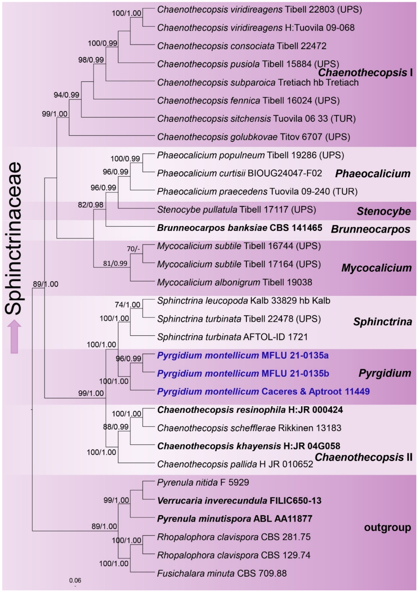
3.1. Phylogenetic Analyses
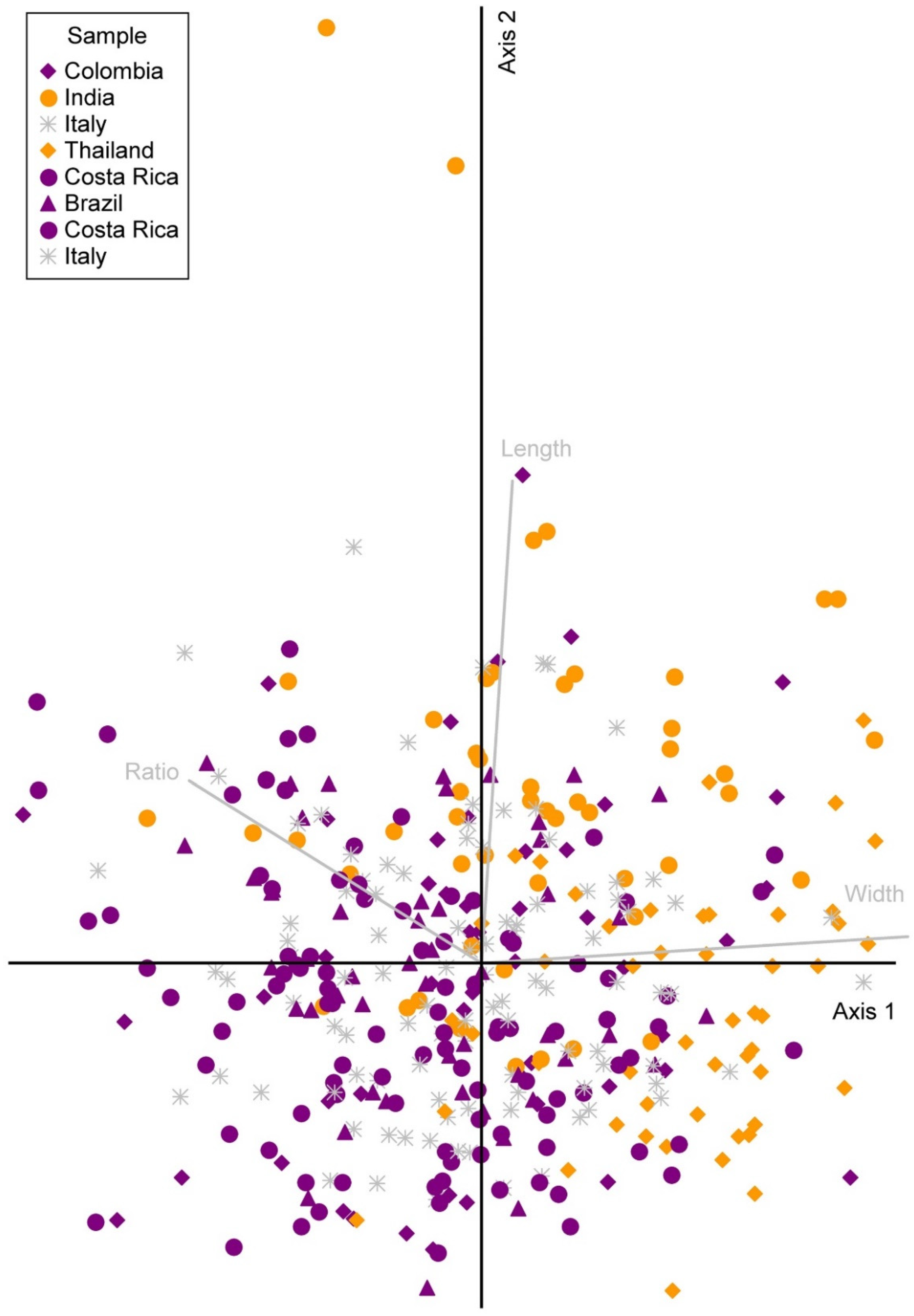
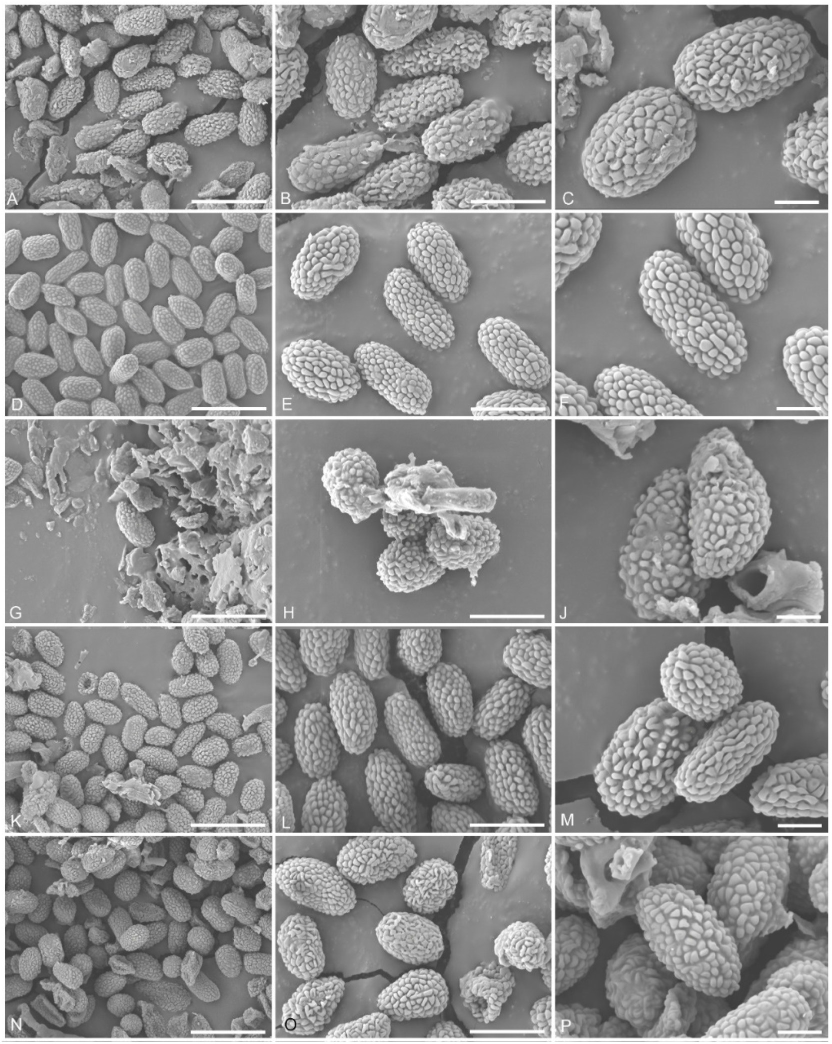
The final LSU–ITS dataset comprised 32 taxa with 1532 aligned characters, including gaps (LSU: 894; ITS: 638). The best-scoring ML tree was selected to represent the relationships between the taxa, with the final ML optimization likelihood value of –10603.813268 (Figure 1). The parameters for the GTR + I + G model of the combined LSU and ITS data were as follows: the estimated base frequencies A = 0.240252, C = 0.243881, G = 0.287821, T = 0.228046, and the substitution rates AC = 1.442589, AG = 2.659004, AT = 1.892966, CG = 1.070779, CT = 7.537779 and GT = 1.000000. Bayesian posterior probabilities from the MCMC were evaluated with the final average standard deviation of split frequencies = 0.001450. The topologies of the ML and the Bayesian tree were manually compared and were largely congruent.
The genera of Mycocaliciales were resolved as monophyletic clades, except for Chaenothecopsis, which appears to be polyphyletic. The genus Pyrgidium formed a sister clade with Sphinctrina, and both clades were strongly supported. Pyrgidium itself formed two clades, one with a single specimen from Brazil and the other with two sequences from Thailand, the latter two clustering with a high level of statistical support.

3.2. PCA

The PCA indicated a homogeneous, unimodal distribution of the ascospore size measurements, with only one large cluster, although the data on the ascospore width included two outliers (Figure 2). There was some tendency of the samples to differentiate according to region, especially regarding the ascospore width; the samples from the Neotropics clustered more towards the left and those from the Paleotropics more towards the right of the first axis, which largely corresponded to the ascospore width. Mean values for the ascospore length were 6.30 µm (Neotropics), 6.42 µm (Europe) and 6.77 µm (Paleotropics). The mean values for the ascospore width were 3.45 µm (Neotropics), 3.52 µm (Europe) and 3.97 µm (Paleotropics). The mean values for the Q value (ratio) were 1.84 (Neotropics), 1.81 µm (Europe) and 1.75 µm (Paleotropics).
This tendency was significant in terms of the ascospore width and length (ascospore length ANOVA: f-ratio = 13.8751, p < 0.00001; ascospore width ANOVA: f-ratio = 51.2113, p < 0.00001) but not for the Q value or the length:width ratio (ANOVA: f-ratio = 2.6403, p = 0.0726). The length and width differed significantly between regions 1 and 2 (Neotropics, Europe), on one hand, and region 3 (Paleotropics), on the other, but not between regions 1 and 2 (length according to post hoc Tukey HSD: 1 vs. 2: Q = 1.77, p = 0.4230; 1 vs. 3: Q = 7.04, p = 0.0000; 2 vs. 3: Q = 5.27 (p = 0.0007; width according to post hoc Tukey HSD: 1 vs. 2: Q = 2.18, p = 0.2736; 1 vs. 3: Q = 13.36, p = 0.0000; 2 vs. 3: Q = 11.19, p = 0.0000).

4. Taxonomy

4.1. Sphinctrinaceae M. Choisy, Bull. Mens. Soc. Linn. Soc. Bot. Lyon 19: 65 (1950)

Type genus: Sphinctrina Fr.
Syn.: Mycocaliciaceae A.F.W. Schmidt, Mitt. Staatsinst. Allg. Bot. Hamburg 13: 127 (1970).
Type genus: Mycocalicium Vain.
Notes: With the inclusion of Mycocaliciaceae, Sphinctrinaceae comprises seven genera, viz., Brunneocarpos, Chaenothecopsis, Mycocalicium, Phaeocalicium, Pyrgidium, Sphinctrina and Stenocybe [23]. Apparently, this is the only “discomycetous” family in Eurotiomycetes [66]. Several taxa in this family also produce coelomycetous and/or hyphomycetous anamorphs [25,27,29].

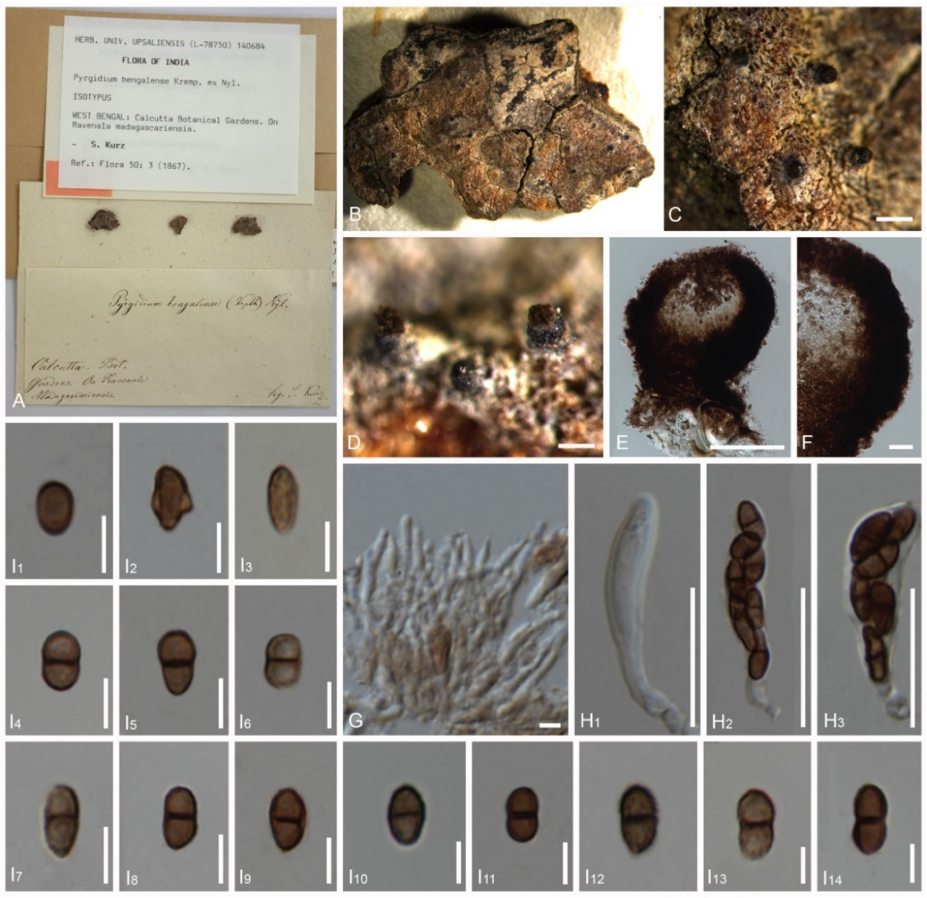
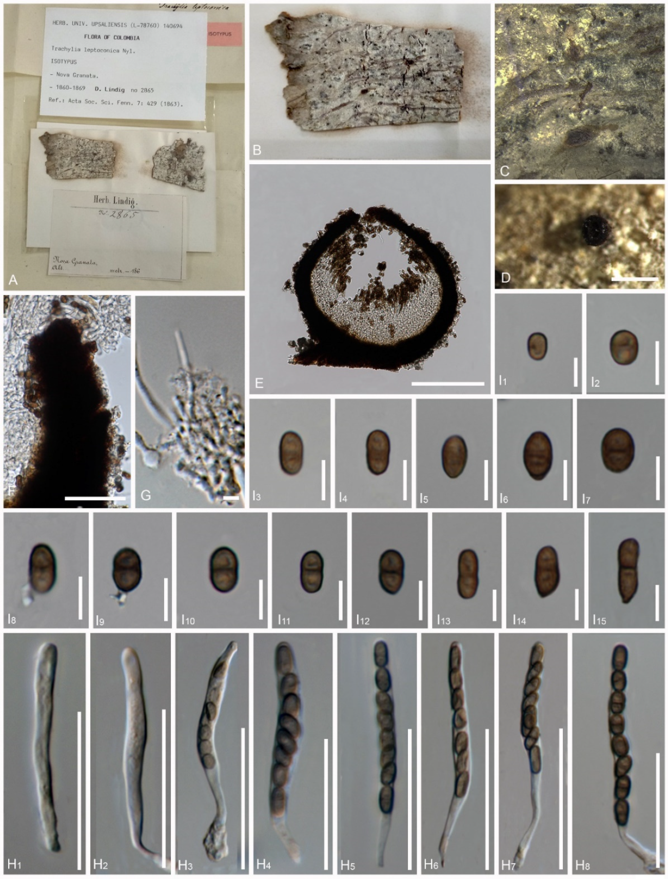
4.2. Pyrgidium Nyl., Flora, Regensburg 50: 3 (1867)

Index Fungorum number: IF 4617; faces of fungi number: FoF 12620.
Type species: Pyrgidium montellicum (Beltr.) Tibell, lichenologist 14(3): 239 (1982).
Notes: Pyrgidium was previously assigned to Sphinctrinaceae, without molecular data, and the present molecular study supports this placement. According to Tibell [5], the genus comprises a single species, P. montellicum, found mainly in the neotropics but also known from other tropical regions. Pyrgidium is characterized by its sessile to stalked ascomata, blackish brown and sclerotized exciple, either simple or 1-septate, and broadly ellipsoid to oval, dark brown, ornamented ascospores [5].

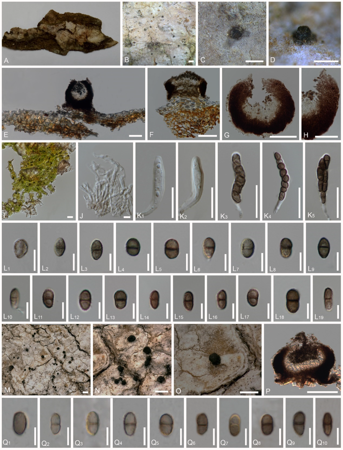
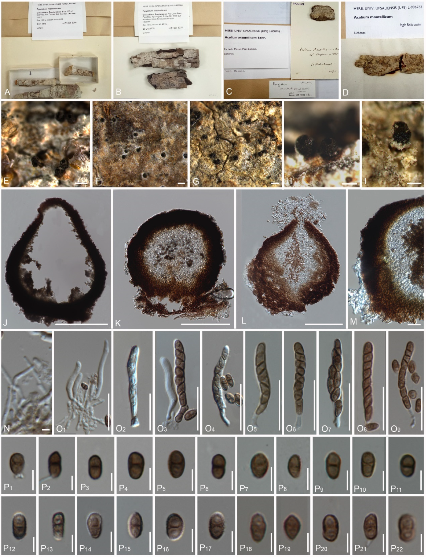
4.3. Pyrgidium montellicum (Beltr.) Tibell, Lichenologist 14(3): 239 (1982) (Figure 3 and Figure 4)

Index Fungorum number: IF 110065; faces of fungi number: FoF 10274.
Basionym: Acolium montellicum Beltr. 1858 [37].
Index Fungorum number: IF 110065; type: Italy. Bazzano, Beltramini s.n., ex Hb. Massalongo (UPS-Syntype).
Synonyms:
Pyrgidium leptoconium (Nyl.) Nádv., Stud. Bot. Čechoslov. 5: 125 (1942).
Index Fungorum number: IF 369854.
Pyrgidium bengaliense Nyl., Flora, Regensburg 50: 3 (1867) (Tibell 1982) (Figure 5)
Index Fungorum number: IF 403559. Type: India, Calcutta Botanic Gardens, on bark of Ravenala madagascariensis, Kurz 66 (Kurz 1866, UPS-Isotype).
Trachylia leptoconia Nyl., Acta Soc. Sci. fenn. 7(2): 429 (1863) [37]. (Figure 6)
Index Fungorum Number: IF 407801; type: Colombia (Colombia), Nova Granata, Fusagasuga, Lindig. A (Lindig 2865, UPS-Isotype).
Saprobic on bark. Thallus crustose, farinose or absent, and whitish. Prothallus is absent. Photobiont with loosely associated algae, sparsely present in the thallus, and mostly trentepohlioid or absent. Sexual morph: Ascomata are scattered, apothecial, rarely urn-shaped, 180–330 μm diam., 130–290 μm high (M = 255 × 210 μm), sessile to shortly stalked, almost sphaerical, mazaedioid, with a black disc. Excipulum of 10–55 μm thickened laterally, 27–90 μm thickened basally, mostly thickened basally and gradually becoming thinner towards the upper part, sometimes comprising sclerotized hyphae. Mazaedium filling the cavity of the ascoma and more or less projecting beyond the excipular edge. Paraphyses are 0.4–2 μm thick, septate, and unbranched. Asci of 18–56 × 3–9 μm (M = 37 × 6 μm, n = 30), cylindrical, 8-spored, unitunicate, tip-blunted, not narrowing towards the apex, with the ascus apex thickened in immaturity and reduced or inconspicuous in maturity, and short pedicellate. Ascospores of 4–10 × 2–5 μm (M = 7 × 3.5 μm, n = 40), broadly ellipsoid to oval, uniseriate to biseriate, and light to dark brown, (0-)1-septate, with a dark brown septum, small appendage present at one end in few ascospores, and with guttulates when immature, wall verrucose or with irregular, longitudinally arranged ridges. Asexual morph: unknown.
Notes: Pyrgidium montellicum has thus far been reported in Central and South America (Costa Rica, Colombia, Ecuador, Brazil, Argentina), Eurasia (Italy, Iran, Russia, China), the eastern Paleotropics (India, Sri Lanka, Thailand, Papua New Guinea) and Australasia [5,64,67,68,69,70,71] (https://www.gbif.org/species/3269679, accessed on 15 January 2022). The new collections of Pyrgidium montellicum studied here also confirm the previously reported presence of this taxon in Brazil, where it is known to be present in the Amazon [72], and the subtropical coast of Rio de Janeiro [5] and Thailand [69], which is now, for the first time, supported by molecular data. The Brazilian specimens were mostly over mature and the asci were difficult to observe.
The collection of Pyrgidium montellicum from Thailand and Brazil clustered together in the phylogenetic analysis, providing strong support for the accurate placement of this lineage. No significant morphological differences were observed between the specimens representing the fresh collections and the types known by three names currently synonymized under the name P. montellicum, excepting the tendency of the paleotropical collections from India and Thailand to produce larger and especially broader ascospores. Base pair comparisons between the Brazilian and the Thai specimens revealed about a 4.7% difference in the ITS markers, correlating with the differences in the ascospore size. Even without evident morphological differences, such sequence divergences have been used to distinguish morphologically cryptic species in other cases [73,74]. Indeed, several studies have been conducted to assess the potentially cryptic nature of mycocalicioid species due to the limited number of taxonomically useful characteristics [75,76]. However, differences in a single molecular marker may not be seen as sufficient for establishing species boundaries in a cryptic lineage, especially if few specimens have been sequenced [74]. Here, a case could be made for the separation of the paleotropical populations into different species, but since only a few specimens have been examined molecularly and morphologically, for the time being, we agree with Tibell [5] that only a single sub-cosmopolitan species should be presently recognized. If the analysis of more material supports the encountered differences, P. bengaliensis could be resurrected for the paleotropical material. Apart from the size of the ascospores, several other characteristics, such as color (brown to dark brown), septation (0-1-septate), ornamentation (verrucose, gattulates in immaturity and irregular ridges, longitudinally arranged), a thicker outer wall, and highly pigmented septa with constrictions at the septum, do not seem to provide any taxonomic value in this case (Table 3).
Material examined: Thailand, Chiang Mai, 128 Moo3, Bahn Pa Dheng, T. Pa Pae, A. Mae Taeng, on the bark of an unidentified tree, 10 September 2020, Vinodhini Thiyagaraja (MFLU 21-0135); Brazil, Rondônia, Porto Velho, Parque Circuito, on the bark of Hevea brasiliensis, 11 March 2012, André Aptroot and Marcela Eugenia da Silva Cáceres (Cáceres and Aptroot ISE 11449).
Description of the fresh material of Pyrgidium montellicum: Saprobic on bark. Thallus crustose, whitish and endoperidermal. Prothallus absent. Photobiont absent or loosely associated algal cells sparsely present in the thallus, and trentepohlioid. Sexual morph: Ascomata scattered, apothecial, 22–330 μm diam., 130–195 μm high M = 277 × 162 μm), sessile to shortly stalked, almost sphaerical, mazaedioid, with a black disc. Excipulum 25–55 μm thick laterally, 30–60 μm thick basally, brown to black, prosoplectenchymatous, with the edge comprised of sclerotized hyphae, the edge of the excipulum turned inward in the topmost part in immature ascomata and eventually turned outward in the topmost part in maturity, laterally and gradually becoming thinner. Mazaedium filling the cavity of the ascoma and more or less projecting beyond the excipular edge. Paraphyses of 1–2 μm thick, septate and unbranched. Asci of 25–40 × 4–7 μm (M = 32 × 5.5 μm, n = 30), cylindrical, shortly pedicellate, 8-spored, unitunicate, tip-blunted and not narrowing towards the apex, with the ascus apex thickened when immature and reduced or inconspicuous when mature. Ascospores of 5–9 × 2.5–4.5 μm (M = 5.5 × 3.5 μm, n = 40), broadly ellipsoid to oval, uniseriate to biseriate, light to dark brown and (0-)1-septate, with a dark brown septum, small appendage present at one end in a few ascospores, with guttulates when immature, wall verrucose or with irregular, longitudinally arranged ridges (Figure 7K–P). Asexual morph: unknown.
The types of the three synonyms of Pyrgidium montellicum are characterized as follows:
Description of the type Acolium montellicum: Saprobic on bark. Thallus crustose, farinose and whitish. Prothallus absent. Photobiont with loosely associated algae, sparsely present in thallus, mostly trentepohlioid or absent. Sexual morph: Ascomata scattered, apothecial, rarely urn-shaped, 180–225 μm diam., 160–290 μm high (M = 202 × 225 μm), sessile to shortly stalked, almost sphaerical, mazaedioid, with a black disc. Excipulum 10–40 μm thickened laterally, 27–46 μm thickened basally, mostly thickened basally and gradually becoming thinner towards the upper part, sometimes comprising sclerotized hyphae. Mazaedium filling the cavity of the ascoma and more or less projecting beyond the excipular edge. Paraphyses 0.5–2 μm thick, septate and unbranched. Asci 25–40 × 2–6 μm (M = 32 × 4 μm, n = 30), cylindrical, 8-spored, unitunicate, tip-blunted and not narrowing towards the apex, with the ascus apex thickened in immaturity and reduced or inconspicuous in maturity, and short pedicellate. Ascospores 4–7 × 2–4 μm (M = 5.5 × 3 μm, n = 40), broadly ellipsoid to oval, uniseriate to biseriate, light to dark brown, (0-)1-septate, with a dark brown septum, small appendage present at one end in few ascospores, with guttulates when immature, wall verrucose or with irregular, longitudinally arranged ridges. Asexual morph: unknown.
Description of the type Pyrgidium bengaliense: Saprobic on bark. Thallus inconspicuous, pruinose around the ascomata. Prothallus absent. Photobiont absent. Sexual morph: Ascomata apothecial, 190–205 μm diam., 180–220 μm high (M = 197 × 200 μm, n = 10), not stalked, mazaedioid, with a black disc, sessile, scattered and almost spherical. Excipulum 30–55 μm thickened laterally, 60–90 μm thickened basally, brown to black, prosoplectenchymatous and comprising some sclerotized hyphae. Mazaedium filling the cavity of the fruit body and more or less projecting beyond the excipular edge. Paraphyses 1.2–1.8 μm thick, septate and simple. Asci 18–33 × 4–9 μm (M = 25.5 × 6.5 μm, n = 30), cylindrical, 8-spored, unitunicate, tip-blunted and not narrowing towards the apex, with the ascus apex thickened in immaturity and reduced or inconspicuous in maturity, and short pedicellate. Ascospores 5.5–10 × 3–5 μm (M = 7.75 × 4 μm, n = 40, ellipsoidal, overlapping bi-seriate, light brown to brown, 0-1-septate, with a slightly dark brown septum, sometimes constricted at the septum, with gattulates when immature, verrucose, irregular ridges that are longitudinally arranged (Figure 7G–J). Asexual morph: undetermined.
Description of the type Trachylia leptoconia: Saprobic on bark. Thallus crustose and farinose. Prothallus absent. Photobiont absent. Sexual morph: Ascomata apothecial, 220–240 μm diam., 220–235 μm high (M = 230 × 227 μm, n = 10), not stalked, mazaedioid, with a black disc, sessile, scattered and almost spherical. Mazaedium filling the cavity of the fruit body and more or less projecting beyond the excipular edge. Excipulum 22–28 μm thickened laterally, 35–55 μm thickened basally, brown to black, prosoplectenchymatous, hardly compromise any sclerotized hyphae, thickened basally and gradually becoming thinner towards the upper part. Paraphyses 1.2–2 μm thick, septate and simple. Asci 34–56 × 4–5 μm (M = 45 × 4.5 μm, n = 30), cylindrical, 8-spored, unitunicate and tip-blunted, with the ascus apex thickened when immature and reduced or inconspicuous when mature, and short pedicellate. Ascospores 4.5–9 × 2–5 μm (M = 6.75 × 3.5 μm, n = 40), ellipsoidal, uniseriate, overlapping, light brown to brown, 0-1-septate, with a dark brown septum, small appendage present at one end in few ascospores, with gattulates when immature, and verrucose, irregular ridges that are longitudinally arranged (Figure 7D–F). Asexual morph: undetermined.

5. Discussion

Although Pyrgidium montellicum is usually considered a saprotrophic taxon, several studies reported Trebouxia or allied cystococcaceous alga as photobionts, and some defined the taxon as commensal on lecanoralean lichens [6,37]. In addition, P. montellicum may serve as a host for lichenicolous fungi, such as Chaenothecopsis rubina Tibell [37]. In the material assessed in the present study, including both the fresh and historical specimens, no obligate and stable association with a particular photobiont was observed, but some specimens showed a weak association with trentepohlioid algae. Nádvorník [36] reported the presence of a Trentepohlia photobiont in the original material of Pyrgidium leptoconium, but this was not seen in the isotype material examined here. These findings support the notion that algal associations are accidental or facultative in the case of P. montellicum and that the taxon is primarily saprotrophic [37]. A similar situation can be observed with other borderline lichenized fungi, such as Arthopyrenia salicis A. Massal., Cresporhaphis macrospora (Eitner) M.B. Aguirre, Requienella seminuda (Pers.) Boise and Splanchnonema lichenisatum Aptroot and K.H. Moon, which are facultatively associated with various photobionts but were also recorded without any algal associations [77,78,79]. Notably, these lineages are found in predominantly non-lichenized clades, such as Dothideomycetes and Sordariomycetes [77,78,79,80], suggesting initial evolutionary attempts at lichenization in these lineages. On the other hand, several bark-inhabiting fungi are known to have emerged from largely lichenized clades, and their saprotrophic mode evolved secondarily from their lichenized ancestors [81,82,83].
Eurotiomycetes comprise lichenized and lichenicolous lineages mainly in the orders of Pyrenulales and Verrucariales within the subclass of Chaetothyriomycetidae [14]. Among these, Pyrenula coryli A. Massal. has been recorded as non-lichenized [84]. Verrucariales largely encompass lichenized species or mycophycobioses [85,86]. In contrast, saprotrophic species are mainly found in Mycocaliciales within the subclass Mycocaliciomycetidae, in the genera Brunneocarpos, Chaenothecopsis, Mycocalicium, Phaeocalicium, Stenocybe and Strongyleuma. Chaenothecopsis also comprise lichenicolous taxa, and one species, C. pusilla (Ach.) A.F.W. Schmidt, was found to be facultatively associated with algae [87,88].
Ascospore characteristics, such as color, size, septation and ornamentation, have been used for the generic and species delineation of mycocalicioid fungi [5,37]. Ascospores have been mostly recorded as 1-septate in Pyrgidium [5,37], although aseptate ascospores were also frequently observed in the material examined in this study. Sometimes, appendages were observed at one end of the ascospores (Tibell 8306; Tibell 8232; Lindig 2865; MFLU 21-0135;). The ascospores of Pyrgidium have small warts or ridges that were visible under the scanning electron microscope. Nylander [35] defined the ascospore dimensions as 5–9 × 3–4 μm for Pyrgidium bengaliense, and the re-examination of the isotype material resulted in dimensions of 5.5–10 × 3–5 μm. The ascospore dimensions of the original material of P. leptoconia were given as 6–8 × 4–4.5 μm, whereas the isotype revealed measurements of 4.5–9 × 2–5 μm. Pyrgidium montellicum was originally described as having 5.5–7 × 3–4.5 μm large ascospores, while our measurements were 4–7 × 2–4 μm for the four non-type specimens. Variations in then ascospore size revealed limited clustering tendencies according to the geographic region, especially regarding the width, and both the length and width showed minor but significant differences between the groups from the Neotropics and Europe, on one hand, and the Paleotropics, on the other. However, due to the limited material examined, we refrained from dividing P. montellicum into more than one species at this point in time.
Tibell [37] described P. montellicum as having perithecial, urn-shaped ascomata, but later studies described them as apothecial [5,88]. The re-examination of several collections revealed both urn-shaped perithecial and apothecial ascomata, suggesting that these morphologies intergrade during ontogeny. The asci of all mycocalicioid fungi are known to arise from croziers [89,90], and careful microscopic observations revealed the presence of croziers in all the studied specimens of Pyrgidium. The ascus apex was almost reduced in maturity and could only be observed in the immature stage or after adding 5% KOH. Overall, considering all these characteristics, there are no clear-cut differences between the examined specimens, which, at present, supports the argument of Tibell [5] that we should consider Pygridium as a monospecific genus. However, the variation in the ITS associated with geography (Brazil vs. Thailand) warrants further attention and should be investigated using more material so as to assess the potential cryptic speciation through vicariance.

Author Contributions

Conceptualization, V.T., D.E. and R.L.; methodology, V.T., D.E., R.L., D.N.W., A.A., M.E.d.S.C., R.C., W.T. and K.D.H.; software, V.T., D.E., R.L. and D.N.W.; validation, D.E., R.L., D.N.W., A.A., M.E.d.S.C., R.C. and K.D.H.; formal analysis, V.T. and D.N.W.; investigation, V.T., D.E., R.L., D.N.W., A.A., M.E.d.S.C., R.C. and K.D.H.; resources, D.E., A.A. and M.E.d.S.C.; data curation, D.E., R.L., D.N.W., A.A., M.E.d.S.C. and K.D.H.; writing—original draft preparation, V.T.; writing—review and editing, V.T., D.E., R.L. and D.N.W.; visualization, V.T., D.E., R.L., D.N.W., A.A., M.E.d.S.C., R.C. and K.D.H.; supervision, D.E., R.L., D.N.W., K.D.H. and R.C; project administration, V.T., D.E., R.L., R.C., W.T. and K.D.H.; funding acquisition, R.C. All authors have read and agreed to the published version of the manuscript.

Funding

Vinodhini Thiyagaraja would like to express thanks for the postdoctoral fellowship of 2022 from Chiang Mai University, Thailand (Reinventing University).

Institutional Review Board Statement

Not applicable.

Informed Consent Statement

Not applicable.

Data Availability Statement

Not applicable.

Acknowledgments

Vinodhini Thiyagaraja would like to express thanks for the postdoctoral fellowship of 2022 from Chiang Mai University, Thailand (Reinventing University). We are grateful for the grants from the Thailand Research Fund (“The future of specialist fungi in a changing climate: baseline data for generalist and specialist fungi associated with ants, Rhododendron species and Dracaena species DBG6080013” and “Impact of climate change on fungal diversity and biogeography in the Greater Mekong Sub-region RDG6130001”) for the funding of this research. Kevin D. Hyde thanks Chiang Mai University for the award of visiting professor. We also thank Udeni Jayalal, Nalin Wijayawardene and S.C. Karunarathna for their valuable help with this research. We thank Tinnakorn Bang-ngoen and Diana Sandamali for taking the SEM photographs. We thank Saranyaphat Boonmee for loaning herbarium specimens. We also thank CAS President’s International Fellowship Initiative (PIFI) for their grant for young staff (number: 2020FYC0002) and the National Science Foundation of China (NSFC), under the project code 31851110759, as well as the CAS President’s International Fellowship Initiative (PIFI), under the grant: 2018PC0006, and the National Science Foundation of China (NSFC), project code 31851110759. Dhanushka Wanasinghe would like to thank CAS President’s International Fellowship Initiative (PIFI) for funding his postdoctoral research (grant number: 2021FYB0005) and expresses thanks for the Postdoctoral Fund from Human Resources and Social Security Bureau of Yunnan Province and the National Science Foundation of China and Chinese Academy of Sciences (grant no. 41761144055). The CNPq—Conselho Nacional de Desenvolvimento Científico e Tecnológico is thanked for the research grant awarded to Marcela E. S. Cáceres (501633/2009-0) and for covering the costs for the collection expeditions (CNPq-Simbiota 563342/2010-2).

Conflicts of Interest

The authors declare no conflict of interest.

References

  1. Prieto, M.; Baloch, E.; Tehler, A.; Wedin, M. Mazaedium evolution in the Ascomycota (Fungi) and the classification of mazaediate groups of formerly unclear relationship. Cladistics 2013, 29, 296–308. [Google Scholar] [CrossRef] [PubMed]
  2. Rikkinen, J.; Meinke, S.K.L.; Grabenhorst, H.; Gröhn, C.; Kobbert, M.; Wunderlich, J.; Schmidt, A.R. Calicioid lichens and fungi in amber–Tracing extant lineages back to the Paleogene. Geobios 2018, 51, 469–479. [Google Scholar] [CrossRef]
  3. Fries, E. Lichenum dianome nova. Lund 1817, 107, 1403–1418. [Google Scholar]
  4. Wedin, M.; Tibell, L. Phylogeny and evolution of Caliciaceae, Mycocaliciaceae, and Sphinctrinaceae (Ascomycota), with notes on the evolution of the prototunicate ascus. Canad. J. Bot. 1997, 75, 1236–1242. [Google Scholar] [CrossRef]
  5. Tibell, L. Caliciales—Flora Neotropica, Monograph 69; New York Botanical Garden: New York, NY, USA, 1996. [Google Scholar]
  6. Tibell, L.; Wedin, M. Mycocaliciales, a new order for nonlichenized calicioid fungi. Mycologia 2000, 92, 577–581. [Google Scholar] [CrossRef]
  7. Temu, S.G.; Tibell, S.; Tibuhwa, D.D.; Tibell, L. Crustose Calicioid Lichens and Fungi in Mountain Cloud Forests of Tanzania. Microorganisms 2019, 7, 491. [Google Scholar] [CrossRef]
  8. Vainio, E.A. Lichenographia fennica III, Coniocarpeae. Acta Soc. Pro Fauna Flora Fenn. 1927, 57, 1–138. [Google Scholar]
  9. Schmidt, A. Anatomisch-taxonomische Untersu- chungen an Europaischen Arten der Flechtenfamilie Caliciaceae. Mitteilung zur Staatsinstitut fuXr Aligemeine Botanik, Hamburg. Hambg. Staatsinst. Allg. Bot. Mitt. 1970, 13, 111–166. [Google Scholar]
  10. Tibell, L.B. Distribution patterns and dispersal strategies of Caliciales. Bot. J. Lin. Soc. 1994, 116, 159–202. [Google Scholar] [CrossRef]
  11. Tibell, L. Phaeocalicium (Mycocaliciaceae, Ascomycetes) in Northern Europe. Annales Botanici Fennici 1996, 33, 205–221. [Google Scholar]
  12. Tibell, L.B. A reappraisal of the taxonomy of Caliciales. Beih. Nova Hedwig. 1984, 79, 597–713. [Google Scholar]
  13. Prieto, M.; Wedin, M. Phylogeny, taxonomy and diversification events in the Caliciaceae. Fung Divers. 2017, 82, 221–238. [Google Scholar] [CrossRef]
  14. Geiser, D.M.; Gueidan, C.; Miadlikowska, J.; Lutzoni, F.; Kauff, F.; Hofstetter, V.; Fraker, E.; Schoch, C.L.; Tibell, L.; Untereiner, W.A.; et al. Eurotiomycetes: Eurotiomycetidae and Chaetothyriomycetidae. Mycologia 2006, 98, 1053–1064. [Google Scholar] [CrossRef] [PubMed]
  15. Hibbett, D.S.; Binder, M.; Bischoff, J.F.; Blackwell, M.; Cannon, P.F.; Eriksson, O.E.; Huhndorf, S.; James, T.; Kirk, P.M.; Lücking, R. A higher-level phylogenetic classification of the Fungi. Mycol. Res. 2007, 111, 509–547. [Google Scholar] [CrossRef] [PubMed]
  16. Lumbsch, H.T.; Huhndorf, S.M. Outline of Ascomycota–2007. Myconet 2007, 13, 1–58. [Google Scholar]
  17. Schmitt, I. 8 Fruiting Body Evolution in the Ascomycota: A Molecular perspective integrating lichenized and non-lichenized groups. In Evolution of Fungi and Fung. Like Organisms; Springer: Berlin, Germany, 2011; pp. 187–204. [Google Scholar]
  18. Sadowski, E.M.; Beimforde, C.; Gube, M.; Rikkinen, J.; Singh, H.; Seyfullah, L.J.; Heinrichs, J.; Nascimbene, P.C.; Reitner, J.; Schmidt, A.R. The anamorphic genus Monotosporella (Ascomycota) from Eocene amber and from modern Agathis resin. Fung Biol. 2012, 116, 1099–1110. [Google Scholar] [CrossRef]
  19. Gueidan, C.; Aptroot, A.; da Silva Cáceres, M.E.; Badali, H.; Stenroos, S. A reappraisal of orders and families within the subclass Chaetothyriomycetidae (Eurotiomycetes, Ascomycota). Mycol. Prog. 2014, 13, 1027–1039. [Google Scholar] [CrossRef]
  20. Geiser, D.M.; LoBuglio, K.F.; Gueidan, C. 5 Pezizomycotina: Eurotiomycetes. Syst. Evol. 2015, 7B, 121–141. [Google Scholar]
  21. Wijayawardene, N.N.; Hyde, K.D.; Rajeshkumar, K.C.; Hawksworth, D.L.; Madrid, H.; Kirk, P.M.; Braun, U.; Singh, R.V.; Crous, P.W.; Kukwa, M.; et al. Notes for genera–Ascomycota. Fung Divers. 2017, 86, 1–594. [Google Scholar]
  22. Wijayawardene, N.N.; Hyde, K.D.; Divakar, P.K.; Rajeshkumar, K.C.; Weerahewa, D.; Delgado, G.; Wang, Y.; Fu, L. Notes for genera update—Ascomycota: 6616-6821. Mycosphere 2018, 9, 115–140. [Google Scholar] [CrossRef]
  23. Wijayawardene, N.; Hyde, K.; Al-Ani, L.; Tedersoo, L.; Haelewaters, D.; Rajeshkumar, K.C.; Zhao, R.-L.; Aptroot, A.; Saxena, R.; Tokarev, Y.; et al. Outline of Fungi and fungus-like taxa. Mycosphere 2020, 11, 1160–1456. [Google Scholar] [CrossRef]
  24. Wijayawardene, N.N.; Hyde, K.D.; Dai, D.Q.; Sánchez-García, M.; Goto, B.T.; Saxena, R.K.; Erdogdu, M.; Selçuk, F.; Rajeshkumar, K.C.; Aptroot, A.; et al. Outline of Fungi and fungus-like taxa-2021. Mycosphere 2022, 13, 53–453. [Google Scholar] [CrossRef]
  25. Titov, A.; Tibell, L. Chaenothecopsis in the Russian Far East. Nord. J. Bot. 1993, 13, 313–329. [Google Scholar] [CrossRef]
  26. Tibell, L.; Titov, A. Species of Chaenothecopsis and Mycocalicium (Caliciales) on exudate. Bryologist 1995, 98, 550–560. [Google Scholar] [CrossRef]
  27. Tuovila, H.; Davey, M.L.; Yan, L.; Huhtinen, S.; Rikkinen, J. New resinicolous Chaenothecopsis species from China. Mycologia 2014, 106, 989–1003. [Google Scholar] [CrossRef]
  28. Ariyawansa, H.A.; Hyde, K.D.; Jayasiri, S.C.; Buyck, B.; Chethana, K.W.T.; Dai, D.Q.; Dai, Y.C.; Daranagama, D.A.; Jayawardena, R.S.; Lücking, R.; et al. Fungal diversity notes 111–252—Taxonomic and phylogenetic contributions to fungal taxa. Fung. Divers. 2015, 75, 27–274. [Google Scholar] [CrossRef]
  29. Crous, P.W.; Wingfield, M.J.; Richardson, D.M.; Le Roux, J.J.; Strasberg, D.; Edwards, J.; Roets, F.; Hubka, V.; Taylor, P.W.J.; Heykoop, M.; et al. Fungal Planet description sheets: 400–68. Persoonia 2016, 36, 316–458. [Google Scholar] [CrossRef]
  30. Choisy, M. Catalogue des lichens de la region Lyonnaise. Bull. Mens. Soc. Linn. Lyon 1950, 19, 57–72. [Google Scholar]
  31. Muniz, D.; Llop, E.; Hladun, N.L. Sphinctrina paramerae, a new Mediterranean lichenicolous species with non-septate spores. Lichenologist 2013, 45, 137–143. [Google Scholar] [CrossRef]
  32. Jaklitsch, W.; Baral, H.-O.; Lücking, R.; Lumbsch, H.T.; Frey, W. Syllabus of Plant Families–Adolf Engler’s Syllabus der Pflanzenfamilien; Borntraeger: Stuttgart, Germany, 2016; p. 288. [Google Scholar]
  33. Lücking, R.; Hodkinson, B.P.; Leavitt, S.D. Corrections and amendments to the 2016 classification of lichenized fungi in the Ascomycota and Basidiomycota. Bryologist 2017, 120, 58–69. [Google Scholar] [CrossRef]
  34. Falswal, A.; Bhandari, B.S. A New Lichenicolous Fungus from Garhwal Himalayan Region of Uttarakhand, India. Acta Bot. 2021, 63, 297–302. [Google Scholar] [CrossRef]
  35. Nylander, W. Addenda nova ad Lichenographiam Europaeam. Continuatio quarta. Flora 1867, 50, 177–180. [Google Scholar]
  36. Nádvorník, J. Systematische Übersicht der mitteleuropäischen Arten der Flechtenfamilie Caliciaceae. Stud. Bot. Cech. 1942, 5, 6–46. [Google Scholar]
  37. Tibell, L. Caliciales of Costa Rica. Lichenologist 1982, 14, 219–254. [Google Scholar] [CrossRef]
  38. Index Fungorum. 2022. Available online: http://www.indexfungorum.org/names/Names.asp (accessed on 15 May 2022).
  39. Mycobank. 2022. Available online: https://www.mycobank.org/page/Simple%20names%20search (accessed on 15 January 2022).
  40. Jayasiri, S.C.; Hyde, K.D.; Ariyawansa, H.A.; Bhat, J.; Buyck, B.; Cai, L.; Dai, Y.C.; Abd-Elsalam, K.A.; Ertz, D.; Hidayat, I. The Faces of Fungi database: Fungal names linked with morphology, phylogeny and human impacts. Fung. Divers. 2015, 74, 3–18. [Google Scholar] [CrossRef]
  41. Dissanayake, A.J.; Bhunjun, C.S.; Maharachchikumbura, S.S.N.; Liu, J.K. Applied aspects of methods to infer phylogenetic relationships amongst fungi. Mycosphere 2020, 11, 2652–2676. [Google Scholar] [CrossRef]
  42. White, T.J.; Bruns, T.; Lee, S.J.W.T.; Taylor, J.W. Amplification and direct sequencing of fungal ribosomal RNA genes for phylogenetics. PCR Protoc. Guide Methods Appl. 1990, 18, 315–322. [Google Scholar]
  43. Vilgalys, R.; Hester, M. Rapid genetic identification and mapping of enzymatically amplified ribosomal DNA from several Cryptococcus species. J. Bacteriol. 1990, 172, 4238–4246. [Google Scholar] [CrossRef]
  44. Messuti, M.I.; Vidal-Russell, R.; Amico, G.C.; Lorenzo, L.E. Chaenothecopsis quintralis, a new species of calicioid fungus. Mycologia 2012, 104, 1222–1228. [Google Scholar] [CrossRef]
  45. Schoch, C.L.; Sung, G.H.; López-Giráldez, F.; Townsend, J.P.; Miadlikowska, J.; Hofstetter, V.; Robbertse, B.; Matheny, P.B.; Kauff, F.; Wang, Z.; et al. The Ascomycota tree of life: A phylum-wide phylogeny clarifies the origin and evolution of fundamental reproductive and ecological traits. Syst. Boil. 2009, 58, 224–239. [Google Scholar] [CrossRef]
  46. Zoller, S.; Scheidegger, C.; Sperisen, C. PCR primers for the amplification of mitochondrial small subunit ribosomal DNA of lichen-forming ascomycetes. Lichenologist 1999, 31, 511–516. [Google Scholar] [CrossRef]
  47. Swindell, S.R.; Plasterer, T.N. Seqman. In Sequence Data Analysis Guidebook; Springer: Berlin, Germany, 1991; pp. 75–89. [Google Scholar]
  48. Tibell, L.; Vinuesa, M. Chaenothecopsis in a molecular phylogeny based on nuclear rDNA ITS and LSU sequences. Taxon 2005, 54, 427–442. [Google Scholar] [CrossRef]
  49. Katoh, K.; Rozewicki, J.; Yamada, K.D. MAFFT online service: Multiple sequence alignment, interactive sequence choice and visualization. Brief. Bioinf. 2019, 20, 1160–1166. [Google Scholar] [CrossRef] [PubMed]
  50. Hall, T.A. BioEdit: A user–friendly biological sequence alignment editor and analysis program for Windows 95/98/NT. Nucl. Ac. Symp. Ser. 1999, 41, 95–98. [Google Scholar]
  51. Glez-Peña, D.; Gómez-Blanco, D.; Reboiro-Jato, M.; Fdez-Riverola, F.; Posada, D. ALTER: Program-oriented conversion of DNA and protein alignments. Nucl. Acids Res. 2010, 38, 14–18. [Google Scholar] [CrossRef]
  52. Stamatakis, A. RAxML version 8: A tool for phylogenetic analysis and post–analysis of large phylogenies. Bioinformatics 2014, 30, 1312–1313. [Google Scholar] [CrossRef]
  53. Miller, M.A.; Pfeiffer, W.; Schwartz, T. Creating the CIPRES Science Gateway for Inference of Large Phylogenetic Trees. In Proceedings of the SC10 Workshop on Gateway Computing Environments (GCE10), New Orleans, LA, USA, 5–28 September 2010. [Google Scholar]
  54. Huelsenbeck, J.P.; Ronquist, F. MRBAYES: Bayesian inference of phylogenetic trees. Bioinformation 2001, 17, 754–755. [Google Scholar] [CrossRef]
  55. Nylander, J.A.A. MrModeltest 2.0. Program Distributed by the Author; Evolutionary Biology Centre; Uppsala University: Uppsala, Sweden, 2004. [Google Scholar]
  56. Rambaut, A. FigTree. Version 1.4. 2; University of Edinburgh: Edinburgh, UK, 2014. [Google Scholar]
  57. Schoch, C.L.; Robbertse, B.; Robert, V.; Vu, D.; Cardinali, G.; Irinyi, L.; Meyer, W.; Nilsson, R.H.; Hughes, K.; Miller, A.N.; et al. Finding needles in haystacks: Linking scientific names, reference specimens and molecular data for Fungi. Database 2014, 2014, bau061. [Google Scholar] [CrossRef]
  58. Tuovila, H.; Schmidt, A.R.; Beimforde, C.; Dörfelt, H.; Grabenhorst, H.; Rikkinen, J. Stuck in time—A new Chaenothecopsis species with proliferating ascomata from Cunninghamia resin and its fossil ancestors in European amber. Fung. Divers. 2013, 58, 199–213. [Google Scholar] [CrossRef]
  59. Beimforde, C.; Tuovila, H.; Schmidt, A.R.; Lee, W.G.; Gube, M.; Rikkinen, J. Chaenothecopsis schefflerae (Ascomycota: Mycocaliciales): A widespread fungus on semi-hardened exudates of endemic New Zealand Araliaceae. N. Zeal. J. Bot. 2017, 55, 387–406. [Google Scholar] [CrossRef]
  60. Réblová, M.; Untereiner, W.A.; Štěpánek, V.; Gams, W. Disentangling Phialophora section Catenulatae: Disposition of taxa with pigmented conidiophores and recognition of a new subclass, Sclerococcomycetidae (Eurotiomycetes). Mycol. Progr. 2017, 16, 27–46. [Google Scholar] [CrossRef]
  61. Telfer, A.C.; Young, M.R.; Quinn, J.; Perez, K.; Sobel, C.N.; Sones, J.E.; Levesque-Beaudin, V.; Derbyshire, R.; Fernandez-Triana, J.; Rougerie, R.; et al. Biodiversity inventories in high gear: DNA barcoding facilitates a rapid biotic survey of a temperate nature reserve. Biodivers. Data J. 2015, 3, e6313. [Google Scholar] [CrossRef]
  62. Gueidan, C.; Aptroot, A.; da Silva Cáceres, M.E.; Binh, N.Q. Molecular phylogeny of the tropical lichen family Pyrenulaceae: Contribution from dried herbarium specimens and FTA card samples. Mycol. Prog. 2016, 15, 7. [Google Scholar] [CrossRef]
  63. Del Prado, R.; Schmitt, I.; Kautz, S.; Palice, Z.; Lücking, R.; Lumbsch, H.T. Molecular data place Trypetheliaceae in Dothideomycetes. Mycol. Res. 2006, 110, 511–520. [Google Scholar] [CrossRef] [PubMed]
  64. Weerakoon, G.; Wolseley, P.A.; Arachchige, O.; da Silva Cáceres, M.E.; Jayalal, U.; Aptroot, A. Eight new lichen species and 88 new records from Sri Lanka. Bryologist 2016, 119, 131–142. [Google Scholar] [CrossRef]
  65. Pykälä, J.; Launis, A.; Myllys, L. Taxonomy of the Verrucaria kalenskyi-V. xyloxena species complex in Finland. Nova Hedw. 2019, 109, 489–511. [Google Scholar] [CrossRef]
  66. Ekanayaka, A.H.; Ariyawansa, H.A.; Hyde, K.D.; Jones, E.B.G.; Daranagama, D.A.; Phillips, A.J.L.; Hongsanan, S.; Jayasiri, S.C.; Zhao, Q. Discomycetes: The apothecial representatives of the phylum Ascomycota. Fung. Divers. 2017, 87, 237–298. [Google Scholar] [CrossRef]
  67. Titov, A. Further notes on calicioid lichens and fungi from the Gongga Mountains (Sichuan, China). Lichenologist 2001, 33, 303–314. [Google Scholar] [CrossRef]
  68. Nayaka, S.; Upreti, D.K. Status of lichen diversity in Western Ghats, India. Sahyadri E-News, Western Ghats BioDivers. Infor. Sys. 2005, 16, 1–28. [Google Scholar]
  69. Aptroot, A.; Saipunkaew, W.; Sipman, H.J.M.; Sparrius, L.B.; Wolseley, P.A. New lichens from Thailand, mainly microlichens from Chiang Mai. Fung. Divers. 2007, 24, 75–134. [Google Scholar]
  70. Seaward, M.R.D.; Sipman, H.J.M.; Sohrabi, M. A revised checklist of lichenized, lichenicolous and allied fungi for Iran. Sauteria 2008, 15, 459–520. [Google Scholar]
  71. Nascimbene, J.; Marini, L. Oak forest exploitation and black-locust invasion caused severe shifts in epiphytic lichen communities in Northern Italy. Sci. Tot. Env. 2010, 408, 5506–5512. [Google Scholar] [CrossRef] [PubMed]
  72. da Silva Cáceres, M.E.; de Lima, E.L.; Nascimento, A.A.; Lücking, R. Liquens brasileiros: Novas descobertas evidenciam a riqueza no Norte e Nordeste do país. Bol. Mus. Biol. Mello Leitão. 2014, 35, 101–119. [Google Scholar]
  73. Crespo, A.; Lumbsch, H.T. Cryptic species in lichen-forming fungi. IMA Fungus 2010, 1, 167–170. [Google Scholar] [CrossRef]
  74. Sato, H.; Ohta, R.; Murakami, N. Molecular prospecting for cryptic species of the Hypholoma fasciculare complex: Toward the effective and practical delimitation of cryptic macrofungal species. Sci. Rep. 2020, 10, 1–13. [Google Scholar] [CrossRef]
  75. Vinuesa, M.D.L.A.; Sanches-Puelles, J.M.; Tibell, L. Intraspecific variation in Mycocalicium subtile (Mycocaliciaceae) elucidated by morphology and the sequences of the ITS1-5.8 S-ITS2 region of rDNA. Mycol. Res. 2001, 105, 323–330. [Google Scholar]
  76. Nilsson, R.H.; Kristiansson, E.; Ryberg, M.; Hallenberg, N.; Larsson, K.H. Intraspecific ITS variability in the kingdom Fungi as expressed in the international sequence databases and its implications for molecular species identification. Evol. Bioinfor. 2008, 4, 193–201. [Google Scholar] [CrossRef]
  77. Ellis, J.B.; Everhart, B.M. New species of fungi from various localities. J. Myc. 1888, 4, 62–65. [Google Scholar] [CrossRef]
  78. Barr, M.E. Redisposition of some taxa described by J. B. Ellis. Mycotaxon 1993, 46, 45–76. [Google Scholar]
  79. Jaklitsch, W.M.; Gardiennet, A.; Voglmayr, H. Resolution of morphology-based taxonomic delusions: Acrocordiella, Basiseptospora, Blogiascospora, Clypeosphaeria, Hymenopleella, Lepteutypa, Pseudapiospora, Requienella, Seiridium and Strickeria. Pers. Mol. Phyl. Evol. Fungi 2016, 37, 82. [Google Scholar] [CrossRef]
  80. Voglmayr, H.; Aguirre-Hudson, M.B.; Wagner, H.G.; Tello, S.; Jaklitsch, W.M. Lichens or endophytes? The enigmatic genus Leptosillia in the Leptosilliaceae fam. nov. (Xylariales), and Furfurella gen. nov. (Delonicicolaceae). Pers. Mol. Phyl. Evol. Fungi 2019, 42, 228. [Google Scholar] [CrossRef] [PubMed]
  81. Hyde, K.D.; Hongsanan, S.; Jeewon, R.; Bhat, D.J.; McKenzie, E.H.C.; Jones, E.B.G.; Phookamsak, R.; Ariyawansa, H.A.; Boonmee, S.; Zhao, Q.; et al. Fungal diversity notes 367–490: Taxonomic and phylogenetic contributions to fungal taxa. Fung. Divers. 2016, 80, 1–270. [Google Scholar] [CrossRef]
  82. Thiyagaraja, V.; Lücking, R.; Ertz, D.; Wanasinghe, D.N.; Karunarathna, S.C.; Camporesi, E.; Hyde, K.D. Evolution of non-lichenized, saprotrophic species of Arthonia (Ascomycota, Arthoniales) and resurrection of Naevia, with notes on Mycoporum. Fung. Divers. 2020, 102, 205–224. [Google Scholar] [CrossRef]
  83. Thiyagaraja, V.; Lücking, R.; Ertz, D.; Karunarathna, S.C.; Wanasinghe, D.N.; Lumyong, S.; Hyde, K.D. The evolution of life modes in Stictidaceae, with three novel taxa. J. Fung. 2021, 7, 105. [Google Scholar] [CrossRef] [PubMed]
  84. Kossowska, M.; Faltynowicz, W.; Dimos-Zych, M.; Faltynowicz, H.; Patejuk, K.; Piegdon, A.; Buksakowska, M.; Jarema, P. Additions to the lichen biota of the Sudety Mountains. I. Records from the Karkonosze Mountains. Acta Mycologica 2018, 53, 1–10. [Google Scholar] [CrossRef]
  85. Hawksworth, D.L. The variety of fungal-algal symbioses, their evolutionary significance, and the nature of lichens. Bot. J. Lin. Soc. 1988, 96, 3–20. [Google Scholar] [CrossRef]
  86. Kohlmeyer, J.; Volkmann-Kohlmeyer, B. A new marine Xylomyces on Rhizophora from the Caribbean and Hawaii. Fung. Divers. 1998, 1, 159–164. [Google Scholar]
  87. Wirth, V. Die Flechten Baden-Württembergs. Teil 1; Ulmer: Stuttgart, Germany, 1995. [Google Scholar]
  88. Groner, U. The genus Chaenothecopsis (Mycocaliciaceae) in Switzerland, and a key to the European species. Lichenol. 2006, 38, 395–406. [Google Scholar] [CrossRef]
  89. Nimis, P.L.; Puntillo, D. Keys to the Lichens of Italy-08) Calicioid Species (Both Lichenized and Non-Lichenized). Available online: https://italic.units.it/flora/index.php?procedure=ext_key_home&key_id=1619 (accessed on 24 November 2021).
  90. Tuovila, H.; Huhtinen, S. New methods for mycocalicioid fungi. Lichenologist 2020, 52, 403–413. [Google Scholar] [CrossRef]
Figure 1. Best-scoring ML tree based on the analysis of the combined LSU and ITS sequence data. Bootstrap support values equal to or greater than 70% and Bayesian posterior probabilities (BP) equal to or greater than 0.95 are given as ML/BP above the branches next to the nodes. Ex-type strains of genera other than Pyrgidium are displayed in bold, and the new sequences generated in this study are indicated in blue. The tree was rooted with Fusichalara minuta (CBS 709.88), Pyrenula minutispora (ABL AA11877), P. nitida (F 5929), Rhopalophora clavispora (CBS 129.74), R. clavispora (CBS 281.75) and Verrucaria inverecundula (FILIC650-13), following Tibell and Vinuesa [48].
Jof 08 00966 g001
Figure 2. PCA plot of the ascospore length and width. Measurements were taken for eight specimens, which included fresh and herbarium specimens from 50 ascospores of each specimen. Brazil: Cáceres and Aptroot 11449; Colombia: Lindig 2865; Costa Rica: Tibell 8232, Tibell 8306; India: Kurz 1866; Italy: Beltramini s.n. (L-008798, L-996762); Thailand: MFLU 21-0135.
Jof 08 00966 g002
Figure 3. Pyrgidium montellicum (Thailand: MFLU 21-0135, Brazil: Cáceres and Aptroot 11449), (AD). Ascomata on substrate (MFLU 21-0135), (EG). Vertical section through the ascoma (MFLU 21-0135), (H). Vertical section through the exciple (MFLU 21-0135), (I). Loosely associated algae (MFLU 21-0135), (J). Paraphyses (MFLU 21-0135), (K1K5). Asci (MFLU 21-0135), (L1L10). Ascospores in water (MFLU 21-0135), (L11L19). Ascospores in 5% KOH (MFLU 21-0135), (MO). Ascomata on substrate (Cáceres & Aptroot 11449), (P). Vertical section through the ascoma (Cáceres and Aptroot 11449). (Q1Q10). Ascospores in water (Cáceres and Aptroot 11449). Scale bars: (BD), (MO) = 500 μm, (C,D) = 200 μm, (EG), (P) = 100 μm, (H) = 50 μm, (I) = 10 μm, (J) = 5 μm, (K1K5) = 20 μm, (L1) = 10 μm, (L2L19), (Q1Q10) = 5 μm.
Jof 08 00966 g003
Figure 4. Pyrgidium montellicum (type materials of Acolium montellicum (L-008798, L-996762) and non-type materials of P. montellicum (Tibell 8306, Tibell 8232)), (AD). Details of herbarium specimens (A). Tibell 8306, (B). Tibell 8232, (C). L-008798, (D). L-996762, (EI). Ascomata on substrate (E,H). Tibell 8306, (F). Tibell 8232, (G,I). L-008798, (JL). Vertical section through the exciple, (J). Tibell 8306, (K). Tibell 8232, (L). L-996762, (M). Tibell 8232, (N). L-996762, (O1O9). Asci, (O1O4,O7). L-996762, (O5,O6). L-008798, (O8,O9). Tibell 8232. Ascospores ((P1P22), (P1P6). Tibell 8306, (P7P8). L-008798, (P12P16). 1981 L-996762, (P17P22). Tibell 8232)). Scale bars: (E,F) = 1000 μm, (G,H) = 500 μm, (JL) = 100 μm, (M) = 20 μm, (K) = 5 μm, (O1O9) = 20 μm, (P1P22) = 5 μm.
Jof 08 00966 g004
Figure 5. Pyrgidium montellicum (type material of P. bengaliense) (Kurz 1866, Isotype) (A). Details of the herbarium specimen, (BD). Ascomata on substrate, (E). Vertical section through the ascoma, (F). Vertical section through the exciple, (G). Paraphyses, (H1H3). Asci, (I1I4). Ascospores (in water), (I5I14). Ascospores (in 5% KOH). Scale bars: (C) = 500 μm, (D) = 200 μm, (E) = 100 μm, (F) = 20 μm, (G) = 5 μm, (H1H3) = 20 μm, (I1I14) = 5 μm.
Jof 08 00966 g005
Figure 6. Pyrgidium montellicum (type material of Trachylia leptoconia) (Lindig 2865, isotype) (A). Details of the herbarium specimen, (BD). Ascomata on substrate, (E). Vertical section through the ascoma, (F). Vertical section through the exciple, (G). Paraphyses, (H1H6). Asci, (I1I13,I15). Ascospores (in water), (I14). Ascospore (in 5% KOH). Scale bars: (D) = 500 μm, (E) = 100 μm, (F) = 20 μm, (G) = 5 μm, (H1H6) = 30 μm, (I1I15) = 5 μm.
Jof 08 00966 g006
Figure 7. Ascospores of Pyrgidium species under SEM. (AC). Kurz 1866, (DF). Lindig 2865, (GJ). MFLU 21-0135, (K). L-996762, (L,M). Tibell 8306, (N). Tibell 8232, (O). L-008798, (P). L-996762. Scale bars: (A,D,G,K,N) = 10 μm, (B,E,H,L,O) = 5 μm, (C,F,J,M,P) = 2 μm.
Jof 08 00966 g007
Table 1. Gene regions, respective primer pairs and PCR conditions used in the study.
Gene RegionPrimersPCR ConditionReferences
ITSITS4 and ITS595 °C: 4 min, (94 °C: 1 min, 54 °C: 1 min, 72 °C: 45 s) × 35 cycles 72 °C: 5 min[7,42]
LSULROR and LR594 °C: 5 min, (94 °C: 40 s, 52 °C: 40 s, 72 °C: 40 s) × 35 cycles 72 °C: 10 min[43,44]
SSUNS1 and NS495 °C: 15 min, (95 °C: 27 s, 54–56 °C: 30 s, 72 °C: 1 min) × 35 cycles 72 °C: 5 min[42,45]
mtSSUmtSSU1 and mtSSU3R94 °C: 3 min, (94 °C: 3 min, 52 °C: 1 min, 72 °C: 1 min) × 35 cycles 72 °C: 10 min[46]
Table 2. Taxa names, strain numbers and corresponding GenBank accession numbers of the LSU and ITS sequences used in the phylogenetic analyses. The newly generated sequences are shown in bold face.
TaxaStrainGenBank Accessions
LSUITSReferences
Brunneocarpos banksiaeCBS 141465NG_066277-[29]
Chaenothecopsis consociataTibell 22472DQ008999AY795851[48]
Chaenothecopsis khayensisH:JR 04G058-NR_120165[57]
Chaenothecopsis resinophilaH:JR 000424JX122782JX122780[58]
Chaenothecopsis scheffleraeRikkinen 13183KY499967KY499965[59]
Chaenothecopsis subparoicaTretiach (hb. Tretiach)-AY795869[48]
Chaenothecopsis viridireagensH:Tuovila 09-068JX119117JX119108[58]
Chaenothecopsis pallidaH:JR 010652JX122781JX122779[58]
Chaenothecopsis pusiolaTibell 15884 (UPS)-AY795865[48]
Chaenothecopsis fennicaTibell 16024 (UPS)AY795995AY795857[48]
Chaenothecopsis sitchensisTuovila 06-33 (TUR)KF157988-[59]
Chaenothecopsis golubkovaeTitov 6707 (UPS)AY795996AY795859[48]
Chaenothecopsis viridireagensTibell 22803 (UPS)DQ013257AY795872[48]
Fusichalara minutaCBS 709.88KX537758KX537754[60]
Mycocalicium subtileTibell 16744 (UPS)AY796004-[48]
Mycocalicium subtileTibell 17164 (UPS)AY796005-[48]
Mycocalicium albonigrumTibell 19038AY796001AF223966[48]
Phaeocalicium curtisiiBIOUG24047-F02-KT695401[61]
Phaeocalicium populneumTibell 19286 (UPS)AY796009AY795874[48]
Phaeocalicium praecedensTuovila 09-240 (TUR)KC590486KC590481[27]
Pyrenula minutisporaABL AA11877-KT820119[62]
Pyrenula nitidaF 5929DQ329023JQ927458[63,64]
Pyrgidium montellicumMFLU 21-0135aON979678ON979674This study
Pyrgidium montellicumMFLU 21-0135b-OP094605This study
Pyrgidium montellicumCáceres and Aptroot 11449OP077215ON979667This study
Rhopalophora clavisporaCBS 129.74MH872573KX537751[60]
Rhopalophora clavisporaCBS 281.75KX537756KX537752[50]
Sphinctrina leucopodaKalb 33829 (hb. Kalb)AY796006AY795875[48]
Sphinctrina turbinataAFTOL-ID 1721EF413632-[14]
Sphinctrina turbinataTibell 22478 (UPS)-AY795876[14]
Stenocybe pullatulaTibell 17117 (UPS)AY796008AY795878[48]
Verrucaria inverecundulaFILIC650-13-MK138796[65]
Table 3. Morphological comparison of the herbarium and fresh specimens based on this study.
P.bengaliense
(Kurz 1866; Isotype UPS)
Trachylia leptoconia (Lindig 2865; Isotype UPS)Acolium montellicum (L-008798; Syntype UPS)Acolium montellicum
(L-996762)
P. montellicum
(Tibell 8306; Non-Type UPS)
P. montellicm
Tibel (8232; Non-Type UPS)
P. montellicum (Cáceres and Aptroot 11449)P. montellicum
(MFLU 21-0135)
ThallusAbsentFarinoseFarinoseFarinoseFarinoseAbsentAbsentFarinose
Ascomata width (μm)190–205220–240215–225185–190180–195190–205225–235180–330
Ascomata height (μm)180–220220–235160–170175–190210–290170–190130–145135–195
Exciple (lateral) (μm)30–5522–2826–3125–4011–3030–3930–4525–55
Exciple (base) (μm)60–9035–5533–4628–4027–3827–4234–4630–60
Paraphyses width (μm)0.9–21–20.8–1.61–20.7–1.70.4–1.61–2
Asci length (μm)18–3334–5631–3629–3928–3225–4025–40
Asci width (μm)4–94–53.5–53–63–42–55–7
Ascospore length (μm)5.5–104.5–94–5.55–74.5–74.5–75–86–9
Ascospore width (μm)3–52–52.6–3.82–42.5–42–42.7–42.5–4.5
Q value 1.941.821.761.851.731.981.811.55
No. of septates per ascospore0–10–10–10–10–10–10–10–1
Geographical occurrenceIndiaColombiaItalyItalyCosta RicaCosta RicaBrazilThailand
Publisher’s Note: MDPI stays neutral with regard to jurisdictional claims in published maps and institutional affiliations.

