Article

Re-Valuation of the Taxonomic Status of Species within the Inocybe similis Complex

1
Independent Researcher, 15029 Solero, Italy
2
Independent Researcher, Panoramastraße 47, 69257 Wiesenbach, Germany
3
Staatliches Museum für Naturkunde Stuttgart, Rosenstein 1, 70191 Stuttgart, Germany
4
Department of Biology and Geology, Physics and Inorganic Chemistry, Rey Juan Carlos University, C/Tulipán s/n, Móstoles, 28933 Madrid, Spain
5
Società Veneziana di Micologia, c/o Museo di Storia Naturale di Venezia, Fontego dei Turchi, S. Croce 1730, 30135 Venezia, Italy
6
Associazione Micologica Bassa Friulana, Via Vespucci 7, 33052 Cervignano del Friuli, Italy
7
Botany Unit, Department of Life Sciences, University of Alcalá, Alcalá de Henares, 28805 Madrid, Spain
*
Author to whom correspondence should be addressed.
J. Fungi 2023, 9(6), 679; https://doi.org/10.3390/jof9060679
Submission received: 24 May 2023 / Revised: 11 June 2023 / Accepted: 12 June 2023 / Published: 16 June 2023
(This article belongs to the Section Fungal Evolution, Biodiversity and Systematics)

Abstract

:
The taxonomy of Inocybe similis and closely allied species is addressed using morphological and molecular data (nrITS and nrLSU DNA). The holotypes of I. chondrospora and I. vulpinella and the isotype of I. immigrans were studied and sequenced. Our results suggest the synonymy between I. similis and I. vulpinella as well as that between I. chondrospora and I. immigrans.

1. Introduction

The family Inocybaceae includes about 1050 species and represents one of the species-richest families within the Agaricales [1]. The ectomycorrhizal fungi of the family Inocybaceae are common poisonous mushrooms and are sometimes confused with edible mushrooms. The latter often leads to poisoning [2]. These poisonings are associated with the fact that members of this family are producers of different toxic substances (muscarinic, psilocybin, and psilocin) [3,4].
The genus Inocybe includes species that show moderate variability of the macro-morphological features but extreme variability of the microscopic features, hence the large number of species described. Several molecular-based studies pointed out that the recognition and identification of the different species is sometimes difficult due to the more or less partial overlap of the features of similar species or in the presence of cryptic species [5,6,7,8].
Here, we focus on Inocybe similis Bres. and its lookalikes as an example of the difficulties in recognising species based on morphology alone. Critical issues regarding correct identification of the species within the I. similis complex were already highlighted by Einhellinger in 1994 [9]. Inocybe similis is a smooth-spored species described from the area near Trento in Italy in 1905 [10]. Since the study of Dovana et al. [11], in which molecular data from the type collection were used to interpret recently collected specimens, this species is now well understood, and its morphological variability was revealed.
According to Kuyper [12], I. similis can only be confused with I. vulpinella Bruyl. [13], from which it differs by the presence of a cortina, caulocystidia only present in the upper part of the stipe, basidiospores without an obtuse apex, and thinner-walled pleurocystidia.
Kuyper [12] also proposed the synonymy of I. chondrospora Einhell. & Stangl [14], I. vulpinella, and I. immigrans Malloch [15], species described from Germany, Belgium, and Canada, respectively.
In our previous study [11] of the variability on I. similis, it was pointed out that the presence of caulocystidia on different positions of the stipe surface is a variable characteristic probably affected by environmental conditions and the development of the velipellis, and the presence of a cortina as reported by Kuyper [12] was not observed in the samples analyzed in Dovana et al. [11]. This observation raised some doubts about the use of these characteristics to separate I. similis from I. vulpinella and, more importantly, the status of both taxa.
In order to better clarify the relationships between these mentioned taxa and find morphological characteristics useful to distinguish them, the holotypes of I. chondrospora, I. similis, I. vulpinella, and the isotype of I. immigrans as well as recent collections were analyzed to better understand the morphological and genetic variability among them. The main aim of this study is to evaluate the synonymies previously proposed by Kuyper [12] for I. similis and I. vulpinella using a molecular approach based on the study of the nrITS and nrLSU DNA regions.

2. Materials and Methods
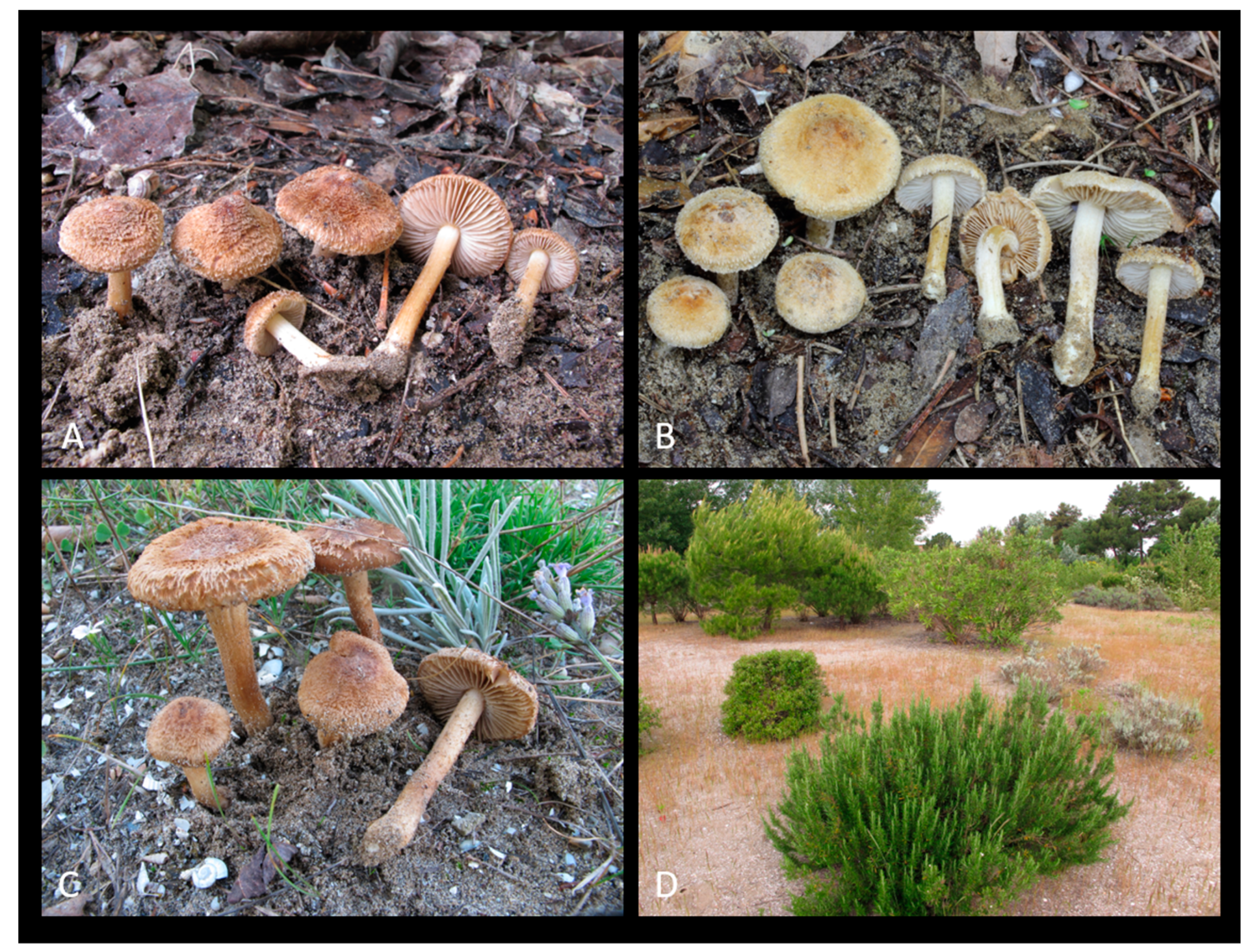
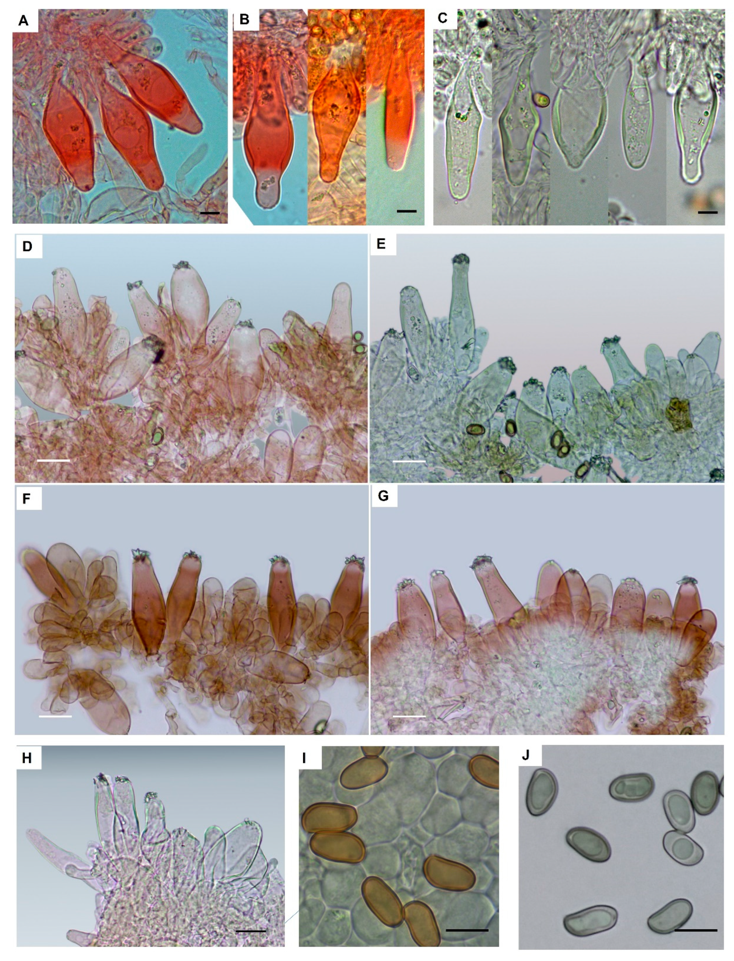
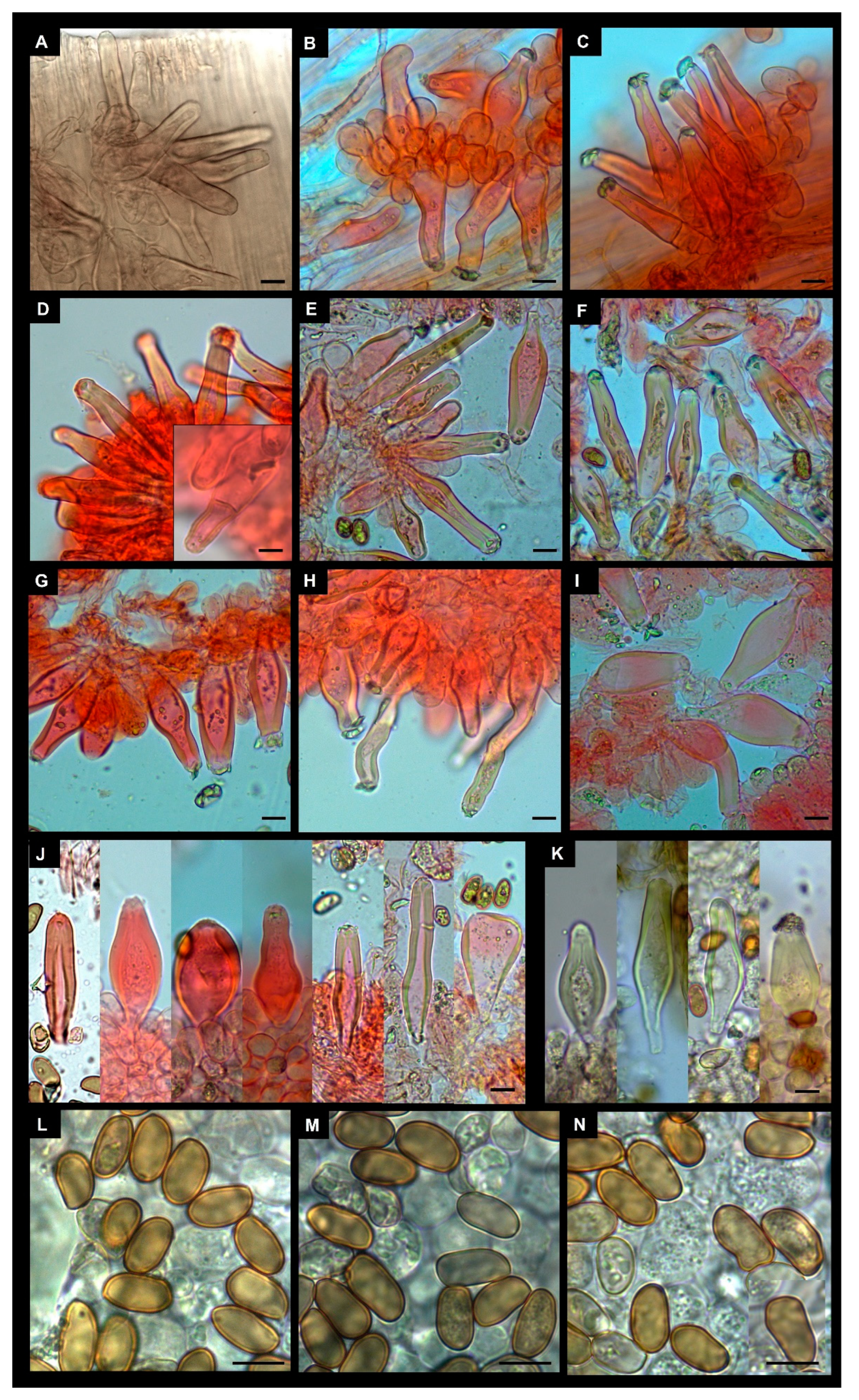
2.1. Morphology

The macroscopic descriptions were based on fresh material collected in Belgium, Germany, Italy, Slovenia, and Spain. Micromorphological features were observed on fresh and dried material; sections were rehydrated in ammonia 10%, water, or KOH (5% aqueous solution) and then mounted in aqueous Congo red, ammoniacal Congo red, aqueous ammonia, or KOH separately. The terminology follows Kuyper [12]. Cystidia were measured without crystals and basidia without sterigmata. A minimum of 30 basidiospores was measured for each collection. Basidiospore dimensions are expressed as (a) b–c–d (e), where (a) = minimum value, b = average—standard deviation, c = average, d = average + standard deviation, and (e) = maximum value. Q indicates the quotient of length and width of the basidiospores in side view.

2.2. Molecular Phylogeny

Genomic DNA was isolated from dry fragments of four freshly collected specimens using the CTAB procedure by Doyle and Doyle [16] or DNA extraction method using NaOH as employed by Dovana et al. [17]. The type specimens of I. chondrospora and I. vulpinella were extracted following the PTB DNA extraction protocol described by Stielow et al. [18] and that of I. immigrans with the Qiagen Puregene kit (Hilden, Germany) following the protocol given in Cripps et al. [7]. The nrITS region was amplified with primers ITS1F-ITS1-58SF/ ITS2-ITS4 [19,20,21], the nrLSU region with primers LR0R/LR5-LR7 [22] and both nrITS and nrLSU regions with ITS1F/TW13 [20]. The sequences obtained in this study were checked and assembled using Geneious vs. R 11.1.5 [23] or Sequencher vs. 4.9 (Genecodes, Ann Arbor, MI, USA) and compared to those available in GenBank (https://www.ncbi.nlm.nih.gov/genbank/) (accessed on 28 July 2022) and UNITE (accessed on 28 July 2022) databases (https://unite.ut.ee/) by using the BLASTN algorithm [24]. Our nrITS-nrLSU dataset includes Inocybe sequences selected based on (I) previously molecular studies focused on species belonging to section Marginatae subsections Praetervisae comprising I. diabolica and I. similis [25,26,27,28,29,30,31] and on species related to I. vulpinella sensu auct. [32] (sequences retrieved from clade: Inocybe_III, Inocybe_IV, and Inocybe_V), (II) the results in BLASTN, and (III) the result of nrITS-nrLSU maximum likelihood tree including 250 best BLAST hits of I. immigrans and I. similis (tree not shown). The list of sequences used in our dataset is given in Table 1.
Sequences were aligned using MAFFT vs. 7.450 [33] with E-INS-i parameters in Geneious vs. 11.1.5. The nrITS and nrLSU alignment were later automatically trimmed using TrimAl vs. 1.3 software [34] with the option “automated1”, as available in PHYLEMON 2.0 [35].
Maximum likelihood (ML) was inferred with IQ-TREE 2 [36]. The best models were selected using ModelFinder [37], and a total of 1000 ultrafast bootstrap (ufb) replicates were used [38]. The Bayesian inference (BI) was performed with MrBayes vs.3.2 [39]. Partition Finder 2 [40] was used to estimate the best partitioning schemes and evolution models for each subset with the Mrbayes option. Newly generated sequences were submitted to GenBank.

2.3. Statistical Analyses
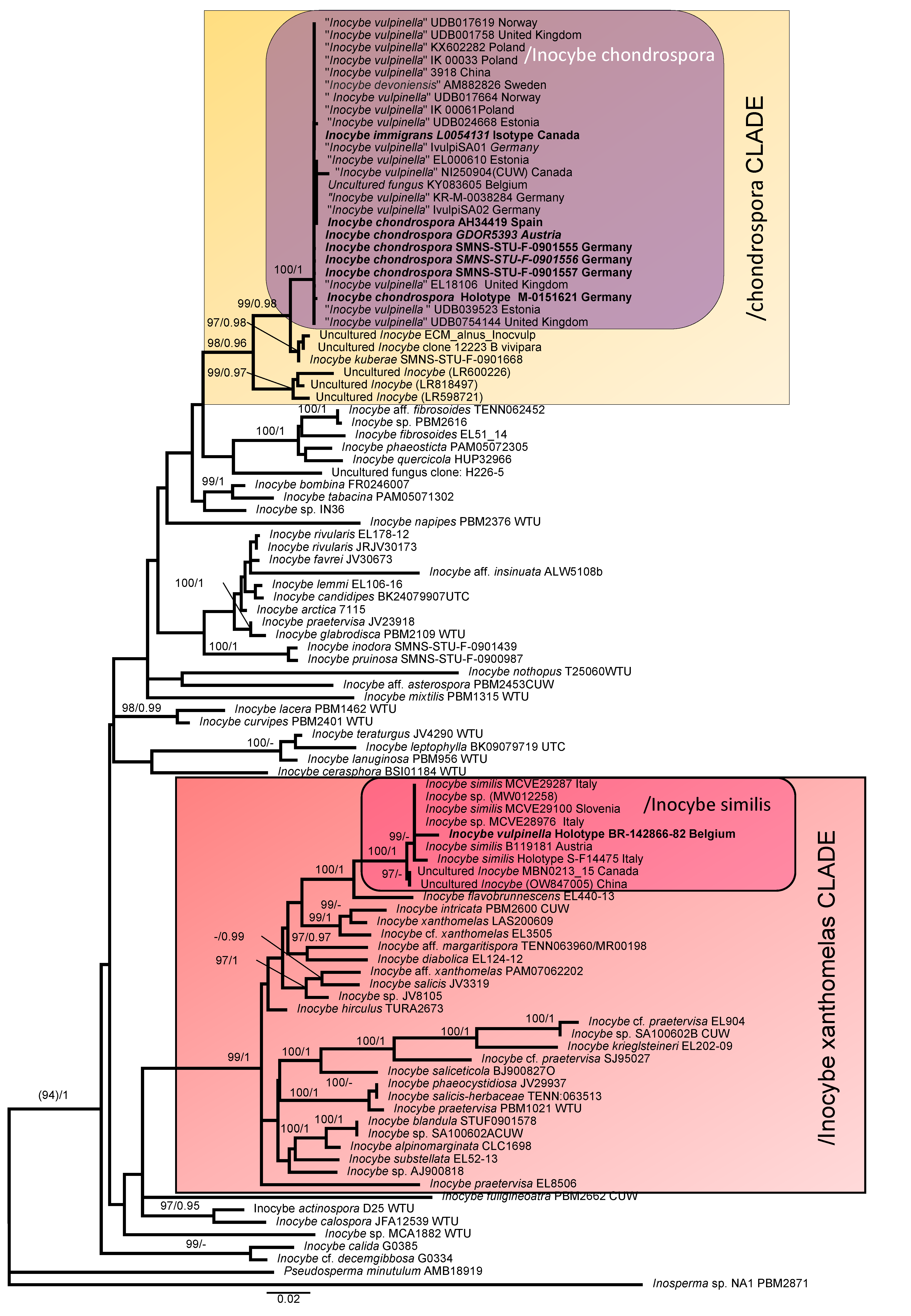
Length, width, and Q variability of the basidiospores of the collections studied are represented by using boxplots drawn in R vs. 3.1.2. The Shapiro–Wilk test was used to test the normality of the sporal morphology data. In the absence of a normal distribution, Kruskal–Wallis tests were performed in order to test at 0.05 significance level whether the length, width, and Q of the basidiospores of the different collections have identical data distributions. In the case of significant differences in the Kruskal–Wallis test, a subsequent Duncan–Waller post hoc test was conducted using the “agricolae” package implemented in R [41].

3. Results

3.1. Molecular Phylogeny

The nrITS and nrLSU DNA combined datasets comprised 1388 characteristics and consisted of 102 sequences. In Figure 1, we present the best tree from the ML analysis of the nrITS + nrLSU dataset, with bootstrap values ≥ 95% and posterior probabilities ≥ 0.95. We found two supported clades that included our sequences: /Inocybe chondropsora (BPP = 1; ufb = 100) in the/chondrospora clade and/Inocybe similis (BPP = 1; ufb = 100) in the /Inocybe xanthomelas clade (Figure 1). The sequences of the holotypes of I. chondrospora and the isotype of I. immigrans grouped in the same clade (/Inocybe chondrospora) with other 23 sequences, 18 retrieved from GenBank and UNITE databases (17 sequences identified as I. vulpinella and a sequence identified as Inocybe devoniensis), and 7 sequences newly generated in this study (Figure 1). Inocybe chondrospora is sister to Inocybe kuberae Bandini & B. Oertel (BPP = 0.98; ufb = 99), and both with other environmental sequences belong to the /chondrospora clade (BPP = 0.96; ufb = 98). Holotypes of I. similis and I. vulpinella grouped in the same clade (/Inocybe similis: BPP = 1; ufb = 100) with another seven sequences retrieved from GenBank./Inocybe similis is the sister clade of I. flavobrunnescens Esteve-Rav., G. Moreno & Bizio, and both are grouped within species belonging to Inocybe section Marginatae Kühner and I. diabolica Vauras in the/Inocybe xanthomelas clade.

3.2. Statistical Analysis
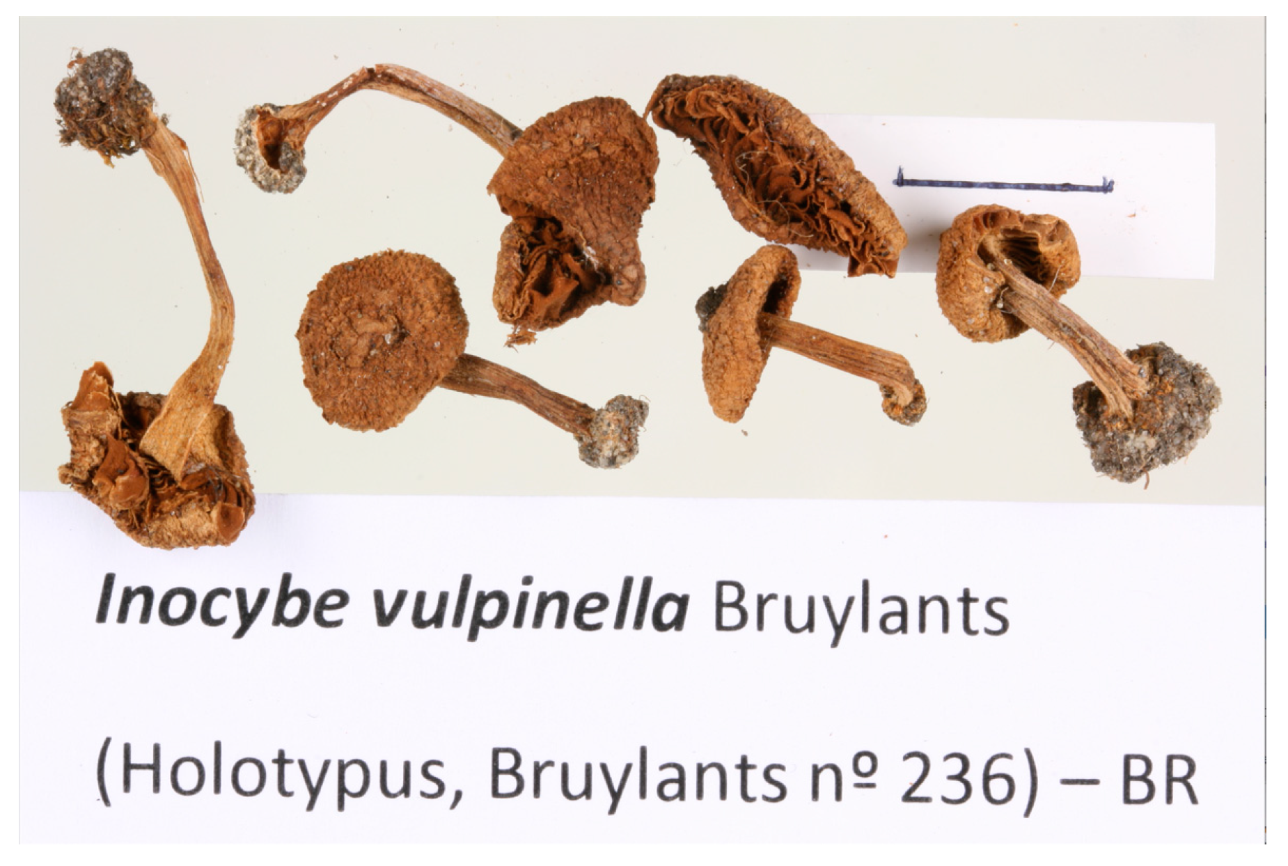
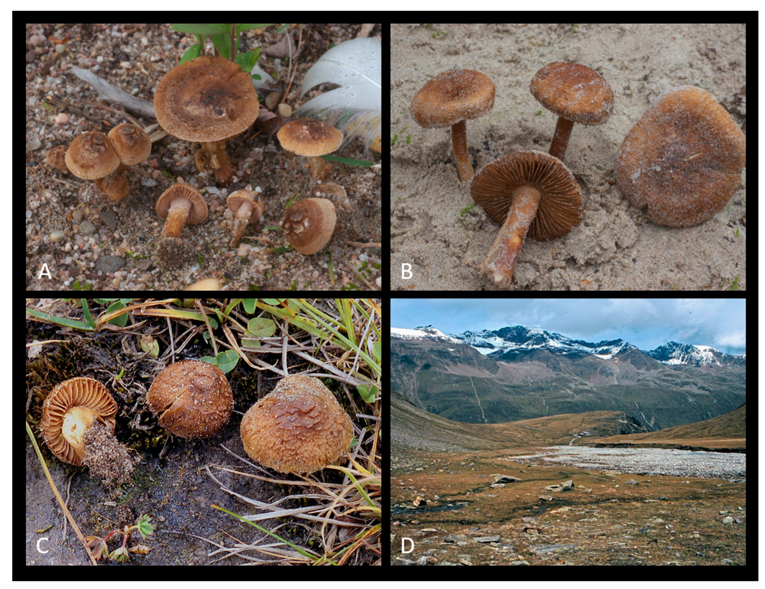
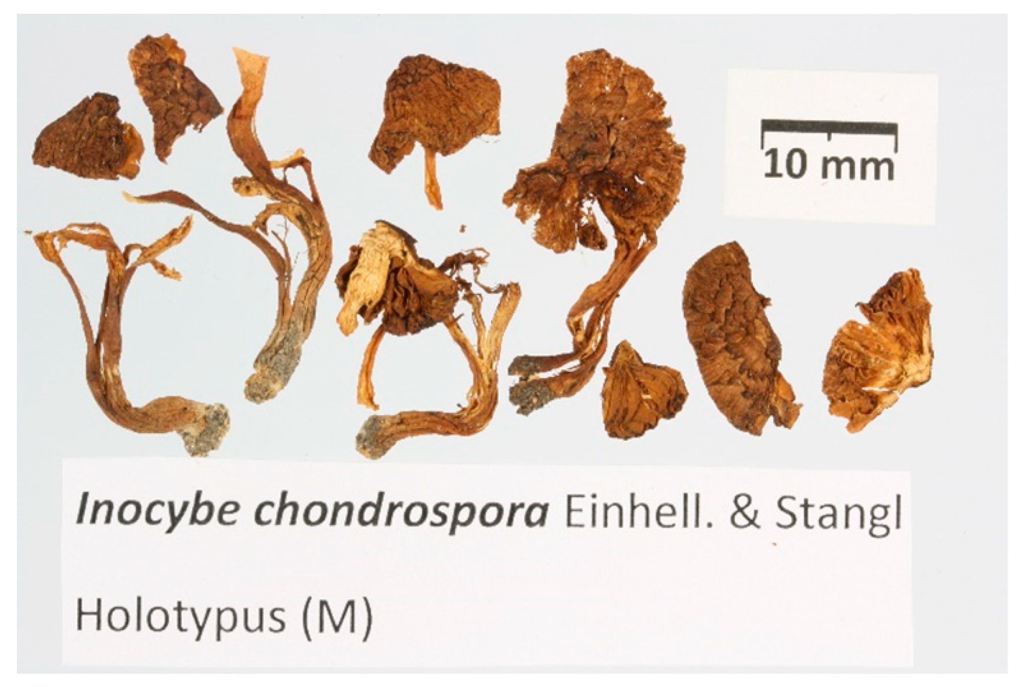
Analyses of “pooled data” (includes all the measurements of the different collections belonging to the same species) show a larger size of the basidiospores of I. chondrospora than those of I. similis, while the Q value does not show significant differences (Figure 2). The average basidiospore length of I. chondrospora is 13.6 μm, which is 9% greater than the basidiospore average length of I. similis (average value: 12.5 μm), while the width average of I. chondrospora is 7.8 μm, which is 9% greater than the basidiospore width of I. similis (average value: 7.1 μm). The length of the basidiospores of the isotype of I. immigrans shows no difference compared to the holotype of I. vulpinella, while it is greater than the holotype of I. chondrospora and the holotype of I. similis (Figure 2); five out of eight collections of I. similis (including the holotype of I. vulpinella) show a shorter basidiospore length than all the I. chondrospora collections, and six out of eight collections examined show smaller widths. Length, width, and Q of the individual collections have significant differences not only between the two species considered in this study but also show significant differences within the same species.

3.3. Taxonomy

Inocybe similis Bres., Ann. Mycol. 3 (2): 161 (1905) Figure 3, Figure 4 and Figure 5.
= Inocybe vulpinella Bruyl., Bull. trimest. soc. mycol. Fr 85: 341 (1970).
The description is based on nine recent collections and the holotypes of I. similis and I. vulpinella: Pileus 20–35 mm, initially campanulate or hemispherical, then convex, and finally plano-convex, with broad and low central umbo, pileus surface woolly-fibrillose, opaque, dry, from extremely fine to coarsely squamulose and often with quadrangular or fringed squamules or fibre bundles, at margin not rimulose and not striate, ochraceous, yellowish-brown to cinnamon-brown, generally lighter at the disk in younger specimens due to the presence of a greyish-white veil strongly sticking to the cuticle. Cortina not observed. Lamellae moderately crowded, adnate to emarginate, pale cream to ochraceous when young, finally brown to olivaceous-brown, edge fimbriate, whitish. Stipe 50–65 × 4–5 mm, central, firm, solid, generally inserted in the sandy substrate for about one-third of its length, equal to sub-bulbous at base, sometimes ending in a small napiform bulb, usually completely pruinose and sometimes somewhat sparsely in the lower part, probably due to abrasion through sand, sometimes longitudinally striate, whitish when young, often with the middle part ochraceous, then concolourous with pileus. Context fibrous, compact, whitish in the cap, whitish to yellowish in the stipe, smell absent.
Basidiospores (10.5)11.4–12.5–13.6(–16.0) × (6.0–)6.6–7.1–7.7(–9.0) μm, Q = (1.40–)1.61–1.76–1.90(–2.47); smooth, regular to sub-phaseoliform, sometimes with a largely obtuse apex but in several collections subangular. Basidia 30–45 × 10–18 μm, clavate, 4-spored. Pleurocystidia 45–110 × 14–28 μm, highly variable in shape, subcylindrical, narrowly utriform, fusiform or clavate, not markedly lageniform, thick-walled; wall up to 2.0–3.0 μm (rarely up to 5.5 μm in the apex) thick, occasionally very thin in the apical portion, bright yellow or hyaline in KOH solution, generally with crystals at apex. Cheilocystidia 50–70 × 17–25 μm, similar to pleurocystidia. Paracystidia up to 30 × 15 μm, abundant, clavate to pyriform. Caulocystidia present along the whole length of stipe or in upper half only, their distribution over the stipe surface very variable among collections, solitary or mainly in clusters, similar to pleurocystidia but more irregular in size and shape. Few cauloparacystidia present in lower part of stipe. Pileipellis a cutis, hyphae 7–5 m wide; terminal elements clavate; pigment ochraceous, parietal, and intracellular, sometimes slightly encrusting and parietal. Clamp-connections present in all tissues.
Habit, habitat, and distribution: in groups, growing on sandy soil with the presence of species belonging to the families Pinaceae and Salicaceae, present in Asia, Canada, and Europe.
Specimens examined:
Austria. Tyrol, Reutte, Forchach, bank of river Lech, in sand and gravel, with Salix sp., 11 September 2018, leg. D. Bandini (DB11-9-18-1); ibidem, Salix sp., 11 September 2018, leg. D. Bandini (DB11-9-18-4); Tyrol, Reutte, Rieden, Lechaue, ÖK25V 2215-West, alt. 870 m, Salix sp., Pinus sylvestris, 19 September 2018, leg. D. Bandini (DB19-9-18-23).
BELGIUM. Antwerp, in arena conchyliosa fluminis Escaut”, 18 June 1955, Julia Bruylants n°236 (holotype of I. vulpinella) (Figure 5). Measurements conducted on the holotype of I. vulpinella. Basidiospores: (11.3–)12.8–13.8–14.8(–15.7) × (6.8–)7.1–7.7–8.3(–9.0) μm, Q = (1.49–)1.63–1.79–1.95 (–2.09). Pleurocystidia: 47–66 × 14–22 μm (average data = 57 × 17 μm).
Germany. Bavaria, Schwaben, Ostallgäu, Füssen, TK25 8430/1, alt. 820 m, shore of river Lech with Salix sp., 22 September 2016, leg. D. Bandini (DB22-9-16-18); ibidem, at some distance from former location, 12 October 2016, leg. D. Bandini (DB12-10-16-19); ibidem, at some distance from the former location, alt. 800 m, Salix sp., 20 September 2018, leg. D. Bandini (DB20-9-18-9).
Italy, Trentino Alto Adige, Trento, place called “desert” by Bresadola, May 1900, G. Bresadola, Holotype F-S14475. Friuli Venezia Giulia, Grado, Municipal park consisting of a flat sand ground, in the presence of Populus tremula and Pinus halepensis, 13 July 2014, G. Ferisin, MCVE 28976; ibidem, 1 May 2014, MCVE29287.
Slovenia. Goriška, Tolmin, close to the levee of Isonzo river, on alluvial sandy-gravelly soil, near Populus tremula and Salix sp., 11 October 2008, leg. E. Bizio and A. Aiardi, MCVE29100.
Inocybe chondrospora Einhell. & Stangl, Z. Mykol. 45(2): 163 (1979) Figure 6, Figure 7 and Figure 8.
= Inocybe immigrans Malloch, Can. J. Bot. 60(1): 40 (1982).
The general description is based on five new collections, the holotype of I. chondrospora. and the isotype of I. immigrans.
Pileus 8–50 mm, initially campanulate or hemispherical, finally convex or plano-convex, sometimes becoming slightly depressed at centre, generally with a broad and prominent central umbo, initially radially fibrillose or rarely nearly glabrous, then conspicuously tomentose-scaly, finally often cracking toward the centre, with scales often becoming dark cinereous or silvery and appearing frosted or brownish orange to greyish or yellowish brown or dark brown when young and fresh, darkening in late stage of maturation through brown to dark brown or reddish brown, generally remarkably lighter at the margin in younger specimens but also sometimes observable in old samples. Lamellae more or less crowded, adnate to emarginate, pale cream, yellowish grey, greyish yellow to ochraceous when young, finally brown to brown olivaceous; edge fimbriate, whitish. Stipe 10–70 × 1.5–9 mm, central, firm, solid, equal or slightly swollen at base to sub-bulbous at base (up to 10 mm), yellowish white when young, then greyish orange to orange-grey, then from light brown to dark brown, at apex minutely flocculose and whitish, often whitish also at the base. Cortina not observed. Context fibrous, compact, whitish to yellowish, rarely darker in the pileus, lacking a distinctive odour.
Basidiospores (10.5–)12.3–13.6–14.9(–19.0) × (6.5–)7.3–7.8–8.3(–9.5) μm, Q = (1.40–)1.57–1.75–1.93(–2.22) variable in shape between the same basidioma and between the different collections, generally smooth, ellipsoid to oblong, rarely subcylindrical, seldom also sub-ovoid, sometimes slightly irregular to rarely slightly gibbous. Basidia 25–50 × 8–18 μm, clavate, four-spored. Pleurocystidia 38–80 × 11–28 μm, highly variable in shape, subcylindrical, narrowly utriform, fusiform, ventricose or clavate, thick-walled up to 6 μm (sometimes filling the whole lumen of the upper, narrow part of the cystidia), bright yellow or light yellow in KOH solution, generally crystalliferous at apex. Cheilocystidia similar to pleurocystidia but more variable in shape even in the same basidioma (see Figure 7E–I), sometimes fusiform with long narrow necks, scattered to abundant, sometimes septate, thick-walled; wall up to 6 μm thick, often of equal thickness over the whole length of the cystidia. Caulocystidia present along the whole length of the stipe, less abundant in the lower half, partly similar to cheilocystidia, partly more irregular in shape (Figure 7A–C), thick-walled with walls up to 5 μm thick, mixed with cauloparacystidia. Pileipellis a cutis, 8–15 µm wide, with clavate terminal elements, pigment ochraceous, parietal, and intracellular, sometimes indistinctly encrusted. Clamp connections present.
Habit, habitat, and distribution: in groups, growing on sandy soil with presence of species belonging to the families Betulaceae, Orchidaceae, Pinaceae, and Salicaceae, present in Asia, Canada, and Europe.
Specimens examined:
Austria. Similaun, Rotmoos, sandy soil in association with Salix herbacea, leg. E. Bizio, GDOR5393; CANADA. Ontario, Hastings Co., Faraday Township, Bow Lake, Madawaska Mines, 16 June 1979, leg. D. Malloch (isotype of I. immigrans) (L0054131). Measurements conducted on the isotype of I. immigrans. Basidiospores: (11.5–)13.0–14.0–15.1(–1.8) × (7.0–)7.7–8.2–8.6(–9.2) μm, Q = (1.44–)1.59–1.72–1.85 (–1.97). Pleurocystidia: 38–65 × 14–19 μm (Average data = 52 × 16 μm).
Germany. Bavaria, Murnauer Moor, MTB 8333, 10 Km west, 31 May 1966, leg. A. Einhellinger M-0151821 (holotype of I. chondrospora). Measurements conducted on the holotype of I. chondrospora. Basidiospores: (11.5–)12.5–13.5–14.5(–16.0) × (7.0–)7.5–7.9–8.4(–9.0) μm; Q = (1.52–)1.61–1.70–1.79 (–1.95). Pleurocystidia: 45–68 × 14–22 μm (average data = 55 × 17 μm). Rheinland-Pfalz, Rhein-Pfalz-Kreis, Rheinauen, Altrip, TK25 6516/4, alt. 90 m, sandy soil with Salix sp., Populus tremula, 4 May 2013, leg. D. Bandini & B. Oertel (DB4-5-13-2); Schleswig-Holstein, Ostholstein, Malente, Sieversdorfer Kiesgrube, TK25 1729/3, alt. 50 m, sandy soil with Populus tremula; Salix sp., Pinus sylvestris, Betula sp., 26 September 2017, leg. G. Schmidt-Stohn & B. Oertel (DB26-9-17-7b).
Netherlands, Friesland, Ameland, Hollum, alt. 0 m, white dunes with Salix repens, 2 September 2012, leg. D. Bandini (DB2-9-12-4); SPAIN. Cantabria, Liencres, Piélagos, 10 October 2004, littoral sand dunes with presence of Salix cf. repens, leg. F. Prieto & M.A. González, AH 34419 (in [42], as I. vulpinella).

4. Discussion

The phylogenetic analyses of the ITS and LSU data from type collections in the I. similis group showed the conspecificity between I. similis and I. vulpinella and confirmed that I. chondrospora and I. immigrans belong to a single taxon not related to I. vulpinella.
Several authors have considered I. similis and I. vulpinella as independent species that are separable based on macroscopic and microscopic characteristics [12,43]. Both Kuyper [12] and Stangl [43] placed I. similis into the “supersection cortinatae” and I. vulpinella within the “supersection marginatae” based on the presence/absence of cortina and distribution of caulocystidia. However, Kuyper [12] did not point out “Marginatae” and “Cortinatae” as formal taxonomic-level units. Neither were the presence of a cortina nor the arrangement of the caulocystidia on the stipe mentioned by Bresadola [10] in his original description of I. similis; however, in more recent morphological classifications, these are two fundamental characteristics [12,43].
Moreover, differently from Kuyper’s opinion, those I. similis collections examined by Dovana et al. [11] did not have a cortina and showed a variable distribution of the caulocystidia on the stipe in the different collections. This last characteristic is often influenced by the development of the velipellis. Kuyper stated that I. vulpinella is morphologically closest to I. similis, although it must belong to a different “supersection”. He also noted that the former species differs from I. similis in that its basidiospores show an applanate apex and thicker-walled pleurocystidia.
The molecular analysis suggested that these differences represent variability within the same species. The basidiospore measurements reported in Kuyper for I. vulpinella (12.0–18.0 × 7.0–9.0 μm) are larger than those of I. similis (11.5–16.0 × 7.0–8.5 μm), even though this has not been considered by the same author as a characteristic to distinguish the two species from each other [12]. Our statistical analyses of the basidiospore measurements showed that the basidiospores of the holotype of I. vulpinella are significantly larger than those of the holotype of I. similis and the other I. similis collections (Figure 2). In this study, we did not consider I. vulpinella var. fuscolamellata Bon, which is a taxon described by Marcel Bon and considered a synonym of I. vulpinella [12]; however, the possible conspecificity of this taxon with I. similis or I. chondrospora would not have priority at the species rank.
The molecular analysis supported the synonymy of I. chondrospora and I. immigrans, but unlike what Kuyper [12] considered, our phylogenetic analyses place both in a lineage independent and distant from I. similis/vulpinella.
Inocybe chondrospora is found to be the oldest name for this taxon, as it was published and used in 1979, three years before I. immigrans. Sixteen sequences that grouped in the /chondrospora clade were erroneously identified as I. vulpinella. This fact is not surprising considering that these three species have been accepted as synonymous by most mycologists since Kuyper [12].
In the same clade, there is a sequence from Sweden (GenBank: AM882826 [25]) named “Inocybe devoniensis”. Inocybe devoniensis P.D. Orton [44] shares some features (for example, large basidiospores of comparable size) with I. chondrospora, but it differs mainly in the presence of (sub)rimose, smooth pileus, sub-lageniform cystidia with much thinner wall, typically ellipsoidal basidiospores, and caulocystidia only in the upper part of the stipe [12,44]. The slightly angulose outline of the basidiospores has not been observed in the study of I. devoniensis either. These differences were also confirmed in the microscopic analysis conducted on the I. devoniensis holotype by one of us (Esteve-Raventós, unpublished data).
Subsequent molecular studies on the holotype of I. devoniensis will be required to definitively clarify its position; if it is found to be the same taxon as I. chondrospora, the name I. devoniensis would have priority, but this possible conspecificity is unlikely.
In the microscopy table attached to the protologue of I. chondrospora, a caulocystidium with an intermediate septum is presented, although this characteristic is not reported in the text. Septa within cheilocystidia and pleurocystidia are not unusual in I. chondrospora (see Figure 7). Although it does not appear to be a constant characteristic, this type of septum has not been observed in I. similis and could be an additional characteristic to separate the two species.
A remarkable feature of I. chondrospora is the great variability of the basidiospore dimensions both within the individual basidiomes and among the different collections. In the original description of I. chondrospora, Einhellinger and Stangl reported values of 11–19 × 8–10 µm for the holotype [14], and Malloch, when describing I. immigrans, indicated pooled data values of (8.9–)10.0–16.0(–20.0) × (6.0–)6.5–8.5(–9.8) µm, and for single collections, the average basidiospore length was reported as 10.9–14.9 µm [15]. Summarising, the differences between I. similis and I. chondrospora are not always well defined in the single collections, and it is also possible to find basidiomata with intermediate characteristics. In most of the studied collections, the basidiospores of I. chondrospora have larger dimensions than those of I. similis even if the basidiospore dimensions of some collections (e.g., basidiospores of I. vulpinella holotype) are similar to those of I. immigrans. In conclusion, I. chondrospora and I. similis represent two species with similar variability in morphological characteristics and distribution areas (Asia, Canada, and Europe). They are also associated with the same plants, mainly in habitats where Pinaceae and Salicaceae are present, although I. chondrospora can also associate with Liparis loeselii (Orchidaceae) and probably some Betulaceae (see Table S1). In typical collections of both species, (I) the pileus surface is rougher scaly with larger fibre bundles in I. similis compared to I. chondrospora; (II) usually in I. similis, there is no such colour contrast in the pileus, as is often observed with I. chondrospora; (III) generally, there are smaller basidiospores in I. similis (11.4–12.5–13.6 × 6.6–7.1–7.7 μm) versus I. chondrospora (12.3–13.6–14.9 × 7.3–7.8–8.3 μm); (IV) there are longer hymenial cystidia in I. similis (pleurocystidia 45–110 × 14–28 μm) versus I. chondrospora (pleurocystidia 38–80 × 11–28 μm) (average values of pleurocystidia in German collections: 67 × 19 µm in I. similis vs. 55 × 16 µm in I. chondrospora), and (V) there are thicker-walled cystidia in I. chondrospora.
In the case of non-typical collections (for example, in the holotype of I. vulpinella), which have intermediate characteristics, the best way to tell the species apart is to use DNA data even considering that they can share a similar habitat.

Supplementary Materials

The following supporting information can be downloaded at: https://www.mdpi.com/article/10.3390/jof9060679/s1, Table S1: Ecological data referring to host plants of I. chondrospora and I. similis.

Author Contributions

Conceptualization, F.D., D.B. and F.E.-R.; field sampling, D.B., E.B., G.F. and F.E.-R.; type studies, D.B. and F.E.-R.; molecular data, F.D, I.O. and U.E.; phylogenetic data analysis, F.D.; statistical analysis, F.D.; microscopic analyses, F.D, D.B., E.B., G.F. and F.E.-R.; writing—original draft preparation, F.D.; writing—review and editing, all authors. All authors have read and agreed to the published version of the manuscript.

Funding

Sequencing of type specimens of I. similis, I. vulpinella, and I. chondrospora was financially supported by grant CGL2017-86450-P (Ministerio de Ciencia, Innovación y Universidades, Spain; principal investigators F. Esteve-Raventós and G. Moreno).

Institutional Review Board Statement

Not applicable.

Informed Consent Statement

Not applicable.

Data Availability Statement

Publicly available datasets were analyzed in this study. These data can be found here: https://www.ncbi.nlm.nih.gov.

Acknowledgments

We thank three anonymous reviewers for the suggestions provided.

Conflicts of Interest

The authors declare no conflict of interest. The funders had no role in the design of the study; in the collection, analyses, or interpretation of data; in the writing of the manuscript; or in the decision to publish the results.

References

  1. Matheny, P.B.; Hobbs, A.M.; Esteve-Raventós, F. Genera of Inocybaceae: New skin for the old ceremony. Mycologia 2019, 112, 83–120. [Google Scholar] [CrossRef]
  2. Lurie, Y.; Wasser, S.P.; Taha, M.; Shehade, H.; Nijim, J.; Hoffmann, Y.; Basis, F.; Vardi, M.; Lavon, O.; Suaed, S.; et al. Mushroom poisoning from species of genus Inocybe (fiber head mushroom): A case series with exact species identification. Clin. Toxicol. 2009, 47, 562–565. [Google Scholar] [CrossRef]
  3. Patocka, J.; Wu, R.; Nepovimova, E.; Valis, M.; Wu, W.; Kuca, K. Chemistry and Toxicology of Major Bioactive Substances in Inocybe Mushrooms. Int. J. Mol. Sci. 2021, 22, 2218. [Google Scholar] [CrossRef]
  4. Kosentka, P.; Sprague, S.L.; Ryberg, M.; Gartz, J.; May, A.L.; Campagna, S.R.; Matheny, P.B. Evolution of the toxins muscarine and psilocybin in a family of mushroom-forming fungi. PLoS ONE 2013, 8, e64646. [Google Scholar] [CrossRef] [PubMed] [Green Version]
  5. Vizzini, A.; Della Maggiora, M.; Tolaini, F.; Ercole, E. A new cryptic species in the genus Tubariomyces (Inocybaceae, Agaricales). Mycol. Prog. 2013, 12, 375–381. [Google Scholar] [CrossRef]
  6. Matheny, P.B.; Bougher, N.L. Fungi of Australia Inocybaceae; ABRS & CSIRO Publishing: Canberra/Melbourne, Australia, 2017; pp. 1–592. [Google Scholar]
  7. Cripps, C.L.; Larsson, E.; Vauras, J. Nodulose-spored Inocybe from the Rocky Mountain alpine zone molecularly linked to European and type specimens. Mycologia 2019, 112, 133–153. [Google Scholar] [CrossRef]
  8. Bandini, D.; Oertel, B.; Eberhardt, U. A fresh outlook on the smooth-spored species of Inocybe: Type studies and 18 new species. Mycol. Prog. 2021, 20, 1019–1114. [Google Scholar] [CrossRef]
  9. Einhellinger, A. Problems of Inocybe similis Bres., vulpinella Bruylants (incl. Inocybe chondrospora Einh. et Stangl) and its variety fuscolamellata Bon. Z. Mykol. 1994, 60, 365–372. [Google Scholar]
  10. Bresadola, J. Hymenomycetes novi vel minus cogniti. Ann. Mycol. 1905, 3, 159–164. [Google Scholar]
  11. Dovana, F.; Ferisin, G.; Bizio, E.; Bandini, D.; Olariaga, I.; Esteve-Raventós, F. A morphological and phylogenetic characterisation of Inocybe similis (Agaricales, Inocybaceae), a rare species described by Bresadola in 1905. Phytotaxa 2020, 474, 71–80. [Google Scholar] [CrossRef]
  12. Kuyper, T.W. A revision of the genus Inocybe in Europe 1. Subgenus Inosperma and the smooth-spored species of subgenus Inocybe. Pers. Suppl. 1986, 3, 1–247. [Google Scholar]
  13. Bruylants, J. Inocybe vulpinella sp. nov. Bull. Soc. Mycol. Fr. 1979, 85, 340–344. [Google Scholar]
  14. Einhellinger, A.; Stangl, J. Inocybe chondrospora Einhellinger & Stangl spec. nov. Z Mykol. 1979, 45, 163–165. [Google Scholar]
  15. Malloch, D. An undescribed species of Inocybe from mine wastes in Ontario. Can. J. Bot. 1982, 60, 40–45. [Google Scholar] [CrossRef]
  16. Doyle, J.J.; Doyle, J.L. A rapid DNA isolation procedure for small quantities of fresh leaf material. Phytochem. Bull. 1987, 19, 11–15. [Google Scholar]
  17. Dovana, F.; Contu, M.; Angeli, P.; Brandi, A.; Mucciarelli, M. Leucoagaricus ariminensis sp. nov., a lilac species from Italy. Mycotaxon 2017, 132, 205–216. [Google Scholar] [CrossRef]
  18. Stielow, B.; Hensel, G.; Strobelt, D.; Makonde, H.M.; Rohde, M.; Dijksterhuis, J.; Klenk, H.P.; Göker, M. Hoffmannoscypha, a novel genus of brightly coloured, cupulate Pyronemataceae closely related to Tricharina and Geopora. Mycol. Prog. 2013, 12, 675–686. [Google Scholar] [CrossRef]
  19. Gardes, M.; Bruns, T.D. ITS primers with enhanced specificity for basidiomycetes—Application to the identification of mycorrhizae and rusts. Mol. Ecol. 1993, 2, 113–118. [Google Scholar] [CrossRef]
  20. White, T.J.; Bruns, T.; Lee, S.; Taylor, J. Amplification and direct sequencing of fungal ribosomal RNA genes for phylogenetics. In PCR Protocols; Innis, M.A., Gelfand, D.H., Sninsky, J.J., White, T.J., Eds.; Academic Press: San Diego, CA, USA, 1990; pp. 315–322. [Google Scholar]
  21. Tedersoo, L.; Mett, M.; Ishida, T.A.; Bahram, M. Phylogenetic relationships among host plants explain differences in fungal species richness and community composition in ectomycorrhizal symbiosis. New Phytol. 2013, 199, 822–831. [Google Scholar] [CrossRef] [PubMed]
  22. Vilgalys, R.; Hester, M. Rapid genetic identification and mapping of enzymatically amplified ribosomal DNA from several Cryptococcus species. J. Bacteriol. 1990, 172, 4239–4246. [Google Scholar] [CrossRef] [Green Version]
  23. Kearse, M.; Moir, R.; Wilson, A.; Stones-Havas, S.; Cheung, M.; Sturrock, S.; Buxton, S.; Cooper, A.; Markowitz, S.; Duran, C.; et al. Geneious Basic: An integrated and extendable desktop software platform for the organization and analysis of sequence data. Bioinformatics 2012, 28, 1647–1649. [Google Scholar] [CrossRef] [Green Version]
  24. Altschul, S.F.; Gish, W.; Miller, W.; Myers, E.W.; Lipman, D.J. Basic local alignment search tool. J. Mol. Biol. 1990, 215, 403–410. [Google Scholar] [CrossRef]
  25. Ryberg, M.; Nilsson, R.H.; Kristiansson, E.; Töpel, M.; Jacobsson, S.; Larsson, E. Mining metadata from unidentified ITS sequences in GenBank: A case study in Inocybe (Basidiomycota). BMC Evol. Biol. 2008, 8, 50. [Google Scholar] [CrossRef] [PubMed] [Green Version]
  26. Vauras, J.; Kokkonen, K. Finnish records on the genus Inocybe. The new species Inocybe saliceticola. Karstenia 2009, 48, 57–67. [Google Scholar] [CrossRef] [Green Version]
  27. Osmundson, T.W.; Robert, V.A.; Schoch, C.L.; Baker, L.J.; Smith, A.; Robich, G.; Mizzan, L.; Garbelotto, M.M. Filling gaps in biodiversity knowledge for macrofungi: Contributions and assessment of an herbarium collection DNA barcode sequencing project. PLoS ONE 2013, 8, E62419. [Google Scholar] [CrossRef] [Green Version]
  28. Esteve-Raventós, F.; Moreno, G.; Bizio, E.; Alvarado, P. Inocybe flavobrunnescens, a new species in section Marginatae. Mycol. Prog. 2015, 14, 14. [Google Scholar] [CrossRef]
  29. Vauras, J.; Larsson, E. Inocybe caprimulgi and I. lacunarum, two new nodulose-spored species from Fennoscandia. Karstenia 2015, 55, 14. [Google Scholar] [CrossRef] [Green Version]
  30. Esteve-Raventós, F.; Alvarado, P.; Olariaga, I. Unraveling the Inocybe praetervisa group through type studies and ITS data: Inocybe praetervisoides sp. nov. from the Mediterranean region. Mycologia 2016, 108, 123–134. [Google Scholar] [CrossRef] [Green Version]
  31. Bandini, D.; Oertel, B.; Moreau, P.-A.; Thines, M.; Ploch, S. Three new hygrophilous species of Inocybe, subgenus Inocybe. Mycol. Prog. 2019, 18, 1101–1119. [Google Scholar] [CrossRef]
  32. Kropp, B.R.; Matheny, P.B.; Nanagyulyan, S.G. Phylogenetic taxonomy of the Inocybe splendens group and evolution of supersection “Marginatae”. Mycologia 2010, 102, 560–573. Available online: http://www.jstor.org/stable/27811069 (accessed on 20 January 2017). [CrossRef] [Green Version]
  33. Katoh, K.; Toh, H. Recent developments in the MAFFT multiple sequence alignment program. Brief. Bioinform. 2008, 9, 286–298. [Google Scholar] [CrossRef] [Green Version]
  34. Capella-Gutiérrez, S.; Silla-Martínez, J.M.; Gabaldón, T. trimAl: A tool for automated alignment trimming in large-scale phylogenetic analyses. Bioinformation 2009, 25, 1972–1973. [Google Scholar] [CrossRef] [PubMed] [Green Version]
  35. Sánchez, R.; Serra, F.; Tárraga, J.; Medina, I.; Carbonell, J.; Pulido, L.; de María, A.; Capella-Gutierrez, S.; Huerta-Cepas, J.; Gabaldón, T.; et al. Phylemon 2.0: A suite of web-tools for molecular evolution, phylogenetics, phylogenomics and hypotheses testing. Nucl. Acids Res. 2011, 39, W470–W474. [Google Scholar] [CrossRef] [Green Version]
  36. Minh, B.Q.; Schmidt, H.A.; Chernomor, O.; Schrempf, D.; Woodhams, M.D.; von Haeseler, A.; Lanfear, R. IQ-TREE 2: New models and efficient methods for phylogenetic inference in the genomic era. Mol. Biol. Evol. 2020, 37, 1530–1534. [Google Scholar] [CrossRef] [PubMed] [Green Version]
  37. Kalyaanamoorthy, S.; Minh, B.Q.; Wong, T.F.K.; von Haeseler, A.; Jermiin, L.S. ModelFinder: Fast model selection for accurate phylogenetic estimates. Nat. Methods 2015, 14, 587–589. [Google Scholar] [CrossRef] [PubMed] [Green Version]
  38. Hoang, D.T.; Chernomor, O.; von Haeseler, A.; Minh, B.Q.; Le Vinh, S. UFBoot2: Improving the ultrafast bootstrap approximation. Mol. Biol. Evol. 2018, 35, 518–522. [Google Scholar] [CrossRef]
  39. Ronquist, F.; Huelsenbeck, J.P.; Teslenko, M. MrBayes Version 3.2 Manual: Tutorials and Model Summaries. 2011. Available online: Mrbayes.sourceforge.net/mb3.2_manual.pdf (accessed on 19 October 2021).
  40. Lanfear, R.; Frandsen, P.B.; Wright, A.M.; Senfeld, T.; Calcott, B. PartitionFinder 2: New Methods for Selecting Partitioned Models of Evolution for Molecular and Morphological Phylogenetic Analyses. Mol. Biol. Evol. 2016, 34, 772–773. [Google Scholar] [CrossRef] [PubMed] [Green Version]
  41. Mendiburu, F.D.; Yaseen, M. Agricolae: Statistical Procedures for Agricultural Research. R Package Version 1.4.0. Available online: https://myaseen208.github.io/agricolae/https://cran.r-project.org/package=agricolae (accessed on 10 December 2020).
  42. Esteve-Raventós, F. Inocybe vulpinella Bruyl. (Inocybaceae, Agaricomycetes), primer registro para la Península Ibérica. Zizak 2008, 5, 27–33. [Google Scholar]
  43. Stangl, J. Die Gattung Inocybe in Bayern. Hoppea 1989, 46, 1–409. [Google Scholar]
  44. Orton, P.D. New check list of British agarics and boleti. Part III. Notes on genera and species in the list. Trans. Br. Mycol. Soc. 1960, 43, 274. [Google Scholar] [CrossRef]
Figure 1. Best tree from the ML analysis of the nrITS + nrLSU dataset. Bootstrap values (ufb) ≥ 95% and posterior probabilities (BPP) ≥ 0.95 are indicated on or below the branches. The GenBank code has been added in brackets when the collection code is not available.
Jof 09 00679 g001
Figure 2. Boxplots comparing length (FIG2A), width (FIG2B), and Q value (FIG2C) of I. chondrospora basidiospores (A, I. immigrans isotype; B, I. chondrospora holotype; C, DB4-5-13-2; D, DB26-9-17-7b; E, DB2-9-12-4; F, AH34419; G, GDOR5393; P_i, pooled value of I. chondrospora) and I. similis (H, I. similis holotype; I, I. vulpinella holotype; J, MCVE29100; K, BAN11-9-18-1; L, BAN1718; M, BAN2609; N, MCVE28976; O, MCVE29287; Q_s, pooled value of I. similis) basidiospores. The small letters above the boxes indicate significant differences between collections according to Duncan–Waller post hoc test p < 0.05.
Jof 09 00679 g002
Figure 3. (AC) Fresh basidiomata of Inocybe similis ((A) MCVE29287; (B) MCVE 28976; (C) MCVE29287). (D) Italy, Grado, where MCVE28976 and MCVE29287 specimens were collected.
Jof 09 00679 g003
Figure 4. Inocybe similis. (A,DG) Cheilocystidia ((A) MCVE29100; (D,E) MCVE28976; (F,G) MCVE29287). (B,C) Pleurocystidia (MCVE29100). (H) Caulocystidia (MCVE29287). (I,J) Basidiospores ((I) MCVE29100; (J) MCVE29287). Scale bar: (AC); (I,J) 10 µm; (DG) 20 µm.
Jof 09 00679 g004
Figure 5. Holotype of Inocybe vulpinella. Scale bar: 1 cm.
Jof 09 00679 g005
Figure 6. (AC) Fresh basidiomata of Inocybe chondrospora ((A) DB4-5-13-; (B) DB-2-9-12-4; (C) GDOR5393); (D) Austria, where the GDOR5393 specimen was collected.
Jof 09 00679 g006
Figure 7. Inocybe chondrospora. (AC) Caulocystidia (AH 34419). (DI) Cheilocystidia ((D) GDOR5393; (EI) AH 34419). (J,K) Pleurocystidia (AH 34419 and GDOR5393). (LN) Basidiospores ((L,M) GDOR5393; (N) AH 34419). Scale bars: 10 μm.
Jof 09 00679 g007
Figure 8. Holotype of Inocybe chondrospora. Scale bar: 1 cm.
Jof 09 00679 g008
Table 1. Inocybe collections used in phylogenetic analyses. Sequences in bold and with * were newly generated for this study, ** sequences belonging to /Inocybe chondrospora, and *** sequences belonging to /Inocybe similis.
TaxonCollectionCountryGenBank/UNITE
ITSnLSU
Inocybe actinosporaD25 WTUArgentinaXAY380363
Inocybe alpinomarginataCLC1698USA: ColoradoMK153647MK153647
Inocybe arctica7115SwedenKY033839KY033839
Inocybe aff. asterosporaPBM2453 CUWUnknownXAY702015
Inocybe blandulaSMNS-STU-F-0901578AustriaMZ144124MZ144124
Inocybe bombinaFR 0246007GermanyNR_173842X
Inocybe calidaG0385HungaryMK278241MK278241
Inocybe calosporaJFA12539WTUSwedenXAY038313
Inocybe candidipesBK24079907 UTCUSA: ArizonaXAY239019
Inocybe cerasphoraBSI01184 WTUChileXAY380370
/Inocybe chondrospora **
Inocybe vulpinella **3918ChinaKR082885X
Inocybe chondrospora **AH34419SpainOR088565 *X
Inocybe vulpinella **EL000610EstoniaAM882825X
Inocybe vulpinella **EL18106United KingdomFN550898FN550898
Inocybe chondrospora **GDOR5393AustriaOR088564 *X
Inocybe vulpinella **IK-00033PolandKX602283X
Inocybe vulpinella **IK-00034PolandKX602282X
Inocybe vulpinella **IK-00061PolandKX602284X
Inocybe vulpinella **IvulpiSA01GermanyMH389771X
Inocybe vulpinella **IvulpiSA02GermanyMH389774X
Inocybe vulpinella **KR-M-0038284GermanyMK929256X
Inocybe immigrans **L0054131 (Isotype)CanadaMW539061 *
Inocybe chondrospora **M-0151621 (Holotype)GermanyOR098441 *OR098446 *
Inocybe vulpinella **NI250904 (CUW)Canada: Ontario EU307834
Uncultured fungus **OTU_127BelgiumKY083605X
Uncultured fungus **saf_F1979Italy: Aosta ValleyMW164608X
Inocybe chondrospora **SMNS-STU-F-0901555GermanyMW539060 *MW539060 *
Inocybe chondrospora **SMNS-STU-F-0901556NetherlandsMW539058 *MW539058 *
Inocybe chondrospora **SMNS-STU-F-0901557GermanyMW539059 *MW539059 *
Inocybe devoniensis **TAA17205SwedenAM882826X
Inocybe vulpinella **UDB001758United KingdomUDB001758X
Inocybe vulpinella **UDB017619NorwayUDB017619X
Inocybe vulpinella **UDB024668EstoniaUDB024668X
Inocybe vulpinella **UDB039523EstoniaUDB039523X
Inocybe vulpinella **UDB0754144United KingdomUDB0754144X
Inocybe curvipesPBM2401 WTUUSA: WashingtonXAY239022
Inocybe cf. decemgibbosaG0334HungaryMK278224MK278224
Inocybe diabolicaEL124-12NorwayKT958913KT958913
Inocybe favreiJV30673NorwayKY033798KY033798
Inocybe aff. fibrosoidesTENN062452USA: TennesseeMT237492MT228850
Inocybe fibrosoidesEL51_14SwedenKY033846KY033846
Inocybe flavobrunnescensEL440-13SpainMK153641MK153641
Inocybe fuligineoatraPBM2662 CUWUSA: TennesseeEU523589EU307831
Inocybe glabrodiscaPBM 2109 (WTU)USA: WashingtonAY239023AY239023
Inocybe glabrodiscaPBM2109 WTUUSA: WashingtonXAY239023
Inocybe hirculusTURA2673FinlandMT241840MT241840
Inocybe inodoraSMNS-STU-F-0901439AustriaMT101875MT101875
Inocybe aff. insinuataALW5108b (WTU)USA: LouisianaXJN975030
Inocybe intricataPBM2600 CUWUSA: TennesseeEU523561EU307835
Inocybe krieglsteineriEL202-09SwedenKT958916KT958916
Inocybe kuberaeSMNS-STU-F-0901668GermanyON003427ON003427
Inocybe laceraPBM1462 WTUUSA: WashingtonXAY038318
Inocybe lanuginosaPBM956 WTUUSA: WashingtonXAY038319
Inocybe lemmiEL106-16SwedenMG574395MG574395
Inocybe leptophyllaBK09079719 UTCUSA: UtahXAY038320
Inocybe aff. margaritisporaTENN:063960/MR00198USA: TennesseeKP308775JN974998
Inocybe mixtilis ceskaePBM1315 WTUUSA: WashingtonXAY380387
Inocybe napipesPBM2376 WTUNorwayXAY239024
Inocybe nothopusTrappe 25060 (WTU)Australia: New South WalesXAY380388
Inocybe phaeocystidiosaJV29937SwedenMK153638MK153638
Inocybe phaeostictaPAM05072305FranceHQ586873HQ641110
Inocybe cf. praetervisaEL904SwedenAM882718AM882718
Inocybe cf. praetervisaSJ95027SwedenAM882721AM882721
Inocybe praetervisaJV23918ItalyKY033785KY033785
Inocybe praetervisaEL8506SwedenFN550892FN550892
Inocybe praetervisaPBM1021 WTUUSA: WashingtonXAY038322
Inocybe pruinosaSMNS-STU-F-0900987GermanyMT101877MT101877
Inocybe quercicolaHUP32966PakistanMK368637MN812170
Inocybe rivularisJRJV30173FinlandKY033813KY033813
Inocybe rivularisEL178-12FinlandKY033823KY033823
Inocybe saliceticolaBJ900827OSwedenAM882717AM882717
Inocybe salicisJV3319 KT958906KT958906
Inocybe salicis-herbaceaeTENN:063513 XKP170999
/Inocybe similis
Inocybe similis ***B11-9-18-1Austria: ForchachMT504413X
Inocybe vulpinella ***BR-142866-82 (Holotype) OR098440 *X
Uncultured Inocybe ***MBN0213_15CanadaKC840624X
Inocybe similis ***MCVE28976Italy: GradoKY848219X
Inocybe similis ***MCVE29100SloveniaKY848218X
Inocybe similis ***MCVE29287ItalyKY848217KY848221
Inocybe similis ***S-F14475 (Holotype)Italy: Trentino Alto AdigeMT704951X
Inocybe sp. ***UnknownFranceMW012258X
Uncultured Inocybe ***UnknownChinaOW847005X
Inocybe sp.IN36USA: IndianaOM473594X
Inocybe sp.JV8105 KT958910KT958910
Inocybe sp.SA100602B CUWSlovakiaXEU307838
Inocybe sp.AJ900818 KT958923KT958923
Inocybe sp.SA100602A CUWSlovakiaKP636858EU307837
Inocybe sp.MCA1882 WTUGuyanaXAY509115
Inocybe sp. PBM 2617PBM 2616USA: TennesseeEU523567X
Inocybe substellataEL52-13SwedenKT958927KT958927
Inocybe tabacinaPAM05071302FranceHQ586865HQ641106
Inocybe teraturgusJV4290 WTU?XAY239027
Inocybe aff. xanthomelasPAM07062202FranceHQ586861HQ641104
Inocybe cf. xanthomelasEL3505NorwayAM882989AM882989
Inocybe xanthomelasLAS200609SwedenFN550895FN550895
Uncultured fungusclone: H226-5China: Huayuan, HunanAB636455X
Uncultured InocybeECM_alnus_InocvulpFranceJQ890277X
Uncultured Inocybeclone 12223_B_viviparaGermanyKF000619X
Uncultured Inocybe ChinaLR598721X
Uncultured Inocybe ChinaLR600226X
Uncultured Inocybe ChinaLR818497X
OUTGROUP
Inosperma sp. NA2PBM2871USA: ConnecticutHQ201348HQ201348
Pseudosperma minutulumAMB 18919ItalyON202637ON202637
Disclaimer/Publisher’s Note: The statements, opinions and data contained in all publications are solely those of the individual author(s) and contributor(s) and not of MDPI and/or the editor(s). MDPI and/or the editor(s) disclaim responsibility for any injury to people or property resulting from any ideas, methods, instructions or products referred to in the content.

